Essential And Newest MCQ ANATOMY - EmergencyPedia
ANATOMYGeneral Questions1. Which is an example of hyaline cartilagea. intervertebral discsb. epiglottisc. articular surface of clavicled. epiphysese. knee menisciF – FibrocartilagenousF – Elastic fibrocartilagenousT – Hyaline cartilage but not the best answerT – Hyaline cartilageF - Fibrocartilagenous2. Hyaline cartilagea. forms glenoid labrumb. does not ossify with agec. relatively vasculard. forms epiphyseal growth platese. forms articular margins of acromioclavicular jointf. unable to be deformedg. regrows in new cartilage? – UnsureF – Does ossify with ageF – avascular so difficult to repairT – yes it does? – UnsureF – able to be deformedF – don’t think so3. An example of a synovial joint is p21 Moorea. intervertebral discb. sternomanubrial jointc. sacroiliac jointd. epiphysese. distal tibulofibular jointF – Fibrocartilagenous secondary cartilagenous jointF – Secondary cartilagenousT – Synovial joint BUT different from most because it has little movementF – Primary cartilaginous jointF – Syndesmosis/fibrous4. An example of a secondary cartilaginous joint p21Moorea. costochondral jointb. intervertebral discc. TMJd. lambdoid suture (head)e. proximal tibial epiphysis5. What type of joint is the 1st sternocostal joint p69 Moorea. Secondary cartilagenousb. Typical synovialc. Primary cartilagenousd. Fibrouse. Secondary synovial6. Which of the following movements are permitted at the jointsnamed p24 Moorea. Plane joint – gliding/sliding movementsb. Hinge joints- multiaxialc. Pivot joint – multi axiald. Saddle joint – multiaxiale. Condyloid joint – biaxialf. Ball and socket joint – biaxialF – Primary cartilaginous (usually temporary union)T – fibrocartilagenous secondary cartilaginous jointF – modified synovial joint p925 Moore’sF – fibrous jointF – primary cartilaginous jointNOTE: Secondary are strong slightly moveable(fibrocartilage –v- primary hyaline cartilage)F – manubriosternal joint, intervertebral discsF – sternocastal joints 2 to 7, costrovertebral joints synovial plane joints.Has joint cavity, articular cartilage and articular capsuleT – costochondral joints, xyphisternal joint, epiphysis and epiphyseal platesF – sutures of skull, radioulnar joints syndesmosis type of fibrous joint,dental joints gomphosisF – ? There are plane, hinge, pivot, saddle, condyloid, ball and socketT – usually uniaxial, gliding or sliding movements AC jointF – uniaxial, permit flexion and extension only elbowF – uniaxial, allows rotation only atlantoaxial jointF – biaxial, permits movements in two different planes first carpometacarpal jointT – biaxial, flexion and extension, abduction and adduction,and circumduction metacarpophalangeal jointF – multiaxial, movement on several axis hip joint1
ANATOMY7. Regarding muscle,a. epimysium covers muscle and collects fluidb. all skeletal muscle is a mix of red and white fibresc. white fibres are slow twitch and aerobicd. Motor unit supplies red and white muscle fibresF – Dense layer of collagen, surrounds skeletal muscle, continuous withtendonsT – best answerF – fast and anaerobic like white lightning!F – a motor unit supplies a motor fibre so you won’t have both types in one8. Regarding cardiac and skeletal muscle (repeat) p31NMa. both striatedb. multinucleatedc. gap junctionsTF - just skeletalF - just cardiac9. Regarding the deep fascia which is incorrecta. It is not present in the faceb. It forms the retinaculaec. It is anchored firmly to the periostiumd. It is well developed in the iliotibial tracte. It is not sensitivef. Can provide attachment for muscleg. Attaches to skin by thin fibrilsT – not present in faceT – it doesT – anchored to bone in some placesT – but unsureF – it is VERY sensitive and is supplied by the skinT – it canT – it does10. Panniculosus adiposusa. not well developed in manb. is a thin layer of musclec. is unlike fatd. contains nerves blood vessels and lymphF – well developed in manF – fat layerF – it is a fat layerT – it does11. Regarding bonea. Periostium covers the articulating surface of bonesb. Harversian canals are the smallest canals in bonec. Bone substance does not receive its nutrition fromthe periostiumd. Periostium is not sensitivee. nutrient artery supplies cortical bone predominantlyf. trabecular network in cancellous bone is capable ofconsiderable re-arrangement with regard to fibrerientationF – hyaline cartilage doesF – Haversian are the largest, canaliculi are smallerF – it does, and via nutrient arteriesF – it is very sensitiveF – but needs to be checkedT – this is how bone ensures good strength in the right direction2
ANATOMYNervous System1. With respect to dermatomal nerve supplyp87 Moore, p 539 and p696 NMa. the umbilicus is supplied by T12b. C7 supplies the index fingerc. anterior axial line divides C6 and C7d. T6 lies at level of the nipplee. heel skin is supplied by S2f. Great toe is L42. A dermatome pg87 Moorea. Is separated from a discontinuous dermatome by anaxial lineb. They do not overlap in the chestc. Is the area of skin and muscle supplied by a singlespinal nerved. They do not overlap at axial linesF – T10T – it doesF – they are contiguousF – T4T – also L5 according to my version of Moore’s, NOT NEW MOORE’sF – L5T – that is the definition of an axial lineF – They overlap in the chestF – pair of spinal nervesT – correct but not the best answer3. Diameter of a motor nerve fibre isa. 1-2 micrometereb. 10 millimetrec. 12-20 micrometresd. 5-7 millimetrese. 20-50 micrometersFFT – this is correctFF4. Regarding parasympathetic nervous systema. supply all viscerab. have connector cells in brainstem and sacrum? – not sureT - craniocaudal3
ANATOMYUpper Limb - Nerves1. Of the Brachial plexus what is INCORRECT?a. Divisions forming behind clavicle and entering anteriorTriangleb. Cords embrace 2nd part axillary arteryc. Cords enter axilla anterior to axillary artery.d. Branches of cords surround 3rd part of axillary arterye. Erbs palsy results in medially rotated arm with elbowflexionf. Ulnar nerve palsy (probably writing as C7/T1)gives interossei weakness and numbness over radialpart of handg. Injury proximal to trunks will not affectsupraspinatus/infraspinatush. Fall onto the shoulder damages C8/T1i. Pec major only muscle that can test all rootsj. suprascapular nerve is C5,6k. nerve to subclavius is C5, 6l. serratus anterior supplied by C6/7/8m. all branches originate from roots, divisions or cordsn. suprascapular nerve comes off the posterior cordo. dorsal scapular nerve comes off C5p. is contained in the anterior triangle of the neckq.r.s.t.u.there are 7 divisions of the trunksthe nerve to subclavius is the only trunkthe radial nerve is derived from C7,8,T1the axillary nerve is derived from the lateral chordthe roots lie between the scalene musclesF – Divisions have noithing to do with itT – named in relation to axillary arteryFT – p709-717F – c5-c6 deltoid, brachioradialis, brachialis and biceps(adducted shoulder, medrotated arm and extended elbow) p716F – gives ulna part of hand p759F – Suprascapular nerve comes off anterior division of superior trunk thereforeinjury proximal to trunks will knock them outFT – C5-T1TTF – C5,6,7F – The early ones come off early eg dorsal scap n comes off venral ramus ofC5FTF - the roots are in the posterior triangle of the neck and leave through thegap between anterior and middle scalene p708F- No 6F - No it is a branch coming off a trunkF - No it is C5-T1F- No it is from the posterior cordT - p 7082. Injury to the middle trunk of the brachial plexusa. will mean C8 sensation will be affectedb. will manifest in the medial chordc. will affect the long thoracic nerved. will affect the median nervee. all of the aboveF - NoF - WrongF - Wrong. It comes off the rootsTF3. In the upper limb, which is CORRECT? P682a. Upper arm recieves supply from T4b. upper arm and forearm supplied by C3,4,5,6,7,8,T1c. upper arm dermatomes are C4,5,8,T1d. elbow flexion is C7,8e. thumb dermatome is C8F - WrongF - Wrong not C3T -C4 is in neck. ? Could this be best answer?F - No. C5,6F - No, C64. Which myotome is incorrect:a. C5 shoulder adduction.F - Adduction is C6,75. Which movement of the arm does not involve C6a. Pronationb. Supinationc. shoulder adductiond. wrist flexione. wrist extensionT – C7 via pronator quadratus and pronator teresF – C6 supinator and biceps brachF – C6,7,8F – C6,7,8 (FCU FCR)F – C6,7,8 (ECRL and brevis and ECU)See 736, 737, 742, 793, 801, 806, 8074
ANATOMY5. Which is a branch of medial corda. Medial pectoral nerveb. Lateral pectoral nervec. Dorsal scapulad. Axillary nervee. Lower subscapularT – C8, TiF – lateral cord c5-c7F – ventral ramus c5F – terminal branch posterior cord c5,6F – anterior branch of posterior cord P711 moores6. Which one of the following statements regarding the dorsalscapular nerve (nerve to the rhomboids) is correctPg 695, 708 to 711 (good table 710)a. it is a branch of C6 from the cervical plexusF - C5 ventral ramus with common contribution from C4b. it passes through scalenus mediusTc. it usually gives a branch to serratus anteriorF - no branches mentionedd. it does not supply levator scapulaeF - occasionally supplies levator scapulaee. it is at risk of injury as it runs superficial to the rhomboids F - enters deep surface of rhomboids7. something medial nerve injury affectsa. all of arm flexors8. If the median nerve is injured at the level of the wrist, whichof these actions CANNOT be performed? Pg 739 Moorea. oppose thumb to little fingerb. flex tip of thumb9. Injury to wrist with impairment of Abduction of thumb,what other lesion is probable p833NMa. Inability to flex DIP joint index fingerb. Inability to flex DIP joint index fingerc. Inability to oppose thumb to little finger10. Which of the following findings makes the diagnosis ofcarpal tunnel syndrome UNLIKELY?a. wasted thenar musclesb. loss of sensation over the thenar eminence11. Regarding the radial nerve p710, 713, 714 p794NMa. it runs with profunda brachii in the radial grooveb. it contains fibres from C 5,6,7,8 onlyc. it has no cutaneous branches in the upper armd. it occupies the whole length of the radial groovee.f.g.h.i.j.Runs with profunda brachii in the radial groovegives off the posterior interosseus in the spiral groovecontains only fibres of C 5,6,7occupies the entire length of the radial groovepasses through the quadrilangular spaceit gives off the posterior interosseous nervein the radial grooveT – as belowT - flexor Pollicus Longus supplied by ant interosseous nerve from mediananterior interosseous nerve supplies pronator quadratus, flexor pollicislongus and FDP non-ulna portion. It is a branch of the MEDIAN n in thdistal part of the cubital fossa)F - The innervation to FDP, FDS is Median nerve (ulna nerve to median part ofFDP) BUT it is ABOVE the wrist (and lumbricals 2,3,4 interossei with still beworking from ulna n)FT - AbdPB and OP are both supplies by Median nerveFT - Correct answer because palmar cutaneous branch comes off before thecarpal tunnelT - pg 83 LastsF - T1 as well) Moore 713F – supplies skin of post aspect of arm-posterior cutaneous nerve of arm- andforearm Moore 713F – lies for most part behind medial head of triceps separating it from bone.Only at lateral edge of humerus is nerve in contact with periosteum of lower endof radial groove) pg 83 LastsTF - No. comes off laterF - No gives C5-T1?F - No. I think it comes through triangular spaceF - No. It gives off PIN at level of lateral epicondyle of the humerus5
ANATOMY12. Ulna digital nerve supply p78 LASTS (Moore page 782, 783, 774)a. digital nerve branches lie superficial to the superficialpalmar archF - No they lie deep to it.b. digital nerve lies dorsal to the digital nerve alongthe fingersTc. common digital nerves lie superficial to superficial arch Fd. palmar nerves only supply palmar surfaceFe. digital nerves are only sensory.Tf. digital nerve lie posterior to digital arteryF - NO. it is NAV palmar to dorsal13. Dorsal scapular nervea. Supplies deep part of rhomboidsb. Branch of cervical plexus – C414. What is supplied by PIN(continuation of deep branch of radial nerve)?a. Extensor carpi radialis longsb. Anconeusc. Extensor carpi ulnaris15. Which nerve does not pass through the muscle showna. radial nerve and brachiradialisb. posterior interosseous nerve and supinatorc. musculocutaneous and coracobrachialsd. ulna nerve and FDSe. median nerve and pronator teresT - pg 695 Moore (and levator scapulae)F - (kinda true but not best answer – arises chiefly from post aspect of ventralramus C5 with frequent contribution from C4) pg 708-mooreF– radial nerve branch above elbow, before PIN given off pg 99 lasts, pg 742MooreF – radial nerve branch that leaves trunk in radial groove)TF - doesn’t go through. It runs btwn brachialis and brachioradialisT - It doesT - It doesF - it passes through FCUT - Yes.16. Regarding the cutaneous nerve supply to arm and forearm (moore 682)a. C3/4 supply pectoral and upper shoulderF - No. C3/C4 supply the neck. The pec is supplied by T1-T5b. Branches of the brachial plexus supply arm and forearm Tc. C4/5/6 T1 supply the majority of the armF - Not really. C7 and C8 supply a lot17. Which is true concerning digital nerves?a. arteries are superficial to them on the palm of the hand F - No NAV from palmar to dorsalb. they are purely sensoryT18. Which mucle is supplied by the posterior interosseous nervein the cubital fossa p742a. Extensor carpi radialis longusb. Anconeusc. Extensor carpi radialis brevisd. Extensor digitorume. SupinatorF - No radial nF - No radial nF - ?radial nT - Yes but ?in cubital fossaF - By deep branch of radial n accord to Moore BUT by PIN accord toLASTS . Ie CORRECT BY LASTS6
ANATOMYUpper Limb - Muscles19. Which muscle initiates shoulder abductiona. the multipennate centre of deltoidb. the anterior and posterior fibres of deltoidc. supraspinatusd. teres minorFFT – first 10degrees but deltoid is chief abductorF – aids lat rot’n20. Which causes lateral rotation of the shoulder ? p792 table 6.13a. Subscapularisb. teres minorc. teres majord. deltoide. serratus anteriorf. Is conducted by muscles supplied by C5g. Is associated with shoulder adductionFT- from BLITZFT - YES deltoid and teres minor are synergists (infraspinatus is main one)FT – but C5 and C6(infrspin, teres, deltoid)F – abduction21. What stabilises the abducted shoulder ? p789a. Capsuleb. long head of tricepsc. glenohumeral ligamentd. coraco-acromial arche. gleno-humeral jointf. Is largely due to the glenoid labrumg. Is mainly due to the glenohumeral ligamentsh. Is due mainly to musculotendinous cuffFT – from BLITZFFFFFF - UNSURE but blitz says triceps22. Rotator cuff includes all the following EXCEPT p698a. Subscapularisb. teres majorc. teres minord. infraspinatuse. supraspinatusFT - All the rest are rotator cuff musclesFFF23. Which muscles directly attach the pectoral girdle( scapula / clavicle) to the thoraxa. pectoralis majorb. pectoralis minorc. subclaviusT – Prox to clavicle and sternum and insertion to humerusTT24. Which pairing is correct regarding scapula movement: CHECKa. Protraction – serratus anteriorT - p752 Mooreb. Rhomboids – depressionF - Retracts scapula and rotates it to depress the glenoid cavityc. Teres minor - arm lateral rotationF - Serratus posterior25. Latimus dorsi p692a. arises from spinous processes of T2 to L5b. externelly rotates humerusc. inserts into lesser tuberosity of humerusd. spirals around the upper border of teres majore. arise from the iliac crestF – T7-T12 pg 692 MooreF – medially rotates humerus – anterior attachment to humerus) pg 691 mooreF – floor of intertubercular groove of humerus) pg 691 MooreF - spirals around lower border of teres majorT26. Teres major table 6.2 p691a. forms the lateral border of the triangular spaceb. largely acts to extend the armc. forms the lower border of the quadrilangular spaced. is supplied by the axillary nervee. arises from the medial border of the scapulaF - forms upper borderF - No adducts and medially rotatesTF - No. C6.C7 lower subscapular nerveF - No. From dorsal surface of inferior angle of scapula7
ANATOMY27. The deltoid p760 NMa. is supplied by the axillary nerveb. has a multipennate arrangement for maximalrange of movementc. inserts into the bicipital grooved. Is unipennatee. Originf. Innervation28. Regarding the subclavius; which is incorrecta. inserts into the first costochondral jointb. is important in stabilising the clavicle with shoulder1. movementc. supplied by the medial pectoral nerve29. Serratus anterior (pg 689)a. Protracts scapulab. Formed by 6 slipsc. Supplied by thoracodorsal nerved.e.f.g.Medially rotates the shoulderis unipennateArises from the upper 6 ribsis supplied by the thoracodorsal artery 30. Pectoralis major (pg 687, 752 moore)a. Only muscle that can be used to test all levels ofbrachial plexusb. Adducts armsc. Attaches to a tuberosityT - p711, 691MooreT - There is a unipennate ant and post part and a ,multipennate middle partp695F - no.proximal attachment is lateral third of clavicle, acromion and scapula, anddistal end is deltoid tuberosity of humerus p691F - mulitpennate in the middle and unipennate posteriorly and anteriorlyFrom deltoid tubercle on humerus to lateral portion clavicle spine ofscapula and acromionAxillary n (C5,6)TTF – by n to subclaviusT - pg 688 MooreFalse – has muscular slips ?how many - 8) p688F – long thoracic nerve supplies serratusanterior, thoracodorsal supplies lat dorsiT - rotates scapulaF - has fleshy slipsF – arises from upper upper 1-8th ribsF - artery is superior thoracicd. Is accessory muscle of respiratione. Abducts armf. Costal part has bone attachmentsTTF – proximal attachment – 2 heads – clavicular head, ant surface of medialhalf of clavicle and Sternocostal head, ant surface of sternum, sup 6 costalcartilages, aponeurosis of ext oblique muscle – distal attachment lateral lip ofintertubercular groove of humerusT – pg 80 moore – when breathing forceful and deepF – adducts and arm and medial rotator of humerusF - attaches proximally to costal cartilagesg. supplied by all branches of the brachial plexusF - it is supplied by all the ROOTS not branches p68h. is quadrilateral in shapei. inserts to the medial lip of bicipital grooveF - More triangular in shapeF - Proximal: Clavicular head: anterior surface of medial ½ of clavicle,Sternocostal head: anterior surface of sternum, superior six costal cartilages,aponeurosis of ext oblique.Distal: Intertubercular groove of humerusT - YES C5-T1F - No I don’t think so. I think it passes over the short head of bicepsF- No the clavicular head arises from the anterior suface of the claviclej. is supplied by all 5 segments of the brachial plexusk. lies between biceps and the humeral shaftl. has a head arising from posterior surface clavicle8
ANATOMY31. Regarding the origins of Triceps Brachii, all are true EXCEPTa. all are below the radial groove and deltoid ridgeb. it has a curved origin32. Tricepsa. blood supply is posterior interosseus arteryb. is supplied by the radial nervec. only has two headsd. stabilises the shoulder in adductione. often has it’s nerve supply compromised by humrealshaft fractures33. Which pair supply Biceps femoris?a. Obturator and Tibial nerveb. Femoral and obturator nervec. Tibial and common peroneal nerved. Common peroneal and femoral nervee. Tibial and femoral nerve34. Which one of the following statements regarding thebiceps muscle of the arm is correct – Pg 722 table 6.5a. the long head arises from the infraglenoid tubercleb. the short head arises from the acromian processc. it is supplied by the musculocutaneous nerved. it inserts into the bicipital tuberosity of the ulnae. it is a powerful pronator of the forearmf. the two heads merge in the upper armg. is supplied by the median nerveh. is a supinator of the forearmi. the short head arises from the acromionj. the long head arises from the greater tuberosityof the humerus35. Regarding brachialis; which is correct pg 722, 723 Mo
ANATOMY 2 7. Regarding muscle, a. epimysium covers muscle and collects fluid F – Dense layer of collagen, surrounds skeletal muscle, continuous with tendons b. all skeletal muscle is a mix of red and white fibres T – best answer c. white fibres are slow twitch and aerobic F – fast and anaerobic like white lightning! d.
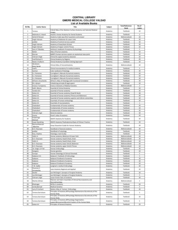
Clinical Anatomy RK Zargar, Sushil Kumar 8. Human Embryology Daksha Dixit 9. Manipal Manual of Anatomy Sampath Madhyastha 10. Exam-Oriented Anatomy Shoukat N Kazi 11. Anatomy and Physiology of Eye AK Khurana, Indu Khurana 12. Surface and Radiological Anatomy A. Halim 13. MCQ in Human Anatomy DK Chopade 14. Exam-Oriented Anatomy for Dental .
39 poddar Handbook of osteology Anatomy Textbook 10 40 Ross ,Pawlina Histology a text & atlas Anatomy Textbook 10 41 Halim A. Human anatomy Abdomen & lower limb Anatomy Referencebook 10 42 B.D. Chaurasia Human anatomy Head & Neck, Brain Anatomy Referencebook 10 43 Halim A. Human anatomy Head & Neck, Brain Anatomy Referencebook 10
Anatomy titles: Atlas of Anatomy (Gilroy) Anatomy for Dental Medicine (Baker) Anatomy: An Essential Textbook (Gilroy) Anatomy: Internal Organs (Schuenke) Anatomy: Head, Neck, and Neuroanatomy (Schuenke) General Anatomy and Musculoskeletal System (Schuenke) Fo
Sample MCQ’s for Nutrition and Meal Planning (20 - 21 batch only) Subject: Nutrition and Meal Planning Semester: III Class: SYBSc Instructions: 1. Attempt any 8 MCQ’s out of 10. 2. Each MCQ carries 1.5 marks 3. Read each statement carefully and mark any one option out of the four Options that you think is the correct answer 4.
MCQ 11.74 When sampling is done with or without replacement, is equal to: MCQ 11.75 If X represent the number of units having the specified characteristic and n is the size of the sample, then popula
AUTHORISATION TO TAKE Australian Medical Council MCQ EXAMINATION WITH PEARSON VUE You have been authorised to take a MCQ examination at a Pearson VUE testing center. Information on the MCQ Examination, the testing rules, and how to schedule your MCQ Examination follows: Awarding Body: Australian
HUMAN ANATOMY AND PHYSIOLOGY Anatomy: Anatomy is a branch of science in which deals with the internal organ structure is called Anatomy. The word “Anatomy” comes from the Greek word “ana” meaning “up” and “tome” meaning “a cutting”. Father of Anatomy is referred as “Andreas Vesalius”. Ph
Pearson Benjamin Cummings Anatomy and Physiology Integrated Anatomy – Gross anatomy, or macroscopic anatomy, examines large, visible structures Surface anatomy: exterior features Regional anatomy: body areas Systemic anatomy: groups of organs working


