Is Dominated By Trachaeophytes Vascular Plants - Cells .
isdominated by Trachaeophytes, or "Vascular Plants".The success of this group is no doubt related to the factthat, in contrast to bryophytes, trachaeophytes posessxylem and phloem. As you remember from previouslectures and lab, xylem - the water conducting tissue contains tracheary elements - cells that are dead atmaturity with thick and lignified secondary cell walls.This tissue is very durable and makes excellent fossils! Asa result, we know much about how early tracheophyteslooked, and it is even is possible to trace the evolution ofincreased complexity in body form through time including the origin of leaves, roots, arborescence, andthe seed.This week, we will examine some of this fossil evidence aswell as current day representatives commonly called:The Pteridophytes - or "Non-Seed Plants" is theterm we apply to vascular plants with more-or-lessprimitive reproduction involving extensive multicellulargametophyte and sporophyte generations. In contrast tobryophytes, pteridophtyes have a dominantsporophyte (diploid) generation producing spores, anda reduced gametophyte generation - either free-livingor dependent on the sporophyte for nutrition. Manypteridophytes have a rhizome. In some, the rhizome ishighly branched and the plant forms dense permanentstands by vegetative propagation and colonialgrowth.The time in lab will be divided between looking at fossilevidence of ancient pteridophytes and analysis of theliving forms. You will also visit the Greenhouse to findadditional examples of different groups. For each groupin our survey, the following suggestions apply:Observe each group in turn.Consult your textbook and lecture notes for adescription of diagnostic features of both externalform and internal anatomy.Study the life cycles of each group. Be sure youunderstand how sexual reproduction works in eachcase.Using fresh or prepared material, observe anddraw what you see.Consult your textbook for useful descriptions,terminology, and pictures. A guide to material in thelab is provided by the links below:
You will also be preparing your own thin-sections of fossilPrimitive Pteridophytes & Psilophytamaterial, called peels. For instructions see:LycophytaSphenophytaPterophytadownload this lab in PDF format
Paleobotany, the study of ancient plants from fossils,has been very useful in our understanding of theevolutionary history and relationships of plants. Givenscant coverage of the topic in typical paleontologydocumentaries on TV (that focus almost exclusively ondinosaur or human bones) few people realize just howmuch detail about ancient plants and their ecology canbe recovered from fossils.Of course, recovering this information requires athorough knowledge of both external morphology andinternal anatomy of plants. Given your work so far inBotany, you are in a good position to see what you cando! In class, you will make acid etch peels of fossil plantmaterial to observe evidence of pteridophytes based ontheir anatomy.As a sourvenir of the course, the peels you makein class are yours to keep!As you know, plants cells produce cellulose cell wallsthat in the case of sclerenchyma can by quite robustand thick. During fossilization, many specimens aresquashed flat forming compressions. In other cases,specimens are permineralized - cell lumina becomefilled with sediment or crystalline precipitate entombingoriginal cell walls within solid three-dimensional rock.Over millions of years, volatile organic molecules arelost from the cell walls, and what remains is mostlyrefractory carbon much like the carbon in coal.However, because the carbon remains in the position ofthe original cell walls in permineralizations, one cansometimes find highly faithful replicas of the originaltissue. These can be interpreted from transverse andlongitudinal section just as one would analyze living planttissues.The trick, of course, is to make thesesections from solid rock!You will be working with material from two differentoccurrences in time:Lower Devonian plants representing members ofthe Trimerophyta (see text) - approximately 386million years old.Obtain a specimen cut into a block. Identify asurface with interesting plant material fromwhich you wish to make a peel.Gently polish the chosen surface on a glass plateusing # 600 carborundum abrasive powder.Rinse.In the sink, etch the surface using 5%
Plants, including extinct lycopsids, horsetails,ferns, and seed plants associated with coal seamsfrom the Carboniferous period - approximately310 million years old.In both cases, the plants have been permineralized withcalcium carbonate (limestone). The key here is thatcalcium carbonate is soluble in acid whereas refractorycarbon is not. Check with your TA for a demonstrationon how to prepare peels. The following are generalinstructions:hydrochloric acid for 30 sec. - 1 min.Thoroughly dry the surface without touching itusing a hair drier.Cut a sheet of cellulose acetate to an appropriatesize.Using acetone as solvent, lay the peel on thesurface. Try as best you can to avoid trapping airbubbles. Allow it to dry.When ready (check with your TA), remove thepeel from the rock. Immediately, cut off excessacetate and press the peel in a book for a fewminutes to make sure it stays flat.Observe under the dissection microscope. Seewhat plant parts you can identify! Ask your TAfor help.
Primitive vascular plants from the Devonian period werevery different from those of today! For the most part,sporophytes probably existed as colonial plants with anextensively branched rhizome and multiple uprightaerial shoots. The shoots were leafless although theymay have been ornamented with epidermalemergences such as trichomes, scales or spines. Theshoots of the sporophytes also bore sporangia,depending on the group, either terminally at the tip ofthe main shoot or branches, or laterally from the mainaxis.A progression of forms are observed in the fossil recordof Trachaeophytes starting in the Lower (early)Devonian with small equally dichotomous forms such asCooksonia. See the model of the plant at the right. By theUpper (late) Devonian, we see the origin of moderntree-sized forms about which you will hear more nextweek.In lab, you will look at some fossil examples from thisremarkable Devonian Period. The fossils come from theBinghamton University Paleobotanical Collection - one ofthe finest collections of Devonian plant fossils in NorthAmerica.Please be extra careful when handling thespecimens! They are delicate and very rare!You will also examine a modern pteridophyte, ofuncertain affinities, with features that are highly
reminiscent of early fossil tracheophytes - perhaps arelict from this early evolutionary radiation.For each group, observe and draw what you see.Evidence suggests that rhyniophytes - or plants verysimilar to them - were the ancestral group of all vascularplants. They appeared during the Silurian Period(439-408my) and died out during the Devonian Period(408-360my). Sporophytes, such as represented by thegenus Aglaophyton, had naked dichotomous brancheswith terminal sporangia, and a centrarch protostele.Gametophytes appear to have been very similar to thesporophytes in morphology, bearing gametangia interminal splash cups - much like some modernliverworts. The best evidence of both sporophytes andgametophytes are permineralized specimens from theRhynie Chert - a justly famous fossil locality inScotland.This group probably evolved from rhyniophytes duringthe Lower Devonian and may have been the ancestors ofmore evolved groups including modern ferns,horsetails, and seed plants. The sporophytes oftrimerophytes were significantly larger than that ofrhyniophytes and exhibited anisotomous branching.Typically there is a main axis with smaller lateralbranches some of which have clusters of terminalsporangia. Researchers believe that these side brancheswere the major photosynthetic organs of the plant andmay ultimately have evolved into leaves. Internally,Trimerophytes had a larger, sometimes lobed protostele.Gametophytes are unknown.This group, consisting of two modern genera Psilotumand Tmesipteris, is very strange indeed! Psilotum lacksroots or leaves but has an underground rhizome andphotosynthetic stems that dichotomously branch.Sporangia are born laterally on distal branches insynangia - fused clusters with typically three sporangia.It is possible that synangia represent lateral branches likethat seen in Trimerophytes that through evolution havebecome fused and highly reduced.Internally, the stems of Psilotum have an exarchprotostele. Gametophytes look like reduced sporophytesand are subterranean. Recent molecular evidencesuggests that the Psilophyta may be related to ferns.However, in overall form of the sporophyte, Psilotumserves as an excellent visual representation of what earlyvascular plants may have looked like. Take a small sample
of Psilotum and make a transverse section, Identify thethe vascular tissues and draw what you see. Also, lookfor synangia on stems of this plant. If you find them,open the sporangia and look for spores.LycophytaSphenophytaPterophyta
Non-fern pteridophytes are sometimes referred to as"fern allies". Although common today in someenvironments, fern allies have only moderate diversity.The fossil record makes it clear, however, that they weremuch more diverse in the past and the modern formsrepresent but a very incomplete sample! In the past,pteridophytes included arborescent species thatprobably served as principal canopy trees in manyancient environments. Pteridophytes were also morediverse reproductively in the past, including severalimportant forms with heterosporous reproduction producing sporangia with two different spore sizes thatgerminate to produce gametophytes of separate sexes.This extinct fossil group from the Lower Devonian isthought to be the ancestors of the Lycophyta.Sporophytes of the group were leafless anddichotomously branched like the rhyniophytes, andpossessed proximal branches that may have functionedas roots. Unlike the rhyniophytes, the zosterophylls hadkidney-shaped & homosporous sporangia bornelaterally on the main axis sometimes in strobili.Internally the group had an exarch protostele.
Gametophytes are unknown.Observe and draw specimens of Sawdonia from theGaspe region of Quebec, available in lab. Note thepresence of stout spines in this plant. It is thought thatthe leaves of the Lycophyta may have evolved fromepidermal emergences called enations such as this.Lycopsids, also called "Club Mosses", appeared asfossils during the Devonian period, and some species alivetoday appear very little changed since that time! Thegroup became the dominant vegetation in the swamps ofthe Carboniferous Period (360-290 million years ago or 'my'), but most went extinct at the end of thePermian Period (290-245my). Some Carboniferousforms grew to the size of trees (up to 100ft in height) andthe decayed remains of these plants are a majorcomponent of coal worldwide.The sporophyte of lycopsids is the first in the fossil recordto have leaves - simple leaves with a single vein calledmicrophylls. Sporangia are located on the adaxial(upper) surface of some leaves. A leaf with asporangium is called a sporophyll . Primitive lycopsidshave equally dichotomous branching whereas derivedforms show anisotomous branching andanisophylly. Starting in the Devonian, two majorsubgroups of lycopsids may be recognized based onreproduction:Homosporous Lycopsids:This group produces only one size of spore - a conditioncalled homospory. Sporophylls usually occur in cone-likeclusters called strobili (singlular strobilus) often nearthe tips of branches. Listen to your TA for instructionsabout available material. On campus more than onespecies of Lycopodium can be found. They are delicateplants and increasingly rare.Observe them in the wild butPLEASE do not disturb!Heterosporous Lycopsids:Sporophytes of this group produce two sizes of spores:megaspores and microspores. Megaspores germinateto produce gametophytes that have only archegoniawhereas microspores produce gametophytes with onlyantheridia. Available in Lab is the genus Selaginella.Notice the overall form of the sporophyte withanisotomous branching. Look for sporophyll andsporangia. Draw what you see. Obtain a prepared slideof the strobilus of Selaginella. Look for sporangia withmegaspores and other sporangia with microspores. Draw
what you see.Another heterosporous hycopsid is the genus Isoetes.This plant is aquatic or semi-aquatic that has a corm(bulb-like flattened stem), with grass-like microphyllseach of which also serves as a sporophyll . Isoetes is, in asense, a living fossil - the last living representatives ofthe great tree-sized heterosporous lycopsids from theCarboniferous! Observe, and compare with the fossilreconstructions.Primitive Pteridophytes & PsilophytaSphenophytaPterophyta
This group, also called "Horsetails" appears in theUpper Devonian and some species also became tree-sizedduring the Carboniferous Period.Sporophytes of the horsetails typically consist of ahorizontal rhizome bearing upright aerial shoots. Themost distinctive character of the horsetails is that allplant organs - leaves, roots, and side shoots - arearranged in whorls. In modern Equisetum, leaves arevery small and fused into a ring-like sheath at eachnode. Observe available specimens for the leaf sheath.Draw what you see. Fossil sphenophytes had largerleaves that were free from each other. Observe and drawthe available examples.Internally, the anatomy of sphenopsids is unique.Vascular tissues are arranges in a ring of vascularbundles analogous, but not homologous, to a eustele inseed plants. In addition, there is an extensive airchannel system in both the pith and cortical regions ofthe stem. Using a sharp razorblade, make a transversesection of Equisetum and observe under both thedissection and compound microscope. Look for anddraw vascular bundles, hollow pith, air canals, and highlysclerified cortex. The outer surface of horsetail stems isoften impregnated with silica (glass), earning the groupanother name: "Scouring Rushes" - in honor of thefact that early settlers used the plant to help wash theirpots & pans.Observe and draw available material of reproductiveportions of the sporophyte. Horsetails have largesporangia borne in clusters on an unbrella-shapedorgan called the sporangiophore ("sporangiumbearer"). Multiple sporangiophores are borne togetherin strobili (singular strobilus). The sporophyte ishomosporous. Within each sporangium, each spore hasfour elators - specialized portions of the outer wall thatmostly detaches and uncoils.
The gametophyte of Equisetum is small andsubterranean. Observe available material and draw whatyou see.Primitive Pteridophytes & PsilophytaLycophytaPterophyta
The Pterophyta or "Ferns" also had their origin in theDevonian Period. Today they are a very successful anddiverse group occupying a wide range of terrestrial andaquatic habitats. The sporophyte typically consists of arhizome or upright stem bearing large compoundleaves. Leaves develop by means of a process calledcircinate vernation - the immature leaf, looking likeand called a fiddlehead, uncoiling as the leaf expands.The compound leaf of a fern has a specializedterminology for the different parts including:petiole - portion below leaf laminarachis - the main axis of the leaf above the petiolepinnae (singular pinna) - subdivisions of leafpinnules - ultimate laminate leafletsObserve the morphology of the compound leaf offerns in both the Greenhouse and Lab. In each case,determine whether the leaf is once, twice, thrice (ormore) compound. Be sure you can identify eachportion of the leaf named above. Draw and label whatyou see. Next look for the main stem often clothed inscales or stout hairs. Look for evidence of circinatevernation and draw what you see.Sporangia are usually borne on the abaxial surface ofthe leaf lamina in clusters called sori (singular sorus).In many species the sorus is partly or completelycovered by sterile tissue called the indusium. Presenceor absense and shape of sori are important charactersused for identification of ferns especially at the level ofgenus. Look for sporangia in several of the livingexamples. Note differences in location of sori andnumber of sporangia borne in each. Draw what you seeincluding shape of indusium if present. Now consult akey for identifying ferns available in lab. See if you canidentify your specimen to genus.
Internally, ferns posess a siphonostele (or morecomplex stelar types derived from siphonosteles) withleaf gaps. Typical transverse sections commonly showC-shaped primary xylem and phloem of the main stemand inverted C-shaped leaf traces. Observe availablematerial of ferns stems and petioles in transversesection. Identify all tissue regions including cortex, pith,primary xylem, primary phloem, and endodermisif present. Identify the leaf gap. Draw what you see.The gametophyte in ferns usually consists of a smallfree-living photosynthetic plant body inhabiting thesurface of damp soil. Observe available examples in Lab.Notice the overall shape of the gametophyte - also calledprothallus - including photosynthetic lamina,apical notch, and rhizoids. The gametophytesproduce archegonia and antheridia, on the lowersurface. Using available live and prepared material, lookfor gametangia. Sporophytes of the next generationdevelop from embryos directly within archegonia. Lookfor evidence of them in either live or prepared material.Compare with figures in your text. Draw and label whatyou see.Primitive Pteridophytes & PsilophytaLycophytaSphenophyta
evidence of ancient pteridophytes and analysis of the living forms. You will also visit the Greenhouse to find additional examples of different groups. For each group in our survey, the following suggestions apply: Observe each group in turn. Consult your textbook and lecture notes
C: 8 MHz Vascular Probe Detection of peripheral vessels and calcified arteries D: EZ8 8 MHz Vascular Probe Easy location of vessels and maintain during cuff inflation or deflation E: 10 MHz Vascular Probe Detection of smaller vessels *Beam shape and depth for illustrative purposes only. 8 MHz Vascular Probe *EZ8 8 MHz Vascular Probe All XS High
development, angiogenesis and vascular permeability [10]. Pericyte dysfunction is observed in vascular diseases such as stroke but also pericyte-induced angiogenesis is an important factor in tumor development and growth [11-13]. Abnormal vascular formation can be observed in many congenital syndromes.
EXPLANATION OF THE CURRICULUM OUTLINE . The SCORE Curriculum Outline for Vascular Surgery (VSCORE) is a list of vascular and core surgery patient care topics to be covered in a vascular surgery training program. The curriculum includes vascular surgical diseases/conditions and operations/procedures and core surgical medical knowledge
Accurate segmentation and quanti cation of vascular struc-tures in medical images is a critical task for clinical surgical planning, and navigation. However, it is highly challenging to extract vascular structures in D and D medical images. e reasons lie in two aspects. On one hand, some vascular
Topic 22. Introduction to Vascular Plants: The Lycophytes Introduction to Vascular Plants Other than liverworts, hornworts, and mosses, all plants have vascular tissues. As discussed earlier, the mosses have cells that serve to conduct water and photosynthate, and these cell
20. List examples of: vascular plants. 21. Why are they called vascular plants? 22. During alternation of generation of vascular plants is the gametophyte or sporophyte predominant? 23. Define waxy cuticle and stomate. 24. Vascular plants are divided into which two main groups? 25. List some examples of
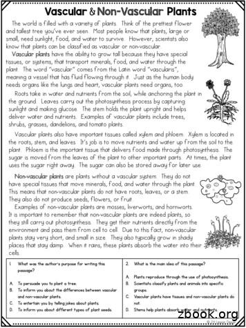
A. Plants reproduce through the use of photosynthesis. B. Scientists classify plants and animals into specific groups. C. Vascular plants have tissues and non-vascular plants do not. D. Stems help plants absorb water and nutrients. Directions: Using the passage
ISO 14001:2015 Standard Overview Understand the environmental management system standard and how to apply the framework in your business. An effective environmental management system takes more than a single software solution or achieving a certificate for the wall. It takes time, energy, commitment and investment. Qualsys’ software and solutions provide your entire organisation with the .


