Biology STAAR EOC Review
Biology STAAR EOC Review(Adapted from Alief ISD Review)Reporting Category 1: Cell Structure and FunctionSTAAR -11 Questions STAAR M-9 Questions4 Readiness Stds5 Supporting StdsBiology is the study of life and living organisms.An organism is a complete, individual, living thing.All organisms are formed from the same basic building block – cells.TEKS(RS)- will betested (65%)(SS)- may betested (35%)Key IdeasCells are the smallest units of living thingsSimple cells are called prokaryotic; Complex cells are called eukaryoticCell Parts or OrganellesCell membraneCytoplasmNucleusMitochondriaEndoplasmic reticulum(smooth or rough)RibosomeGolgi body or complexLysosomevacuoleSurrounds the cell; controls what enters/leaves the cell; recognizes othercells; maintains homeostasisSuspends organelles in a eukaryotic cell; enclosed within the cell membraneControls the cell’s activities; contains chromosomes made of DNABreaks down food to release energyMoves substances within the cell (pipe-like structures)Makes proteins; round structures located in rough endoplasmic reticulumChanges and packages cell productsContains enzymes (proteins that speed up digestion and chemical reactions)Holds material like water; large in a plant cellPlant cells onlyCell wallchloroplastSurrounds the cell membrane; supports and protects plant cellContains chlorophyll (green pigment) for photosynthesis1
Cellular ProcessesI. Homeostasis is a process by which organisms keep internal conditions relatively stable regardless of changes in4BInvestigate andexplain rsions,transport ofmolecules, andsynthesis ofnew materials(RS)the external environment. It is important because the processes that keep the cell alive can only take place undercertain internal conditions. Balanced internal condition of cells Homeostasis is also called equilibrium Maintained by plasma membrane controlling what enters & leaves the cellPlasma or Cell MembraneThe cell membrane is flexible and allows a unicellular organism to moveWhen you transport something, you move it from one place to another. Cells transport materials across the cellmembrane.Functions (what they do) of Plasma or Cell Membrane Protective barrierRegulate transport in & out of cell (selectively permeable- only lets some things and out of the cell like aclub bouncer; Specifically, small molecules and larger hydrophobic molecules move through easily. e.g.O2, CO2, H2O; Ions, hydrophilic molecules larger than water, and large molecules such as proteins do notmove through the membrane on their own.Allow cell recognitionProvide anchoring sites for filaments of cytoskeletonProvide a binding site for enzymesInterlocking surfaces bind cells together (junctions)Contains the cytoplasm (fluid in cell)Structure of the Cell Membrane2
Cell Membrane Polar heads are hydrophilic “water loving”Nonpolar tails are hydrophobic “water fearing”Makes membrane “Selective” in what crosses - “Selectively permeable”II. Types of Transport Across Cell MembranesSimple DiffusionRequires NO energy; Molecules move from area of HIGH to LOW concentrationDIFFUSIONDiffusion is a PASSIVE process which means no energy is used to make the molecules move,they have a natural KINETIC ENERGYEx: Diffusion of LiquidsDiffusion through a MembraneSolute moves DOWN concentration gradient (HIGH to LOW)3
Osmosis- Diffusion of water across a membraneMoves from HIGH water potential (low solute) to LOW water potential (high solute)Diffusion of H2O Across A MembraneCells in Different Solutions (Think of a pimple)Isotonic (Balance flow)NO NET MOVEMENT OF H2O(equal amounts entering & leaving)Hypotonic SolutionCYTOLYSIS (Cell Swells)Hypertonic SolutionPLASMOLYSIS (Cell ---------------4
III. Energy ConversionsThree Forms of Transport Across the MembraneThree Forms of Transport Across the Membrane35A. Passive Diffusion- Simple DiffusionDoesn’t require energyMoves high to low concentrationExample: Oxygen or water diffusing into a cell and carbon dioxide diffusing out.B. Facilitated diffusionDoesn’t require energyUses transport proteins to move high to low concentrationExamples: Glucose or amino acids moving from blood into a cell.C. Active TransportRequires energy or ATPMoves materials from LOW to HIGH concentrationAGAINST concentration gradientExamples: Pumping Na (sodium ions) out and K (potassium ions) in against strong concentration gradients.Called Na -K -------------------------------------Proteins Are Critical to Membrane FunctionTypes of Transport Proteins Channel proteins are embedded in the cell membrane & have a pore for materials to cross5
Carrier proteins can change shape to move material from one side of the membrane to theotherFacilitated DiffusionMolecules will randomly move through the pores in Channel Proteins. Some Carrier proteins do not extend through the membrane.They bond and drag molecules through the lipid bilayer and release them on the opposite ---------------------------------------Carrier Proteins Other carrier proteins change shape to move materials across the cell is - moving things out of the cell Molecules are moved out of the cell by vesicles that fuse with the plasma membrane.This is how many hormones are secreted and how nerve cells communicate with one anotherMoving the “Big Stuff” Out in the Cell6
Endocytosis- Large molecules move materials into the cell by one of three forms of endocytosis.Moving the “Big Stuff” in the cell3 Types of EndocytosisA. Pinocytosis- Most common form of endocytosis; Takes in dissolved molecules as a vesicleCell form an invagination*Materials dissolve in water to be brought into cellB. Phagocytosis-Used to engulf large particles such as food, bacteria, etc. into vesicles;Called “Cell Drinking”Phagocytosis about to occurC. Receptor-Mediated EndocytosisSome integral proteins have receptors on their surface to recognize & take in hormones, cholesterol, etc.7
Homeostasis Energy Conversions MoleculetransportationSummary of Cellular ProcessesRegulation of conditions (like pH or temperature) within a cell whichallows for stable, “normal” internal equilibrium (balance)During photosynthesis, plant cells use energy from the sun to makesugar called glucose; during aerobic cellular respiration, mitochondriarelease energy from molecules like glucoseMolecules move in and out of cells across the cell membrane byvarious means; active transport (like transport proteins) requiresenergy, but passive energy (like diffusion) does notCells can create new molecule from simpler molecules, like whenproteins are made from amino acids 4CCompare thestructures ofviruses to cells,describe viralreproduction,and describethe role ofviruses incausingdiseases suchas humanimmunodeficiency virus (HIV)and influenza(RS)Synthesis of NewMoleculesStructure of VirusesVirus StructureCapsidDNAor RNASPIKESENVELOPEOutside View of a VirusCAPSOMERESViruses and CellsCharacteristicStructureVirusDNA or RNA in capsid, some with envelopeReproductionOnly within a host cellGenetic CodeGrowth and DevelopmentDNA or RNANoObtain and use energyResponse to EnvironmentChange over timeNoNoYes CellCell membrane, cytoplasm,eukaryotes also contain nucleus andmany organellesIndependent cell division, eitherasexually or sexuallyDNAYes; in multicellular organisms, cellsincrease in number and differentiateYes—Eukaryotic ellYesYesA virus is a nonliving particle made up of proteins, nucleic acids, and sometimes lipids.8
Viruses can reproduce only by infecting living cells. A virus consists of a core of DNA or RNAsurrounded by a protein coat called a capsid.Unlike a cell, a virus lacks structures to take in food, break apart food for energy, or synthesize molecules.Because viruses are noncellular and cannot perform most functions of life, scientists classify viruses asnonliving particles. However, viruses are able to perform one life function- reproduction- with the aid ofa host organism.Hosts can be prokaryotes or eukaryotes.Viruses that use prokaryotes as host cells are called bacteriophages or phages.Viral ReproductionViruses reproduce by taking over the host cell. The process begins when a virus attaches to the outside of a cell. Thevirus then injects its genetic material into the cell. After the viral genetic material enters a host cell, one of twoprocesses may occur.A. Steps of Lytic Cycle and Lysogenic cycleStages of the Lytic CycleThe host cell starts making messenger RNA from the viralDNA.Stages of the Lysogenic CycleRole of Viruses in Causing Diseases Such as HIV and InfluenzaViruses can cause disease in humans through the lytic cycle such as HIV and Influenza.HIVandInfluenzaHIVInfluenza or the “flu”9
Human immunodeficiency Virus or HIVCauses AIDS or Acquired immunodeficiency Syndrome;currently there is no cure for AIDS because HIV mutatesand evolves rapidly;HIV infects and destroys immune system cells called helperT cells; Helper T cells play a role in keeping the body freefrom diseaseWhen HIV attacks a helper T cell, it binds to the cellmembrane and enters the cell. Once the virus is inside thecell, it uses the cell’s structures to make new viruses. Thenthe virus destroys the cell and the new viruses arereleased into the bloodstream. They travel throughout theblood, infecting and destroying other helper T cells.As an HIV infection progresses, more helper T cells aredestroyed. Doctors determine the number of helper Tcells in the blood of people with HIV infections to monitorhow far their infections have progressed. The fewer the Tcells in the blood, the more advanced the infection.As the immune system becomes increasingly compromisedby HIV, the body becomes more susceptible to diseasesthat seldom show up in people with a healthy immunesystem; People who have AIDS die from other diseasesbecause their immune system is too weak to fight offinfections (opportunistic diseases)5ADescribe thestages of thecell cycle,includingdeoxyribonucleic acidreplication andmitosis, andthe importanceof cell cycle tothe growth ofthe organism(ReadinessStandard)Influenza or the fluRNA virusInfects the respiratory tract of humans as well as otheranimalsThe death of the infected cells and a person’s immunesystem response causes inflammation which leads to sorethroat and mucus secretionsInfection causes mild to severe illness, including fever,cough, headache, and a general feeling of tirednessInfection lasts for 1 to 2 weeks and can cause a moresevere illnessThe Cell Cycle: Stages in growth & divisionG1 PhaseS PhaseG2 PhaseM Phase(Mitosis)CytokinesisFirst growth stageCopying of all ofDNA’s instructionsTime betweenDNA synthesis &mitosisCell growth & proteinproduction stop(Cell plate forming betweenthe two cells)Occurs after chromosomesseparateCell increases insizeChromosomesduplicatedCell continuesgrowingCell’s energy used tomake 2 daughter cellsForms two, identical daughtercellsNeeded proteinsproducedCalled mitosis orkaryokinesis(nuclear division)Cell prepares tocopy its DNA10
I. DNA ReplicationA process that transforms one DNA molecule into 2 identical copies; enzyme help DNA strands unwind and separates;each DNA strand serves as a template (pattern) for a new, complementary strand to form by matching (pairing)nitrogen bases. As a result, each new DNA molecule contains half of the original molecule Replication Facts Synthesis Phase(S phase)DNA Replication(in a nut shell)DNA has to be copied before a cell dividesDNA is copied during the S or synthesis phase ofinterphaseNew cells will need identical DNA strandsS phase during interphase of the cell cycleOccurs in the Nucleus of eukaryotesBegins at Origins of ReplicationTwo strands open forming Replication Forks (Y-shaped region)New strands grow at the forksthe process used bycells to copy DNA –enzyme unzips DNAand each side of theladder acts as atemplate for thebuilding of the newhalf. Use the N-baseparing rules : A-T ; C-GExample)TACGGAC (old strand)ATGCCTG (new strandAs the 2 DNA strands open at the origin, Replication Bubbles form Prokaryotes (bacteria) have a single bubble Eukaryotic chromosomes have MANY bubbles11
Enzyme Helicase unwinds and separates the 2 DNA strands by breaking the weakhydrogen bonds Single-Strand Binding Proteins attach and keep the 2 DNA strands separatedand untwistedEnzyme Topoisomerase attaches to the 2 forks of the bubble to relieve stress on the DNAmolecule as it separatesBefore new DNA strands can form, there must be RNA primers present to start theaddition of new nucleotides;Primase is the enzyme that synthesizes the RNA Primer;DNA polymerase can then add the new nucleotidesDNA polymerase can only add nucleotides to the 3’ end of the DNAThis causes the NEW strand to be built in a 5’ to 3’ directionDirection of ReplicationDNA replication:The Leading Strand is synthesized as a single strand from the point of origin toward the12
Synthesis of the NewDNA Strandsopening replication forkThe Lagging Strand is synthesized discontinuously against overall direction of replicationThis strand is made in MANY short segments.It is replicated from the replication fork toward the originLagging StrandSegmentsOkazaki Fragments - series of short segments on the lagging strandMust be joined together by an enzymeJoining of OkazakiFragmentsThe enzyme Ligase joins the Okazaki fragments together to make one strandReplication of StrandsSemiconservativeModel of Replication(Watson & Crick)Replication ForkPoint of OriginThe two strands of the parental molecule separate, and each acts as a template for a newcomplementary strandNew DNA consists of 1 PARENTAL (original) and 1 NEW strand of DNA13
Summarize DNA Replication- Explain what happens using the diagram below: II. Mitosis- Life Cycle of a CellMitosis is a cycle with no beginning or end;Mitosis creates diploid cells and is for the purpose of tissue repair and growthin animals] DNA coils to form chromosomes during cell divisionInterphase – Resting Stage Cells carrying on normalactivitiesChromosomes aren’tvisibleCell metabolism isoccurringOccurs before mitosis14
Stages of Mitosis lls Undergoing Mitosis DNA coils tightly & becomes visible as chromosomesNuclear membrane disappearsNuceolus disappearsCentrioles migrate to polesSpindle begins to formSpindle fibers from centrioles attach to each chromosomeCell preparing to separate its chromosomesCell aligns its chromosomes in the middle of the cellMetaphase Anaphase Cell chromosomes are separated Spindle fibers shorten so chromosomes pulled to ends of cellTelophase 15Separation of chromosomes completedCell Plate forms (plants)Cleavage furrow forms(animals)Nucleus & nucleolus reform
Chromosomes uncoilCytokinesis(Cell plate forming betweenthe two cells) Occurs after chromosomes separate Forms two, identical daughter cellsIII. Importance of the cell cycle to the growth of the organism9ACompare thestructures andfunctions ofdifferent typesofbiomolecules,includingcarbohydrates,lipids, proteins,and nucleicacids(ReadinessStandard)Types of BiomoleculesExamples of BiomoleculesProteinsLipidsCarbohydratesNucleic AcidsCopyright Cmassengale1615
BiomoleculeStructureFunctionProteinContains carbon (C), nitrogen (N),oxygen (O), hydrogen (H), andpossibly sulfur (S) atoms; made ofamino acids; large and complexUsed to build cells;(enzyme, hormone)Structural molecule (like keratinin fingernails); enzyme, hormone,transport molecule (likehemoglobin in blood);contractionsExample: NutsExample: MuscleLipids(fats, steroid, wax, oil, fatty acid)Example: CriscoExample: oilsCarbohydratesContains carbon (C), nitrogen (N),and hydrogen (H) atoms; ratio ofhydrogen to oxygen atoms is 2:1(Sugar, starch)Example: Sugar in CokeExample: Starch in pastaNucleic Acids (DNA & RNA)Contains a carbohydrate (sugar)group, phosphate group (PO4-3),and a nitrogen base (adenine,thymine (in DNA only) or uracil(in RNA only), cytosine, andguanine; very large and complexNucleotidePhosphateGroupOO P-OO5CH2ONC1C4Sugar(deoxyribose)C3copyright cmassengaleC2Contains carbon(C), oxygen (O),hydrogen (H), and possibly otheratoms: ratio of hydrogen (H)atoms to oxygen (O) atoms ishigh; insoluble (does not dissolve)in waterSource of energy; cell membranecomponent; protective coating(like wax); chemical messenger(like cholesterol)Example: butterSource of energy (like glucose);structural molecule (cellulose)Ex: Orange (fruits are carbohydratesbecause they have sugar)Carrier of genetic informationand instructions of proteinsynthesisNitrogenous base(A, G, C, or T)29Cells Use ATP from Biochemical energy for: Active transport, Movement, Photosynthesis, Protein Synthesis, Cellularrespiration, and all other cellular reactions17
Structure of BiomoleculesCARBOHYDRATE(Sugar – Glucose)4ACompare andcontrastprokaryoticand eukaryoticcells(SupportingStandard)PROTEIN(One Amino Acid)LIPID (Fat)NUCLEIC ACID(One Nucleotide)DIFFERENCES IN CELLSCells can be grouped according to their similarities and differences. All cells can be divided into twocategories – prokaryotes and eukaryotes.Prokaryotic CellEukaryotic Celllacks a true nucleusdoes not have membrane bound organellesDNA in a prokaryote is a single circular moleculehave no mitochondria, chloroplasts, Golgi bodies,lysosomes, vacuoles, or endoplasmic reticulumhave a cell wall and a cell membraneExample: bacteria cell (below) and blue green algaeHas a well-defined nucleus surrounded by a nuclearmembraneDNA is in the form of complex chromosomesMore complexcells are found in plants, animals, fungi, and protists.Example:Animal cellPlant cellEukaryotic cells also differ between plants and animals. Plant cells contain three structures not found inanimal cells – cell walls, large central vacuoles, and plastids. Centrioles are found in some, but not all types ofplant cells. They are found in all animal cells.18
SummaryCharacteristicProkaryote(Bacteria and Blue green algae)YesYesCell membraneEukaryote(Plant and animal esNucleusOrganellesSpecialized CellsDNA holds the genetic information that controls what the cell can do and what molecules it makes.Ex: White blood cells in animals are specialized to attack pathogens (disease causing agents) like viruses or bacteria5BExaminespecializedcells, includingroots, stems,and leaves ofplants; andanimal cellssuch as blood,muscle, andepithelium (SS)Plant CellsPlant partExamples of specialized plant cells and functionsLeafCells containing chloroplasts (green coloring) for photosynthesiGuard cells control size of stomates (pores) allowing gas transfeGuard CellStemXylem cells move water and minerals;Phloem cells move nutrients like glucose throughout the plantusing pipe like structures (this provides support for leaves,branches, and flowers)Xylem CellRootEpidermis cells on root hairs increase surface area to allowfor the absorption of water and mineral nutrients19
Animal Cells Muscle cells are individual cells that comprise themuscle tissue of the body and executemuscle contraction. There are three types of muscle cells: skeletal, cardiac,and smooth. Each of these types differ in cellularstructure, specific function, and location within the body. Together, the three muscle cell types play specific rolesin supporting the skeletal structure and posture of thebody, assisting in the flow of blood through bloodvessels, aiding in digestion, and driving the heartbeat.Mammals have 3 types of blood
When HIV attacks a helper T cell, it binds to the cell membrane and enters the cell. Once the virus is inside the cell, it uses the cell’s structures to make new viruses. Then the virus destroys the cell and the new viruses are released into the bloodstream. They travel throughout the blood, infecting and destroying other helper T cells.
Algebra 1 EOC, Geometry EOC, Biology EOC, US HistoryEOC NGSSS. 2013-2014; FCAT 2.0 NGSSS. Civics EOC NGSSS; FCAT 2.0 NGSSS. Algebra 1 EOC, Geometry EOC, Biology EOC, US HistoryEOC NGSSS; 2014-2015. PARCC CCSS (M ELA) FCAT 2.0 Science. Civics EOC NGSSS. PARCC CCSS (ELA) Algebra 1 EOC, Geometry EOC CCSS; Biology EOC, US .
THE STAAR MASTER DIFFERENCE RIGOROUS DESIGN STAAR MASTER is the most rigorous STAAR curriculum on the market today . STAAR MASTER content carefully exceeds the level of difficulty of the STAAR to ensure success . STANDARDS ALIGNED STAAR MASTER materials are 100% alig
animation, biology articles, biology ask your doubts, biology at a glance, biology basics, biology books, biology books for pmt, biology botany, biology branches, biology by campbell, biology class 11th, biology coaching, biology coaching in delhi, biology concepts, biology diagrams, biology
Biology EOC Review. 14 Biology EOC Review Work with your neighbors to answer questions at the end of Biology EOC Study Guide: Part 2, Cell Biology. .
CHANGES TO STAAR IN 2017 Only one form of test—paper or online (no more STAAR A or STAAR L) . may need significant support next year. Phase-in Level II: Satisfactory Approaches Grade Level . Grade 5 Grade 8 Biology Hays CISD STAAR Science Scores 2015 2016 2017. STAAR SOCIAL STUDIES 57%
Biology EOC Study Guide . This Study Guide was developed by Volusia County teachers to help our students prepare for the Florida Biology End-Of-Course Exam. Molecular and Cell Biology Classification, Heredity, Evolution Organisms, Populations, Ecosystems 35% of EOC 25% of EOC 40% of EOC The Nature of Science Theories, Laws, Models
Biology EOC Test Strategies to Use the Morning of the Biology EOC Test Strategies to Use During the Biology EOC Test - Review what you have learned from the study guide. - Review general test -taking strategies. - Review content -specific information that shows connections and relationships (lists, diagrams, graphic organizers, etc.).
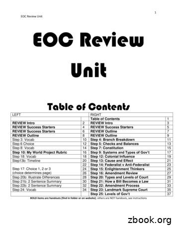
1 EOC Review Unit EOC Review Unit Table of Contents LEFT RIGHT Table of Contents 1 REVIEW Intro 2 REVIEW Intro 3 REVIEW Success Starters 4 REVIEW Success Starters 5 REVIEW Success Starters 6 REVIEW Outline 7 REVIEW Outline 8 REVIEW Outline 9 Step 3: Vocab 10 Step 4: Branch Breakdown 11 Step 6 Choice 12 Step 5: Checks and Balances 13 Step 8: Vocab 14 Step 7: Constitution 15


