MS Cell Division, Reproduction, And Protein Synthesis
MS Cell Division,Reproduction, and ProteinSynthesisJean Brainard, Ph.D.Say Thanks to the AuthorsClick http://www.ck12.org/saythanks(No sign in required)
To access a customizable version of this book, as well as otherinteractive content, visit www.ck12.orgCK-12 Foundation is a non-profit organization with a missionto reduce the cost of textbook materials for the K-12 marketboth in the U.S. and worldwide. Using an open-content, webbased collaborative model termed the FlexBook textbook, CK-12intends to pioneer the generation and distribution of high-qualityeducational content that will serve both as core text as well asprovide an adaptive environment for learning, powered throughthe FlexBook Platform .Copyright 2015 CK-12 Foundation, www.ck12.orgThe names “CK-12” and “CK12” and associated logos and theterms “FlexBook ” and “FlexBook Platform ” (collectively“CK-12 Marks”) are trademarks and service marks of CK-12Foundation and are protected by federal, state, and internationallaws.Any form of reproduction of this book in any format or medium,in whole or in sections must include the referral attribution linkhttp://www.ck12.org/saythanks (placed in a visible location) inaddition to the following terms.Except as otherwise noted, all CK-12 Content (including CK-12Curriculum Material) is made available to Users in accordancewith the Creative Commons Attribution-Non-Commercial 3.0Unported (CC BY-NC 3.0) License (http://creativecommons.org/licenses/by-nc/3.0/), as amended and updated by Creative Commons from time to time (the “CC License”), which is incorporatedherein by this reference.Complete terms can be found at http://www.ck12.org/about/terms-of-use.Printed: March 8, 2015AUTHORJean Brainard, Ph.D.EDITORDouglas Wilkin, Ph.D.
www.ck12.orgChapter 1. MS Cell Division, Reproduction, and Protein SynthesisC HAPTER1MS Cell Division,Reproduction, and Protein SynthesisC HAPTER O UTLINE1.1Cell Division1.2Reproduction1.3Protein Synthesis1.4References1
www.ck12.orgThis baby boy is just a few days old, but his body already consists of billions of cells. By the time he’s as big as hisfather, his body will contain trillions of cells. Like all other organisms, the baby actually started out in life as a singlecell. How do we develop from a single cell into an organism with trillions of cells? The answer is cell division.2
www.ck12.orgChapter 1. MS Cell Division, Reproduction, and Protein Synthesis1.1 Cell DivisionLesson Objectives Outline the process of DNA replication.Compare and contrast cell division in prokaryotic and eukaryotic cells.Describe the four phases of mitosis in eukaryotic cells.Identify the stages of the cell cycle in prokaryotic and eukaryotic cells.Lesson Vocabulary anaphasebinary fissioncell cyclecell divisionchromosomecytokinesisDNA (deoxyribonucleic acid)DNA haseIntroductionCell division is the process in which a cell divides to form two new cells. The original cell is called the parent cell.The two new cells are called daughter cells. All cells contain DNA. DNA is the nucleic acid that stores geneticinformation. Before a cell divides its DNA must be copied. That way, each daughter cell gets a complete copy ofthe parent cell’s genetic material.Copying DNADNA stands for deoxyribonucleic acid. It is a very large molecule. It consists of two strands of smaller moleculescalled nucleotides. Before learning how DNA is copied, it’s a good idea to review its structure.3
1.1. Cell Divisionwww.ck12.orgDNA StructureAs you can see in Figure 1.1, each nucleotide includes a sugar, a phosphate, and a nitrogen base. The sugar in DNAis called deoxyribose. There are four different nitrogen bases in DNA: adenine (A), thymine (T), cytosine (C), andguanine (G). Chemical bonds between the bases hold the two strands of DNA together. Adenine always bonds withthymine, and cytosine always bonds with guanine. These pairs of bases are called complementary base pairs.FIGURE 1.1Structure of DNAChromosomesAs a cell prepares to divide, its DNA first forms one or more structures called chromosomes. A chromosomeconsists of DNA and protein molecules coiled into a definite shape. Chromosomes are circular in prokaryotes androdlike in eukaryotes. You can see an example of a human chromosome in Figure below. The rest of the time, DNAlooks like a tangled mass of strings. In this form, it would be very difficult to copy and divide.DNA ReplicationThe process in which DNA is copied is called DNA replication. You can see how it happens in Figure 1.3. Anenzyme breaks the bonds between the two DNA strands. Another enzyme pairs new, complementary nucleotideswith those in the original chains. Two daughter DNA molecules form. Each contains one new chain and one originalchain.4
www.ck12.orgChapter 1. MS Cell Division, Reproduction, and Protein SynthesisFIGURE 1.2Human chromosome5
1.1. Cell Divisionwww.ck12.orgFIGURE 1.3DNA replicationCell Division in Prokaryotic and Eukaryotic CellsHow cell division proceeds depends on whether a cell has a nucleus. Prokaryotic cells lack a nucleus. Their DNA isin the cytoplasm. It forms just one circular chromosome. Eukaryotic cells have a nucleus holding their DNA. TheirDNA forms multiple rodlike chromosomes, like the one in Figure 5.2. Eukaryotic cells also have other organelles.For these reasons, cell division is more complex in eukaryotic cells.Prokaryotic Cell DivisionYou can see how a prokaryotic cell divides in Figure 1.4. This type of cell division is called binary fission. The cellsimply splits into two equal halves. Binary fission occurs in bacteria and other prokaryotes. It takes place in threecontinuous steps:1. The cell’s chromosome is copied to form two identical chromosomes. This is DNA replication.2. The copies of the chromosome separate from each other. They move to opposite poles, or ends, of the cell.This is called chromosome segregation.6
www.ck12.orgChapter 1. MS Cell Division, Reproduction, and Protein Synthesis3. The cell wall grows toward the center of the cell. The cytoplasm splits apart, and the cell pinches in two. Thisis called cytokinesis.FIGURE 1.4Binary fission in a prokaryotic cellEukaryotic Cell DivisionBefore a eukaryotic cell divides, the nucleus and other organelles must be copied. Only then will each daughter cellhave all the needed structures.1. The first step in eukaryotic cell division, as it is in prokaryotic cell division, is DNA replication. As you can seein Figure 1.5, each chromosome then consists of two identical copies. The two copies are called sister chromatids.They are attached to each other at a point called the centromere.2. The second step in eukaryotic cell division is division of the cell’s nucleus. This includes division of thechromosomes. This step is called mitosis. It is a complex process that occurs in four phases. The phases ofmitosis are described below.3. The third step is the division of the rest of the cell. This is called cytokinesis, as it is in a prokaryotic cell. Duringthis step, the cytoplasm divides, and two daughter cells form.These three steps are shown in Figure 1.6.MitosisMitosis, or division of the nucleus, occurs only in eukaryotic cells. By the time mitosis occurs, the cell’s DNA hasalready replicated. Mitosis occurs in four phases, called prophase, metaphase, anaphase, and telophase. You can seewhat happens in each phase in Figure below. The phases are described below. You can also learn more about thephases of mitosis by watching this video: https://www.youtube.com/watch?v gwcwSZIfKlM .7
1.1. Cell Divisionwww.ck12.orgFIGURE 1.5FIGURE 1.6Cell division in a eukaryotic cellMEDIAClick image to the left or use the URL below.URL: 168
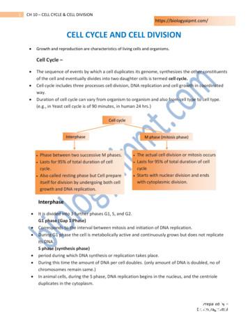
www.ck12.orgChapter 1. MS Cell Division, Reproduction, and Protein SynthesisPhases of mitosis1. Prophase: Chromosomes form, and the nuclear membrane breaks down. In animal cells, the centrioles nearthe nucleus move to opposite poles of the cell. Fibers called spindles form between the centrioles.2. Metaphase: Spindle fibers attach to the centromeres of the sister chromatids. The sister chromatids line up atthe center of the cell.3. Anaphase: Spindle fibers shorten, pulling the sister chromatids toward the opposite poles of the cell. Thisgives each pole a complete set of chromosomes.4. Telophase: The chromosomes uncoil, and the spindle fibers break down. New nuclear membranes form.The Cell CycleCell division is just one of the stages that a cell goes through during its lifetime. All of the stages that a cell goesthrough make up the cell cycle.Prokaryotic Cell CycleThe cell cycle of a prokaryotic cell is simple. The cell grows in size, its DNA replicates, and the cell divides.Eukaryotic Cell CycleIn eukaryotes, the cell cycle is more complicated. The diagram in Figure 1.7 shows the stages that a eukaryotic cellgoes through in its lifetime. There are two main stages: interphase and mitotic phase. They are described below.You can watch a eukaryotic cell going through the phases of the cell cycle at this link: http://www.cellsalive.com/cell cycle.htmFIGURE 1.7Eukaryotic cell cycleInterphase is longer than mitotic phase. Interphase, in turn, is divided into three phases:9
1.1. Cell Divisionwww.ck12.org1. Growth phase 1 (G1): The cell grows rapidly. It also carries out basic cell functions. It makes proteins neededfor DNA replication and copies some of its organelles. A cell usually spends most of its lifetime in this phase.2. Synthesis phase (S): The cell copies its DNA. This is DNA replication.3. Growth phase 2 (G2): The cell gets ready to divide. It makes more proteins and copies the rest of its organelles.Mitotic phase is when the cell divides. It includes mitosis (M) and cytokinesis (C).Lesson Summary DNA is the nucleic acid that stores genetic information. It must be copied before a cell divides. DNAreplication is the process in which DNA is copied. Cell division is the process in which a parent cell divides to form two daughter cells. It occurs by binary fusionin most prokaryotic cells. It is more complex in eukaryotic cells. Mitosis is the process by which the nucleus of a eukaryotic cell divides. It happens in four phases: prophase,metaphase, anaphase, and telophase. Cell division is just one stage of the cell cycle. The cell cycle includes all of the stages in the life of a cell.The cell cycle is more complex in eukaryotic than prokaryotic cells.Lesson Review QuestionsRecall1.2.3.4.What is DNA replication? When and why does it occur?What are chromosomes? When do chromosomes form?Identify the steps of cell division in a prokaryotic cell.List the phases of mitosis and what happens during each phase.Apply Concepts5. A single-celled organism belongs to the Eukarya Domain. Apply lesson concepts to describe how the organism’s cells divide.Think Critically6. Explain why cell division is more complicated in eukaryotic than prokaryotic cells.7. Compare and contrast the cell cycles of prokaryotic and eukaryotic cells.Points to ConsiderCell division is how organisms grow and replace worn out or damaged cells. It’s also how they produce offspring.Producing offspring is known as reproduction. How do you think prokaryotes reproduce? How do you think multicellular eukaryotes reproduce?10
www.ck12.orgChapter 1. MS Cell Division, Reproduction, and Protein Synthesis1.2 ReproductionLesson Objectives Identify three methods of asexual reproduction.Give an overview of sexual reproduction.Explain how meiosis produces haploid gametes.State advantages of asexual and sexual reproduction.Lesson Vocabulary asexual mologous chromosomesmeiosissexual reproductionspermzygoteIntroductionReproduction is how organisms produce offspring. The ability to reproduce is a characteristic of all living things. Insome species, all the offspring are genetically identical to the parent. In other species, each offspring is geneticallyunique. Look at the kittens in Figure 1.8. They are brothers and sisters, but they are all different from each other.Why does this happen in some species but not others? It’s because there are two types of reproduction. Reproductioncan be sexual or asexual.Asexual ReproductionAsexual reproduction is simpler than sexual reproduction. It involves just one parent. The offspring are geneticallyidentical to each other and to the parent. All prokaryotes and some eukaryotes reproduce this way. There are severaldifferent methods of asexual reproduction. They include binary fission, fragmentation, and budding.11
1.2. Reproductionwww.ck12.orgFIGURE 1.8These kittens have the same parents, buteach kitten is unique.Binary FissionBinary fission occurs when a parent cell simply splits into two daughter cells. This method is described in detail inthe lesson "Cell Division." Bacteria reproduce this way. You can see a bacterial cell reproducing by binary fission inFigure 1.9.FIGURE 1.9Binary fission in a bacteriumFragmentationFragmentation occurs when a piece breaks off from a parent organism. Then the piece develops into a new organism.Sea stars, like the one in Figure 1.10, can reproduce this way. In fact, a new sea star can form from a single “arm.”12
www.ck12.orgChapter 1. MS Cell Division, Reproduction, and Protein SynthesisFIGURE 1.10A sea star can reproduce by asexuallyby fragmentation. It can also reproducesexually.BuddingBudding occurs when a parent cell forms a bubble-like bud. The bud stays attached to the parent while it grows anddevelops. It breaks away from the parent only after it is fully formed. Yeasts can reproduce this way. You can seetwo yeast cells budding in Figure 1.11.FIGURE 1.11Budding in yeast cells13
1.2. Reproductionwww.ck12.orgSexual ReproductionSexual reproduction is more complicated. It involves two parents. Special cells called gametes are produced bythe parents. A gamete produced by a female parent is generally called an egg. A gamete produced by a male parentis usually called a sperm. An offspring forms when two gametes unite. The union of the two gametes is calledfertilization. You can see a human sperm and egg uniting in Figure 1.12. The initial cell that forms when twogametes unite is called a zygote.FIGURE 1.12Fertilization: human sperm and eggChromosome NumbersIn species with sexual reproduction, each cell of the body has two copies of each chromosome. For example,human beings have 23 different chromosomes. Each body cell contains two of each chromosome, for a total of 46chromosomes. You can see the 23 pairs of human chromosomes in Figure 1.13. The number of different types ofchromosomes is called the haploid number. In humans, the haploid number is 23. The number of chromosomes innormal body cells is called the diploid number. The diploid number is twice the haploid number. In humans, thediploid number is two times 23, or 46.Homologous ChromosomesThe two members of a given pair of chromosomes are called homologous chromosomes. We get one of eachhomologous pair, or 23 chromosomes, from our father. We get the other one of each pair, or 23 chromosomes, fromour mother. A gamete must have the haploid number of chromosomes. That way, when two gametes unite, thezygote will have the diploid number. How are haploid cells produced? The answer is meiosis.MeiosisMeiosis is a special type of cell division. It produces haploid daughter cells. It occurs when an organism makesgametes. Meiosis is basically mitosis times two. The original diploid cell divides twice. The first time is called14
www.ck12.orgChapter 1. MS Cell Division, Reproduction, and Protein SynthesisFIGURE 1.13Humans have 23 pairs of chromosomesin each body cellmeiosis I. The second time is called meiosis II. However, the DNA replicates only once. It replicates before meiosisI but not before meiosis II. This results in four haploid daughter cells.Meiosis I and meiosis II occurs in the same four phases as mitosis. The phases are prophase, metaphase, anaphase,and telophase. However, meiosis I has an important difference. In meiosis I, homologous chromosomes pair up andthen separate. As a result, each daughter cell has only one chromosome from each homologous pair.Figure 1.14 is a simple model of meiosis. It shows both meiosis I and II. You can read more about the stages below.You can also learn more about them by watching this video: http://www.youtube.com/watch?v toWK0fIyFlY .MEDIAClick image to the left or use the URL below.URL: 17Meiosis IAfter DNA replicates during interphase, the nucleus of the cell undergoes the four phases of meiosis I:1. Prophase I: Chromosomes form, and the nuclear membrane breaks down. Centrioles move to opposite polesof the cell. Spindle fibers form between the centrioles. Here’s what’s special about meiosis: Homologouschromosomes pair up! You can see this in Figure below.2. Metaphase I: Spindle fibers attach to the centromeres of the paired homologous chromosomes. The pairedchromosomes line up at the center of the cell.15
1.2. Reproductionwww.ck12.orgFIGURE 1.14Meiosis occurs in two stages: meiosis Iand meiosis II3. Anaphase I: Spindle fibers shorten, pulling apart the chromosome pairs. The chromosomes are pulled towardopposite poles of the cell. One of each pair goes to one pole. The other of each pair goes to the opposite pole.4. Telophase I: The chromosomes uncoil, and the spindle fibers break down. New nuclear membranes form.Phases of meiosis IMeiosis I is followed by cytokinesis. That’s when the cytoplasm of the cell divides. Two haploid daughter cellsresult. Both of these cells go on to meiosis II.Meiosis IIMeiosis II is just like mitosis.1. Prophase II: Chromosomes form. The nuclear membrane breaks down. Centrioles move to opposite poles.Spindle fibers form.2. Metaphase II: Spindle fibers attach to the centromeres of sister chromatids. Sister chromatids line up at thecenter of the cell.3. Anaphase II: Spindle fibers shorten. They pull the sister chromatids to opposite poles.4. Telophase II: The chromosomes uncoil. The spindle fibers break down. New nuclear membranes form.Meiosis II is also followed by cytokinesis. This time, four haploid daughter cells result. That’s because bothdaughter cells from meiosis I have gone through meiosis II. The four daughter cells must continue to develop beforethey become gametes. For example, in males, the cells must develop tails, among other changes, in order to becomesperm.Advantages of Sexual and Asexual ReproductionBoth types of reproduction have certain advantages.Advantage of Asexual ReproductionAsexual reproduction can happen very quickly. It doesn’t require two parents to meet and mate. Under idealconditions, 100 bacteria can divide to produce millions of bacteria in just a few hours! Most bacteria don’t live16
www.ck12.orgChapter 1. MS Cell Division, Reproduction, and Protein Synthesisunder ideal conditions. Even so, rapid reproduction may allow asexual organisms to be very successful. They maycrowd out other species that reproduce more slowly.Advantage of Sexual ReproductionSexual reproduction is typically slower. However, it also has an advantage. Sexual reproduction results in offspringthat are all genetically different. This can be a big plus for a species. The variation may help it adapt to changes inthe environment.How does genetic variation arise during sexual reproduction? It happens in three ways: crossing over, independentassortment, and the random union of gametes. Crossing over occurs during meiosis I. It happens when homologous chromosomes pair up during prophase I.The paired chromosomes exchange bits of DNA. This recombines their genetic material. You can see wherecrossing over has occurred in Figures 5.15 and 5.16. Independent assortment occurs when chromosomes go to opposite poles of the cell in anaphase I. Whichchromosomes end up together at each pole is a matter of chance. You can see this in Figures 5.15 and 5.16 aswell. In sexual reproduction, two gametes unite to produce an offspring. Which two gametes is a matter of chance.The union of gametes occurs randomly.Due to these sources of variation, each human couple has the potential to produce more than 64 trillion uniqueoffspring. No wonder we are all different!Lesson Summary Asexual reproduction involves just one parent. It produces offspring that are genetically identical to the parent.Methods of asexual reproduction include binary fission, fragmentation, and budding. Sexual reproduction involves two parents. It produces offspring that are all genetically unique. It requires theproduction of haploid gametes. The union of gametes is called fertilization. It results in a diploid zygote. Haploid gametes are produced through meiosis. This is a special type of cell division. The cell divides twice,called meiosis I and meiosis II. However, the DNA replicates just once. Homologous chromosomes separate.The outcome is four haploid cells. Asexual reproduction has the advantage of occurring quickly. Sexual reproduction has the advantage ofcreating genetic variation. This can help a species adapt to environmental change. The genetic variationarises due to crossing over, independent assortment, and the random union of gametes.Lesson Review QuestionsRecall1. What are three methods of asexual reproduction? For each method, give an example of an organism that canreproduce that way.2. Briefly describe sexual reproduction.3. Define haploid and diploid numbers. Which cells are haploid and which are diploid?17
1.2. Reproductionwww.ck12.orgApply Concepts4. If you don’t have an identical twin, how likely is it that a brother or sister would be just like you?Think Critically5. A single-celled organism belongs to the Eukarya Domain. Apply lesson concepts to describe how the organismdivides.”6. Some organisms can reproduce sexually or asexually. Under what conditions might each type of reproductionbe an advantage?Points to ConsiderAll of our cells contain DNA. Meiosis ensures that each gamete receives a copy of each chromosome. Why do cells need DNA? What specific role does DNA play?18
www.ck12.orgChapter 1. MS Cell Division, Reproduction, and Protein Synthesis1.3 Protein SynthesisLesson Objectives Identify the structure and functions of RNA.Describe the genetic code and how to read it.Explain how proteins are made.List causes and effects of mutations.Lesson Vocabulary codongenetic codemutagenmutationprotein synthesisRNA (ribonucleic s, like those pictured in Figure 1.15, contain the instructions for building a house. Your cells also contain“blueprints.” They are encoded in the DNA of your chromosomes.DNA, RNA, and ProteinsDNA and RNA are nucleic acids. DNA stores genetic information. RNA helps build proteins. Proteins, in turn,determine the structure and function of all your cells. Proteins consist of chains of amino acids. A protein’s structureand function depends on the sequence of its amino acids. Instructions for this sequence are encoded in DNA.In eukaryotic cells, chromosomes are contained within the nucleus. But proteins are made in the cytoplasm atstructures called ribosomes. How do the instructions in DNA reach the ribosomes in the cytoplasm? RNA is neededfor this task.Comparing RNA with DNARNA stands for ribonucleic acid. RNA is smaller than DNA. It can squeeze through pores in the membrane thatencloses the nucleus. It copies instructions in DNA and carries them to a ribosome in the cytoplasm. Then it helpsbuild the protein.19
1.3. Protein Synthesiswww.ck12.orgFIGURE 1.15Blueprints for a houseRNA is not only smaller than DNA. It differs from DNA in other ways as well. It consists of one nucleotide chainrather than two chains as in DNA. It also contains the nitrogen base uracil (U) instead of thymine (T). In addition, itcontains the sugar ribose instead of deoxyribose. You can see these differences in Figure 1.16.FIGURE 1.16Comparison of RNA and DNA20
www.ck12.orgChapter 1. MS Cell Division, Reproduction, and Protein SynthesisTypes of RNAThere are three different types of RNA. All three types are needed to make proteins. Messenger RNA (mRNA) copies genetic instructions from DNA in the nucleus. Then it carries the instructionsto a ribosome in the cytoplasm. Ribosomal RNA (rRNA) helps form a ribosome. This is where the protein is made. Transfer RNA (tRNA) brings amino acids to the ribosome. The amino acids are then joined together to makethe protein.The Genetic CodeHow is the information for making proteins encoded in DNA? The answer is the genetic code. The genetic codeis based on the sequence of nitrogen bases in DNA. The four bases make up the “letters” of the code. Groups ofthree bases each make up code “words.” These three-letter code words are called codons. Each codon stands for oneamino acid or else for a start or stop signal.There are 20 amino acids that make up proteins. With three bases per codon, there are 64 possible codons. This ismore than enough to code for the 20 amino acids plus start and stop signals. You can see how to translate the geneticcode in Figure 1.17. Start at the center of the chart for the first base of each three-base codon. Then work your wayout from the center for the second and third bases.FIGURE 1.17Translating the genetic codeFind the codon AUG in Figure 1.17. It codes for the amino acid methionine. It also codes for the start signal. Afteran AUG start codon, the next three letters are read as the second codon. The next three letters after that are read asthe third codon, and so on. You can see how this works in Figure 1.18. The figure shows the bases in a molecule21
1.3. Protein Synthesiswww.ck12.orgof RNA. The codons are read in sequence until a stop codon is reached. UAG, UGA, and UAA are all stop codons.They don’t code for any amino acids.FIGURE 1.18How the genetic code is readCharacteristics of the Genetic CodeThe genetic code has three other important characteristics. The genetic code is the same in all living things. This shows that all organisms are related by descent from acommon ancestor. Each codon codes for just one amino acid (or start or stop). This is necessary so the correct amino acid isalways selected. Most amino acids are encoded by more than one codon. This is helpful. It reduces the risk of the wrong aminoacid being selected if there is a mistake in the code.Protein SynthesisThe process in which proteins are made is called protein synthesis. It occurs in two main steps. The steps aretranscription and translation. Watch this video for a good introduction to both steps of protein synthesis: http://www.youtube.com/watch?v h5mJbP23Buo .22
www.ck12.orgChapter 1. MS Cell Division, Reproduction, and Protein SynthesisMEDIAClick image to the left or use the URL below.URL: 18Transcription: DNA RNATranscription is the first step in protein synthesis. It takes place in the nucleus. During transcription, a strand ofDNA is copied to make a strand of mRNA. How does this happen? It occurs by the following steps, as shown inFigure 1.19.1. An enzyme binds to the DNA. It signals the DNA to unwind.2. After the DNA unwinds, the enzyme can read the bases in one of the DNA strands.3. Using this strand of DNA as a template, nucleotides are joined together to make a complementary strand ofmRNA. The mRNA contains bases that are complementary to the bases in the DNA strand.FIGURE 1.19Transcription step of protein synthesisTranslation is the second step in protein synthesis. It is shown in Figure 1.20. Translation takes place at a ribosomein the cytoplasm. During translation, the genetic code in mRNA is read to make a protein. Here’s how it works:1.2.3.4.5.The molecule of mRNA leaves the nucleus and moves to a ribosome.The ribosome consists of rRNA and proteins. It reads the sequence of codons in mRNA.Molecules of tRNA bring amino acids to the ribosome in the correct sequence.At the ribosome, the amino acids are joined together to form a chain of amino acids.The chain of amino acids keeps growing until a stop codon is reached. Then the chain is released from theribosome.Causes of MutationsMutations have many possible causes. Some mutations occur when a mistake is made during DNA replication ortranscription. Other mutations occur because of environmental factors. Anything in the environment that causes a23
1.3. Protein Synthesiswww.ck12.orgFIGURE 1.20Translation step of protein synthesismutation is known as a mutagen. Examples of mutagens are shown in Figure 1.21. They include ultraviolet rays insunlight, chemicals in cigarette smoke, and certain viruses and bacteria.FIGURE 1.21Examples of mutagensEffects of MutationsMany mutations have no effect on the proteins they encode. These mutations are considered neutral. Occasionally,a mutation may make a protein even better than it was before. Or the protein might help the organism adaptto a new environment. These mutations are considered beneficial. An example is a mutation that helps bacteriaresist antibiotics. Bacteria with the mutation increase in numbers, so the mutation becomes more common. Othermutations are harmful. They may even be deadly. Harmful mutations often result in a protein that no longer can doits job. Some harmful mutations cause cancer or other genetic disorders.Mutations also vary in their effects depending on whether they occur in gametes or in other cells of the body. Mutations that occur in gametes can be passed on to offspring. An offspring that inherits a mutation in a24
www.ck12.orgChapter 1. MS Cell Division, Reproduction, and Protein Synthesisgamete will have the mutation in all of its cells. Mutations that occur in body cells cannot be passed on to offspring. They are confined to just one cell and itsdaughter cells. These mutations may have little effect on
www.ck12.orgChapter 1. MS Cell Division, Reproduction, and Protein Synthesis 1.1 CellDivision Lesson Objectives Outline the process of DNA replication. Compare and contrast cell division in prokaryotic and eukaryotic cells. Describe the four phases of mitosis in eukaryotic cells.
reproduction and the reasons why both reproductive strategies still persist today are also explored. Timeline 00:00:00 Reproduction 00:02:24 Types of asexual reproduction 00:06:12 Sexual reproduction in animals 00:10:10 Sexual reproduction in flowering plants 00:12:33 Asexual and sexual reproduction - advantages and disadvantages
of the cell and eventually divides into two daughter cells is termed cell cycle. Cell cycle includes three processes cell division, DNA replication and cell growth in coordinated way. Duration of cell cycle can vary from organism to organism and also from cell type to cell type. (e.g., in Yeast cell cycle is of 90 minutes, in human 24 hrs.)
The Cell Cycle The cell cycle is the series of events in the growth and division of a cell. In the prokaryotic cell cycle, the cell grows, duplicates its DNA, and divides by pinching in the cell membrane. The eukaryotic cell cycle has four stages (the first three of which are referred to as interphase): In the G 1 phase, the cell grows.
11/17/2015 1 Lesson Overview Cell Growth, Division, and Reproduction Lesson Overview 10.1 Cell Growth, Division, and Reproduction Lesson Overview Cell Growth, Division, and Reproduction THINK ABOUT IT When a living thing grows,
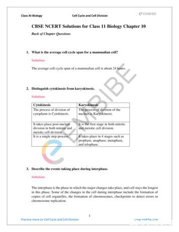
Class-XI-Biology Cell Cycle and Cell Division 1 Practice more on Cell Cycle and Cell Division www.embibe.com CBSE NCERT Solutions for Class 11 Biology Chapter 10 Back of Chapter Questions 1. What is the average cell cycle span for a mammalian cell? Solution: The average cell cycle span o
Chapter 7 The Cell Cycle and Cell Division Key Concepts 7.1 Different Life Cycles Use Different Modes of Cell Reproduction 7.2 Both Binary Fission and Mitosis Produce Genetically Identical Cells 7.3 Cell Reproduction Is Under Precise Control 7.4 Meiosis Halv
of chromosomes. The Cell Cycle The cell cycle is the repeating set of events in the life of a cell. Cell division is one phase of the cycle. The time between cell divisions is called interphase. Interphase is divided into three phases, and cell division is divided into two phases, as shown in
cycles of growth and division allow a single cell to form a structure consisting of millions of cells. 10.1 CELL CYCLE Cell division is a very important process in all living organisms. During the division of a cell, DNA replication and cell growth also take place. All these processes, i.e., cell division, DNA replication, and cell growth .


