PY5019 –Ultramicroscopy
PY5019 – UltramicroscopyTransmission Electron MicroscopyDr Hongzhou Zhanghozhang@tcd.ieOffice: SNIAM 1.06Tel: 896 4655School of PhysicsTrinity College Dublin
There’ss plenty of room at the bottomThereRichard Feynman, 1960 “with the greatest care and effort, itcan only resolve about 10 angstroms.” “ItIt is very easy to answer many ofthese fundamental biologicalquestions; you just look at the thing!” “WhatWhat you should do in order for us tomake more rapid progress is to makethe electron microscope 100 timesbetter” Is there no way to make the electronmicroscope more powerful?
Content Transmission Electron Microscopy Alternative Types of EMs–––– Emission Electron MicroscopyR fl ti ElectronReflectionEl tMiMicroscopyMirror Electron MicroscopyScanning Electron MicroscopyX‐Ray and EELS MicroanalysisScanning‐Probe MicroscopyFocused‐ion BeamHelium‐ion Microscopy
Content IntroductionThe InstrumentElectron‐Specimen InteractionDiffractionDiffraction ContrastPhase ContrastEDXEELSSEM‐HIM
Textbooks David Williams and Barry Carter, Transmission ElectronMicroscopy, 2ndd Edition, Springer Ludwig Reimer, Transmission Electron Microscopy, 5thEdition, Springer L. M. Peng, S. L. Dudarev, M. J. Whelan, High‐energyElection Diffraction and Microscopy, Oxford SciencePublications Brent Fultz, James M. Howe, Transmission ElectronMicroscopy and Diffraction of Materials, Springer J. W. Edington, Practical Electron Microscopy in MaterialsScience, The MacMillan Press, LTD Marc De Graef, Introduction to Conventional TransmissionElectron MicroscopyMicroscopy, Cambridge
Marking Attendance (8%)An essay (20%)– Something about your research and the cutting‐edge electron/ion microscopy– Not limited to the materials we cover in this module Two sets of homework (20%)– Lecture 4 (due at lecture 5): Find a paper – Lecture 8 (due at lecture 9) Interview (20%)– 15 minmin, my office (SNIAM 11.06)06)– You choose a relevant topic (different from your essay) and we discuss it– Please send me the topic and arrange a time with me by Lecture 6, and we willfinish it before Christmas break Q i ((eightQuizi ht 55‐mini open‐bookb k quizzesibbeforefclass,lCCalculators):l l t ) 32%– How fast can you write?– How fast can you find the answer?
Lecture One: Introduction Somethingg about the TEMBrief history of TEM developmentElectrons: the very basics for the TEMIntroduction of TEM modesExamples of TEM CharacterizationApplications of the TEMLimitations of the TEMCCurrentresearchh andd futureftrendsdResources
TEM Extremely Expensive:TEM ‐ Overview– Basic configuration:g 4/eV– With options: 9/eV (FEI Titan: 2.7M)– FE costs twice as much as Thermionic source Different Forms: HRTEM, STEM, AEM, etc– Routine instruments: 100‐200 kV– Medium (Intermediate) –voltage (IVEM): 200‐500 kV– High‐voltage (HVEM): 500 kV‐ 3MV Advantages and applications:lHighh Spatiall and Analyticalll Resolutionlwith Completely Quantitative Understanding–––––Structural and chemical informationA range of spatial range: atomic scale – nano – micrometreAtomic resolutionQuantitative informationIn‐situInsitu capability How can we do it?––––Instrumentation: Fundamental Physics of electrons & Electron OpticsSignalg ggeneration: Electron‐samplep interactionSignal detection: modesOperation and interpretation
Historical DevelopmentE l DEarlyDaysLouis de Broglie:Wave nature ofelectrons271925Knoll&Ruska:Electron MicroscopeSiemens&Halske: RegularProduction of TEM, 7nmKossel/Mollenstedt:ElElectronDiffDiffractioni ini TEMsTEM193233 36 3945Commercial TEM:Metropolitan‐Vickers EM1light microscope Resolutionlimit surpassedDavisson&Germer;; Thomson&ReidElectron diffractionBusch: Electromagnet/Electrostaticfocused electronsHitachi, JEOL,HitachiJEOL PhilipsPhilips, FEIFEI,Carl Zeiss: Widely available
Historical DevelopmentAnalyticalTEM, SAED, e, Hirsch&HowieCs correctedMonochromatorSub‐eV, sub‐AFeynman’s talkEarly Field EmissionSTEM, EELS,HolographKey Events in the History of Electron Microscopy, DOI: 10.1017/S1431927603030113
Resolution 1.0Siemens, UM100Resoolution (nm))0.80.60.4JEM 200CXJEM 100CX0.2JEOL 20100.0Tecnai F30Cs correctedTitan and TEAM1950196019701980Year199020002010
Nobel Prizes Karl Manne Georg Siegbahn: 1924 in Physics– “for his discoveries and research in the field of X‐ray spectroscopy” Denis Gabor: 1971 in Physics– “forfor his invention and development of the holographic methodmethod” Albert Claude, Christian de Duve, George E. Palade: 1974 inPhysiology or Medicine– “for their discoveries concerning the structural and functionalorganization of the cell” David Baltimore, Renato Dulbecco, Howard Martin Temin: 1975 inPhysiology or Medicine– “forfor their discoveries concerning the interaction between tumour virusesand the genetic material of the cell” Aaron Klug: 1982 in Chemistry– “HisHis development of crystallographic electron microscopy and hisstructural elucidation of biologically important nucleic acid‐proteincomplexes” Ernst Ruska; Gerd Binnig, Heinrich Rohrer(STM): 1986 in Physics– “hi“his fundamentalf dl workk ini electronloptics,i andd ffor theh ddesigni off theh fifirstelectron microscope”
Why Electrons? Image resolution/resolving power– Naked eyes: 0.1‐0.2 mm– Magnification (100,000X) – resolution (1 nm)0.61 – Rayleigh criterion (VLM): 550 nm – 300 nm resolution– TEM Atomic‐resolution 100keV – 0.004nm1960s High‐voltage EM 1.22 4pmE 1/ 2(1 nm 10‐9 m)(1pm 10‐12 m)Poor electron lens, but electrons can be focused :–– n sin Electron wavelength:–– Small aperture required: 10‐25 mrad (resolution: 0.1‐0.3nm)Electron Probe (spatial resolution/sensitivity)» Typically 5 nm, at best 0.1nm (Cs)» Higher currents (Cs)Information limit: Partial Spatial& temporal Coherence: E: 0.3‐2eVSpherical &chromatic aberration Corrections (resolution: 0.1Spherical‐&chromatic 0 1 nm),nm) thin sampleFocal Series Reconstruction/holographyElectrons, ionizing radiation: Strong Interactions– Wide range of 2nd signals: Chemical information ((AEM: XEDS,, EELS )‐) Compositionpand distributionEFTEM for band‐gap/chemical bond imaging(Cc)Materials:– Metals, alloys, ceramics, glasses, polymers, semiconductors, composite mixtures, wood,textiles, concrete– Bulk,B lk particles,ti l fibers,fibnanostructurest t– Nanoscale Materials and Devices– Biomaterials, bio‐inorganic interface
The System and the equationThe system: Solid: nuclei and atomic electrons i H t High‐energy electrons‐ A closed system: H T V‐ Time independentp((definite energy):gy) expp i E E t s H E Es ‐ The Hamiltonian:H 2 2 V (r ; r1.rj .; R1.Rn .) H cr2m‐ Distinguishable:Di tii h bl exchangeheffectsff t ignoredid Fermi Velocity of atomic electrons 106 m/s Velocity of beam electrons 0.5c – 0.99 c (108 m/s)1 Z ee V (r ; r .r .;; R .R .)) r R r r 4 22n1j1n0 nnjj ‐ Elastic scattering: the solid does not change its states r ; r1.ri .; R1.Rn . r1.ri .; R1.Rn . r H cr r1.ri .; R1.Rn . Es r1.ri .; R1.Rn . ‐ The one‐body equation for the incident electrons: 2 2 V (r ) r E r 2m V (r ) *V (r ; r1.rj .; R1.Rn .) dri dR j ‐ The relativistic invariance Dirac equation should be used Diffraction: Neglect effects associated withthe spin of electrons The relativistic Klein‐Gordon equationm Z n e 2 1 e 2 (r ' ) dr' n r R 4 0 r r 'n m01 2vc2E 2 k 02 m E E 0 1 2 m 2m0 c 2m E m r H r E 0 1 2 m 2m0 c
Electron‐Optical Refractive Index and the Schrodinger EquationTime‐independent wave equation:Electron‐opticalElectronoptical Refractive Index: 2 r k m2 r 0v 2 n km nkvm m mRelativistic h 22m0 E 1 E2 E0 E0 m0 c 511 kev 2 E V E0 E V 2 n r m 2 EE0 E 2 Non‐relativistickm 1/ 2h2m0 E2 m 2 r 2 h2m0 E V 2m0 E V r 0 22m0 E V AAssume: V(V(r)) E 2 EE0 2VE0 E 2 2 EV V 2 n r 2 EE0 E 2 1/ 2 2( E0 E )V 1 2 2 EE0 E V (r ) 2 E0 E 2 EE0 E 2 r 0 2 r 1 E 2 E0 E 2 c 2 V (r ) E 2 r 2 E0 E 2 E0 E 2 2 r 0E c 2 E0 E E 2E V (r ) 1 2 02 r 0 2 r E 2 E0 E E0 c 2 E0 E E 2m 2 r E V (r ) 1 2 0 r 0 E0 2 E0 E 1 V ( r ) E0 E .E 2 E0 EIn the vacuum: EIn the material: E-V(r)V (r ) E E 2 E V (r ) 1 2 02 r 0 2 r E 1 E0 c 2 E0 1/ 22Z n e 2 e Z effff ( r )1 e 2 ( r ' )dr' n r R 4 r4 0 r r 'n 0 E m0 m m0 1 E0 E0 2 E0 E E0 2 E0 EE E m E E E 1 E E 0 1 2 2 E E0 m 2m0 c E E0 2 E0 E E0 2 E0
Why can we use k and for electrons? k and are not uniquely defined quantities(not observable quantities) p mv eA kA iis not uniquelyil defined:d fi d A AA A’,A’ A’ A’ 0 (B (B A)Interference, phase difference 1 e1 2 1 mv eA dl mv dl A ds S A limited wave packet (r ) A(k ) exp ik rkUse infinite extensionplane wave for thecentre of thespectrum: k k 0 exp ikz A(k ) exp ikz dk
Some comments on Electrons in TEM Single electron– Beam current: I 1 nA 6.25 x 10⁹ electrons/sl/ Up to 0.1‐ 1 uA– Linear density: Assume regular interval emission ne I/v (6.25 x 10⁹ /v) electrons/(nA m)– 100 keV: v 0.5c 1.5x10⁸ m/s; ne 50 electrons/(nA m)– Separating (0.1 uA 100 nA): s 1/(ne I) 1/(50*100) 0.2 mm Stationary atoms and lattice vibration:– 100 nm sample: t 1 x 10‐7 m/(1.6X10m/(1 6X10⁸ m/s ) 5x105x10‐16 s– Lattice vibration: 10‐12 s– Time interval between two electrons (at 1 nA): 0.02m/(1.6x108 m/s) 10‐10 s:the atom have gone through 100 cycles, no correlation between atomicposition for consecutive electronshDuality h 2m0eV 1/ 2 – Non‐relativistic:p– Relativistic ( 100eV):h 1/ 2 eV 2m0 eV 1 2 2m0 c
TEMs Modes Elasticl i– Coherent Diffraction contrast: Bright‐field, Dark field, weak beam PhasePhcontrast: latticel iresolving, High resolutionTEM (HRTEM) Selected Area electronDiffraction (SAD) Large‐angle Convergent‐beam electron diffraction(CBED)?– Incoherent Z‐contrast:Z contrast: HighHigh‐angleangleannular dark‐field (HAADF) Inelastic– Energy‐Filtering TEM(EFTEM): 0.30.3‐0.50.5 nm– Energy‐dispersive x‐rayspectroscopy (EDS)– Electron energy‐lossspectroscopy (EELS) Conventional TEM Scanning Transmission Electron Microscopy(STEM) Lorentz Microscopy Analytical electron microscopy (AEM)– X‐ray Energy‐dispersiveSpectromtry(XEDS) : 0.10 1‐ 1um– Electron energy‐loss spectrometry(EELS) Relatively ‘New’– ElectronlMagnetic Circularl Dichroismh(EMCD)– Electron vortex– Electron Holographyg p y ((no lens):) electronbiprism, Aharonov‐Bohm effect– In‐situ MicroscopyInstrumentation/Techniques/Signal generation and detection/Interpretation
Diffraction ‐ SAEDPhase, crystallographic orientation, order disorder, defects
Diffraction ‐ CBEDSpecimen thicknessFull 3D symmetryLattice‐strainEEnantiomorphismtihi anddpolarity Valence‐electrondistribution, structurefactors, and chemicalbondingg Characterization of lineand planar defects Chapter 20‐21, David Williams and C. Barry Carter
Diffraction Contrast – BF and DFEdington, Practical Electron Microscopy in Materials Science
Phase Contrast ‐ HRTEMAluminiumB: 101 b ½ 110 Shamsuzzoha, M., et al., Scripta Metallurgica et Materialia, 1990. 24(8): p. 1611‐1615.
Applications: R&D Low dimensional materials/objects– Nanostructures– Surfaces and Interfaces Catalysis: Automotive/Petroleum– particle size, shape, surface layers/absorbates, reactions Image overlap: matrix and precipitates Sufficient contrast against background noise Mobile under investigation Pharmaceutical––––Drug discovery: substancesUnderstand & characterize molecular targetsValidate the effectsContaminations Museum/Forensic Z.R. Li, “Industrial Application of Electron Microscopy”, Marcel Dekker, INC, 2003
Automotive Applications Automotive exhaustcatalysts: Pt, Pd, Rh– Reduce : hydrocarbons,y,CO, NOx– Catalyst DeactivationS diStudies Thermal aging:migration/redistributiong/of elements Chemical Poisoning
Semiconductor Industry Failure analysis Dopingp g contrast
Example: Nanotubes/Nanocones
Limitations of the TEM Sampling: small part of your specimenInterpreting TEM images– Projection‐limitation Transmission 2D image: artefacts Average through the thickness– Electron tomography Beam Damage and Safety: Useful viewing time (Cs?)––––Knock‐onRadiolyticdi l i processes: ionisationi i i damagedImage magnification and beam current densitySolutions: Sensitive detector intense sources: dose Cryo‐microscopy Specimen Preparation – Thinner is Better– Thin Sample: electron transparent (beam energy, Z, resolution desired) For 100 keV 100 nm HRTEM: 50 nm, 10 nm– Thinning: artefacts, damages, ElectropolishingIon‐beam etchinggUltromicrotomyCryofixation
Emerging Trends – Now and the Future Atomic Location and Quantitative imaging– Comparison:pexperimentalpand simulation– The Stobbs’ Factor: HRTEM contrast level deviates from simulation Quantitative Intensity: contrast level– Thermal diffuse scattering Detection and correction of Aberrations– Higher order objective aberration – Automatic diffractogram analysis FSR/Off‐axis EH: Cs 1% accuracy3‐fold Astigmatism2nd axial coma(beam tilt)Cs correction: standard imaging conditions– information limit 1.3A– Probe size 1A– Image delocalisation On‐line microscope control Environmental EM with in‐situ sample treatment 4‐dimensional4 diil TEM
Resources Internet Simulation software packages Journals/Proceedingsl/di– Proceedings of the International Conferences onElectronlMicroscopy Groups/Labs/ConsortiumsDavid Williams and Barry Carter, Transmission Electron Microscopy, 2nd Edition, Springer
EM Books "Analytical electron microscopy for materials science". D. Shindo, T. Oikawa. Springer (2002). Excellent, up to date, practical . (ELS, EDX, CBED, Alchemi, Sample prep, holography etc)."High resolution electron microscopy and related techniques". P. Buseck, J.Cowley and L.Eyring, Eds. Oxford Univ Press.(1989). Comprehensive overview.Electron Backscattering Diffraction in Materials Science, A. J. Schwartz, M. Kumar and B. L. Adams (Eds.) Plenum (New York, 2000)Atlas of Backscattering Kikuchi Diffraction Patterns D J Dingley, K Z Baba‐Kishi and V Randle IOP (Bristol, 1995)Introduction to Texture Analysis V Randle and O Engler Gordon and Breach (Amsterdam 2000)Texture and Anisotropypy U F Kocks,, C. N. Tomé and H‐R Wenk Cambridgeg ((Cambridgeg 1998))Elastic and Inelastic Scattering in Electron Diffraction and Imaging Z L Wang Plenum (New York 1995)Introduction to Analytical Electron Microscopy J J Hren, J I Goldstein and D. C Joy (Eds) Plenum (New York 1979)Principles of Analytical Electron Microscopy D C Joy, A D Romig and J I Goldstein (Eds) Pleum (New York 1986)Convergent Beam Electron Diffraction of Alloy Phases J Mansfield (Ed) Adam Hilger (Bristol 1984)Large‐angle convergent beam electron diffraction. J.P. Morniroli. (Society of French Microscopists. Paris). 2002. In english. ISBN 2‐901483‐05‐4Diffraction Physics. J.M.Cowley. North‐Holland. 3rd Edition. 1990.Advanced computing in electron microscopy. E.J.Kirkland. Plenum. New York. 1998."Transmission Electron Microscopypy and Diffractometryy of Materials". B. Fultz and J. Howe. Springer.p g 2001. Excellent coverageg of theoryy and worked examples.p"Fundamentals of HREM". S. Horiuchi. North Holland. 1994."Structural Electron Crystallography" D. L. Dorset, Plenum/Kluwer. 1997. Mainly organics."Transmission electron microscopy: A textbook for materials science". D.B.Williams and C.B.Carter. Plenum Press. 1996. Pedagogically sound introductory text. Indispensible."High Resolution Electron Microscopy". J.C.H.Spence. Oxford Univ Press. 2003. (3rd Edn). How to do HREM.Electron energy loss specrtroscopy in the electron microscope. R.F. Egerton. Plenum. New York. 2nd edition 1996."Convergent beam electron diffraction IV". M.Tanaka, M.Terauchi, K.Tsuda, K.Saitoh. JEOL Ltd. Tokyo. and earlier volumes. Superb collection of CBED patterns."Electron microdiffraction". J.Spence and J.M. Zuo (Plenum, 1992). How to do CBED. Worked example of how to find space‐group of crystal from CBED patterns."Electron Diffraction Techniques". Vols 1 and 2. Oxford/IUCr Press. J.Cowley, ed. 1993."High resolution electron microscopy for materials science". D.Shindo, K.Hiraga.Springer. 1998. Beautiful collection of HREM images and examples of their analysis."Electron Microscopy of thin crystals". P.B.Hirsch et al. Krieger. New York. 1977. Classic text with many worked examples. Indispensible."Electron‐diffraction Analysis of Clay Mineral Structures". B. B Zvyagin. Plenum. 1967"Electron Diffraction Structure Analysis". B. K. Vainshtein. Pergamon. 1964"Intro. to Scanning Transmission Electron Microscopy", R. J. Keyse, A. J. Garratt‐Reed, P.J. Goodhew and G. W. Lorimer, (BIOS Scientific Publishers, Royal Micros. Soc., 1998)"Electron Energy Loss Spectroscopy", Rik Brydson, (BIOS Scientific Publishers, Royal Micros. Soc., 2001)."Transmission Electron Microscopy. 4th edit.", L. Reimer, (Springer‐Verlag 1997). Excellent broad coverage with all the basic physics, including radiation damage. Indispensible."Electron Holography", A. Tonomura, (Springer‐Verlag, 1999)"Introduction to electron holography". E. Voelkl, Ed. (1998). Plenum."Practical Electron Microscopy in Materials Science", J. W. Edington (Van Nostrand Reinhold, 1976)"Electron beam analysis of materials" by M. Loretto. Chapman and Hall. 1984."Electron microscopy in heterogeneous catalysis". P. Gai and E. Boyes. Inst Phys. (2003)."Interpretation of electron diffraction patterns" Andrews, K., Dyson, D., Keown, S. (1971). Plenum New York."Crystallography and crystal defects". Reprinted by Techbooks, 4012 Williamsburg Court, Fairfax, Virginia, USA 22032. Extremely useful. Highly recommended.JCPDS‐ICDD Powder diffraction file. http://www.icdd.com/ . Identify crystalline phases from their diffraction data.Special issue of Zeitschrift Kirstallographie on electron crystallography. 2003/4. U.Kolb.Journal of Microscopy and Microanalysis (mid 2003) Special issue on Quantitative Electron Diffraction. J.C.H. Spence, editor.
Summary TEM: expensive but powerful techniquesElectrons: wave/particle dualityLimitationsi i ioff theh TEMCurrent research and future trendResources
Lecture Two: The Instrument Optics Elements– ElectronEl tGunsG– Lens– Recording system TEM Modes/alignment
– Lecture 8 (due at lecture 9) Interview (20%) – 15 minmin, mymy officeoffice (SNIAM(SNIAM 1 06)1.06) – You choose a relevant topic (different from your essay) and we discuss it – Please send me the topic and arrange a time with me by Lecture 6, and we will finish it before Christmas break
965 7954; e-mail: cowleyj@asu.edu. 1Also at: Center for Solid State Science, Arizona State University, USA. 2Present address: Department of Physics, Taylor Hall, The College of Wooster, Wooster, OH 44691, USA. Ultramicroscopy 72 (1998) 223—232 The contrast of images formed by atomic focusers J.M. Cowley*, R.E. Dunin-Borkowski1, Michele Hayward2
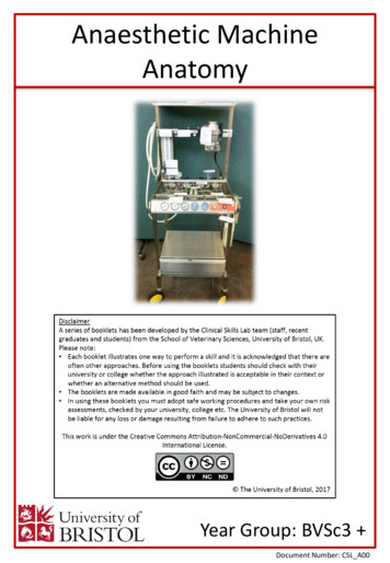
Anaesthetic Machine Anatomy O 2 flow-meter N 2 O flow-meter Link 22. Clinical Skills: 27 28 Vaporisers: This is situated on the back bar of the anaesthetic machine downstream of the flowmeter It contains the volatile liquid anaesthetic agent (e.g. isoflurane, sevoflurane). Gas is passed from the flowmeter through the vaporiser. The gas picks up vapour from the vaporiser to deliver to the .
Artificial Intelligence (AI) is an important and well established area of modern computer science that can often provide a means of tackling computationally large or complex problems in a realistic time-frame. Digital forensics is an area that is becoming increasingly important in computing and often requires the intelligent analysis of large amounts of complex data. It would therefore seem .
The book normally used for the class at UIUC is Bartle and Sherbert, Introduction to Real Analysis third edition [BS]. The structure of the beginning of the book somewhat follows the standard syllabus of UIUC Math 444 and therefore has some similarities with [BS]. A major difference is that we define the Riemann integral using Darboux sums and not tagged partitions. The Darboux approach is .
X707/77/02 Biology Section 1 — Questions TUESDAY, 30 APRIL 1:00 PM – 3:30 PM A/SA. page 02 SECTION 1 — 25 marks Attempt ALL questions 1. Primary cell lines have A a limited number of cell divisions and are sourced from tumours B a limited number of cell divisions and are sourced directly from normal animal tissue C an indefinite number of cell divisions and are sourced from tumours D an .
Following the publication of the UK Sanctions List, information on the Consolidated List has been updated. Notice summary 4. The following entries have been amended and are still subject to an asset freeze: Ntabo Ntaberi SHEKA (Group ID: 12438). Bosco TAGANDA (Group ID: 8736). 2 What you must do 5. You must: i. check whether you maintain any accounts or hold any funds or economic .
This publication was developed through a consultative process led by the International Telecommunication Union (ITU) and UNICEF and benefited from the expertise of a wide range of contributors from leading institutions active in the information and communications technologies (ICT) sector and on child online safety issues. UNICEF Corporate Social Responsibility Unit: Amaya Gorostiaga, Eija .
The LSE Careers website (lse.ac.uk/careers) contains information on different employment sectors, ways of planning your career, and marketing your skills. You will also find a range of reference material in the LSE Careers Resource Centre on Floor 5 of the Saw Swee Hock Student Centre. Visiting the organisation’s website and reading their publications are good places to start to find out .


