Normal Hand Anatomy Ghadiali - General Surgery
PRESENTSDr. Mufa T. Ghadiali is skilled in all aspects of General Surgery.His General Surgery Services include:General SurgeryAdvanced Laparoscopic SurgerySurgical OncologyGastrointestinal SurgeryHernia SurgeryEndoscopyNormal Hand AnatomyMultimedia Health EducationDisclaimerThis movie is an educational resource only and should not be used tomanage Orthopaedic Health. All decisions about management of theelbow must be made in conjunction with your Physician or a licensedhealthcare provider.Mufa T. Ghadiali, M.D., F.A.C.SDiplomate of American Board of Surgery6405 North Federal Hwy., Suite 402Fort Lauderdale, FL 33308Tel.: 954-771-8888Fax: 954- 491-9485www.ghadialisurgery.com
Normal Hand AnatomyMultimedia Health EducationMULTIMEDIA HEALTH EDUCATION MANUALTABLE OF CONTENTSCONTENTSECTION1 . Anatomy of the Hand Introductiona. Skeletal Anatomy2 . Anatomy of the Handa. Soft Tissue Anatomy3 . Biomechanics of the Handa. Normal Movementwww.ghadialisurgery.com
Normal Hand AnatomyMultimedia Health EducationUnit 1:Anatomy of the Hand IntroductionIntroductionThe hand in the human body is made up of the wrist, palm, and fingers. The most flexiblepart of the human skeleton, the hand enables us to perform many of our daily activities.When our hand and wrist are not functioning properly, daily activities such as driving a car,bathing, and cooking can become impossible.(Refer fig.1)(Fig.1)The hand’s complex anatomy consists of27 bones27 joints34 MusclesOver 100 ligaments and tendonsNumerous Blood vessels, nerves,and soft tissueIt is important to understand the normal anatomy of the hand in order to learn aboutdiseases and conditions that can affect our hands.Normal Hand(Refer fig. 2)(Fig.2)www.ghadialisurgery.com
Normal Hand AnatomyMultimedia Health EducationUnit 1:Anatomy of the Hand Introduction27 bones(Refer fig. 3)(Fig. 3)27 joints(Refer fig. 4)(Fig. 4)34 Muscles(Refer fig. 5)(Fig. 5)Over 100 ligaments and tendons(Refer fig. 6)(Fig. 6)www.ghadialisurgery.com
Normal Hand AnatomyMultimedia Health EducationUnit 1:Anatomy of the Hand IntroductionNumerous Blood vessels, nerves,and soft tissue(Refer fig. 7)(Fig. 7)Skeletal AnatomyThe wrist is comprised of 8 bones calledcarpal bones. These wrist bones connectto 5 metacarpal bones that form the palmof the hand. Each metacarpal boneconnects to one finger or a thumb at ajoint called the metacarpophalangealjoint, or MCP joint. This joint is commonlyreferred to as the knuckle joint.(Fig. 8)(Refer fig. 8)The bones in our fingers and thumb arecalled phalanges. Each finger has 3phalanges separated by two joints.The first joint, closest to the knuckle joint,is the proximal interphalangeal joint orPIP joint. The second joint nearer the endof the finger is called the distalinterphalangeal joint, or DIP joint.(Fig. 9)The thumb in the human body only has 2phalanges and one interphalangeal joint.(Refer fig. 9 &10 )(Fig. 10)www.ghadialisurgery.com
Normal Hand AnatomyMultimedia Health EducationUnit 2:Anatomy of the HandSoft Tissue AnatomyOur hand and wrist bones are held in place and supported by various soft tissues.These includeCartilageShiny and smooth, cartilage allowssmooth movement where two bonescome in contact with each other.(Refer fig. 11)(Fig. 11)TendonsTendons are soft tissue that connectsmuscles to bones to provide support.Extensor tendons enable each finger tostraighten.(Refer fig. 12)(Fig. 12)LigamentsLigaments are strong rope like tissue thatconnects bones to other bones and helphold tendons in place providing stabilityto the joints. The volar plate is thestrongest ligament in the hand andprevents hyperextension of the PIP joint.(Refer fig. 13)(Fig. 13)www.ghadialisurgery.com
Normal Hand AnatomyMultimedia Health EducationUnit 2:Anatomy of the HandMusclesMuscles are fibrous tissue capable ofcontracting to cause body movement.Interestingly, the fingers contain nomuscles. Small muscles originating fromthe carpal bones of the wrist areconnected to the finger bones withtendons.(Fig. 13)These muscles are responsible for movement of the thumb and littlefinger enabling the handto hold and grip items by allowing the thumb to move across the palm, a movement referredto as Thumb Opposition. The smallest muscles of the wrist and hand are responsible for finemotor movement of the fingers.(Refer fig. 13)NervesNerves are responsible for carrying signals back and forth from the brain to muscles in ourbody, enabling movement and sensation such as touch, pain, and hot or cold.The three main nerves responsible for hand and wrist movement all originate at theshoulder area and include the following(Fig. 14)(Refer fig. 14)Radial: The radial nerve runs down the thumb side of the forearm and provides sensation tothe back of the hand from the thumb to the third finger.Median: The median nerve travels through the wrist tunnel, also called carpal tunnel,providing sensation to the thumb, index finger, long finger, and part of the ring finger.Ulnar: The ulnar nerve travels through a tunnel in the wrist called Guyon’s tunnel formed bytwo carpal bones and the ligament that connects them together. The ulnar nerve suppliesfeeling to the little finger and half of the ring finger.www.ghadialisurgery.com
Normal Hand AnatomyMultimedia Health EducationUnit 2:Anatomy of the HandBlood VesselsThe two main vessels of the hand and wrist areRadial Artery: The radial artery is thelargest artery supplying the hand andwrist area. Traveling across the front ofthe wrist, nearest the thumb, it is thisartery that is palpated when a pulse iscounted at the wrist.Ulnar Artery: The ulnar artery travelsnext to the ulnar nerve through Guyon’scanal in the wrist. It supplies blood flowto the front of the hand, fingers andthumb.(Fig. 15)(Refer fig. 15)BursaeBursae are small fluid filled sacs thatdecrease friction between tendons andbone or skin. Bursae contain special cellscalled synovial cells that secrete alubricating fluid. When this fluid becomesinfected, a common painful conditionknown as Bursitis can develop.(Fig. 16)(Refer fig. 16)www.ghadialisurgery.com
Normal Hand AnatomyMultimedia Health EducationUnit 3:Biomechanics of the HandNormal MovementBiomechanics is a term to describe movement of the body. Metacarpophalangeal joint(MCP) or knuckle joint The fingers of the hand permit the following movements at themetacarpophalangeal joint (MCP) or knuckle jointMetacarpophalangeal joint (MCP) or knuckle joint The fingers of the hand permit thefollowing movements at the metacarpophalangeal joint (MCP) or knuckle jointFlexionMoving the base of the finger towardsthe palm.(Refer fig. 17)(Fig. 17)ExtensionMoving the base of the fingers away fromthe palm.(Refer fig. 18)(Fig. 18)AdductionMoving the fingers toward the middlefinger.(Refer fig. 19)(Fig. 19)www.ghadialisurgery.com
Normal Hand AnatomyMultimedia Health EducationUnit 3:Biomechanics of the HandAbductionMoving the fingers away from the middlefinger.(Refer fig. 20)(Fig. 20)FlexionMoving the last two segments of thefinger towards the base of the fingers.(Refer fig. 21)(Fig. 21)ExtensionMoving the last two segments of thefinger away from the base of the fingers.(Refer fig. 22)(Fig. 22)Biomechanics of the wrist include the followingFlexionMoving the palm of the hand towards thefront of the forearm.(Refer fig. 23)(Fig. 23)www.ghadialisurgery.com
Normal Hand AnatomyMultimedia Health EducationUnit 3:Biomechanics of the HandExtensionMoving the back of the hand towards theback of the forearm.(Refer fig. 24)(Fig. 24)AdductionMoving the pinky side of the handtoward the outer aspect of the forearm.(Refer fig. 25)(Fig. 25)AbductionMoving the thumb side of the handtoward the inner aspect of the forearm.(Refer fig. 26)(Fig. 26)www.ghadialisurgery.com
Normal Hand AnatomyMultimedia Health EducationUnit 3:Biomechanics of the HandThe thumb performs different movements at three separate joints. The carpometacarpaljoint is where the wrist bones, carpals, meet the metacarpals, the bones in the palm of thehand. At this articulation, the following movements can be performedAbductionMoving the bone below the thumbtowards the palm of the hand.(Refer fig. 27)(Fig. 27)ExtensionMoving the bone below the thumb awayfrom the hand.(Refer fig. 28)(Fig. 28)AdductionMoving the bone below the thumbtowards the back of the wrist.(Refer fig. 29)(Fig. 29)AbductionMoving the bone below the thumbtowards the front of the wrist.(Refer fig. 30)(Fig. 30)www.ghadialisurgery.com
Normal Hand AnatomyMultimedia Health EducationUnit 3:Biomechanics of the HandOppositionMoving the thumb across the palm of thehand touching the other fingers.(Refer fig. 31)(Fig. 31)The following movements occur at the metacarpophalangeal joint or MCP joint at the baseof the thumbFlexionMoving the joint at the base of thethumb towards the heel of the hand.(Refer fig. 32)(Fig. 32)ExtensionMoving the joint at the base of thethumb away from the heel of the hand.(Refer fig. 33)(Fig. 33)AdductionMovement of the thumb base towardsthe back of the hand.(Refer fig. 34)(Fig. 34)www.ghadialisurgery.com
Normal Hand AnatomyMultimedia Health EducationUnit 3:Biomechanics of the HandAbductionMovement of the thumb base away fromthe back of the hand.(Refer fig. 35)(Fig. 35)At the interphalangeal joint of the thumb or IP joint, the following movements can beperformed:FlexionBending the top of the thumb towardsthe base of the thumb.(Refer fig. 36)(Fig. 36)Extension hyperextensionMoving the top of the thumb away fromthe base of the thumb.(Refer fig. 37)(Fig. 37)www.ghadialisurgery.com
Normal Hand AnatomyMultimedia Health EducationUnit 3:2:GastritisLessonsDisclaimerDisclaimerAlthough every effort is made to educate you on normal anatomy of the Hand, there will bespecific information that will not be discussed. Talk to your doctor or health care providerabout any questions you may have.www.ghadialisurgery.com
Normal Hand AnatomyMultimedia Health EducationYOUR SURGERY DATEREAD YOUR BOOK AND MATERIALVIEW YOUR VIDEO /CD / DVD / WEBSITEPRE - HABILITATIONARRANGE FOR BLOODMEDICAL CHECK UPADVANCE MEDICAL DIRECTIVEPRE - ADMISSION TESTINGFAMILY SUPPORT REVIEWPhysician's Name :Patient’s Name :Physician's Signature:Patient’s Signature:Date :Date :www.ghadialisurgery.com
Normal Hand Anatomy The hand’s complex anatomy consists of (Fig.1) (Refer fig.1) 27 bones 27 joints 34 Muscles Over 100 ligaments and tendons Numerous Blood vessels, nerves, and soft tissue It is important to understand the normal anatomy of the hand in order to learn about diseases and conditions that can affect our hands. (Fig.2) Normal Hand
Clinical Anatomy RK Zargar, Sushil Kumar 8. Human Embryology Daksha Dixit 9. Manipal Manual of Anatomy Sampath Madhyastha 10. Exam-Oriented Anatomy Shoukat N Kazi 11. Anatomy and Physiology of Eye AK Khurana, Indu Khurana 12. Surface and Radiological Anatomy A. Halim 13. MCQ in Human Anatomy DK Chopade 14. Exam-Oriented Anatomy for Dental .
39 poddar Handbook of osteology Anatomy Textbook 10 40 Ross ,Pawlina Histology a text & atlas Anatomy Textbook 10 41 Halim A. Human anatomy Abdomen & lower limb Anatomy Referencebook 10 42 B.D. Chaurasia Human anatomy Head & Neck, Brain Anatomy Referencebook 10 43 Halim A. Human anatomy Head & Neck, Brain Anatomy Referencebook 10
Anatomy Of The Foot And Ankle The gastrocnemius or calf muscle is the largest of these and assists with movement of the foot. Muscle strains occur usually from overuse of the muscle in which the muscle is stretched without being properly warmed up. Unit 1: Anatomy (Fig.16) Muscles (Refer fig.16) (Fig.17) Bursae Bursae Bursae are small fluid .File Size: 579KB
Anatomy titles: Atlas of Anatomy (Gilroy) Anatomy for Dental Medicine (Baker) Anatomy: An Essential Textbook (Gilroy) Anatomy: Internal Organs (Schuenke) Anatomy: Head, Neck, and Neuroanatomy (Schuenke) General Anatomy and Musculoskeletal System (Schuenke) Fo
A. Department of Anatomy: Recommended Books (Anyone latest edition) General Anatomy 1. Text Book of General Anatomy by Vishram Singh 2. Text Book of General Anatomy by Chaurasia 3. Text Book of General Anatomy by A K Datta Gross Anatomy 1. Text Book of Gross
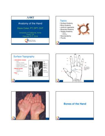
Anatomy of the Hand Diane Coker, PT, DPT, CHT University of California, Irvine Irvine, CA February 9-11, 2018 Topics Surface Anatomy Bony Anatomy Joints & Ligaments Muscular Anatomy Tendon Anatomy Flexors Extensors Neuroanatomy Thumb Surface Topography Joint flexion creases DPC Thenar crease .
Descriptive anatomy, anatomy limited to the verbal description of the parts of an organism, usually applied only to human anatomy. Gross anatomy/Macroscopic anatomy, anatomy dealing with the study of structures so far as it can be seen with the naked eye. Microscopic
RTS performs tree risk assessment in accordance with ANSI A300 (Part 9) - Tree Risk Assessment. Not only because we must as ISA Certified Arborists who are Tree Risk Assessment Qualified (TRAQ), but also because it ensures consistency by providing a standardized and systematic process for assessing tree risk. Risk assessment via TRAQ methodology takes one of three levels, depending on the .


