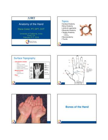
Topics Anatomy Of The Hand - Semantic Scholar
TopicsAnatomy of the HandDiane Coker, PT, DPT, CHTUniversity of California, IrvineIrvine, CAFebruary 9-11, 2018 Surface Anatomy Bony Anatomy Joints & Ligaments Muscular Anatomy Tendon Anatomy Flexors Extensors Neuroanatomy ThumbSurface TopographyP3 Joint flexion creases DPC Thenar crease Hypothenar crease Digital creases Distal wrist creases Muscle groups Thenar Hypothenar Rays 1-5P2DIPP1PIPDPC MP jointvolar crease (prox& distal palmarcrease)IPBones of the Hand
Bony AnatomyMetacarpal Cascade 19 bones distal to thecarpus Metacarpals (5) Numbered Phalanges (12) Proximal (P1) Middle (P2) Distal (P3) Thumb phalanges (2)Structural Units Fixed Unit Distal carpal row Metacarpals 2 & 3 3 mobile units Thumb ray Index finger ray Metacarpals 4 & 5, withlong, ring, & little fingersGreen: Mobile UnitsRed: Fixed UnitsTypes of JointsJoints and Articulations(condyloid)
Joints in the HandFixed and Mobile Units Saddle: Carpometacarpal(CMC) “Ulnar” opposition Ellipsoidal:Metacarpophalangeal(MP or MCP) Hinge: Interphalangeal (IP) Plane: Hamate andTriquetrum Not represented: ball andsocket 20-30 at SF 10-15 at RF Less mobility at MCsII & III thought to be afunctional adaptationto enhance ECRL/B& FCR activityMCP Joints Condyloid(Ellipsoid)Joints flexion/extension abduction/adduction IF sl rotation Motion increases radialto ulnar in digits 0/90‐110⁰Green: Mobile UnitsP1MC Hyperextension variesamong individualRed: Fixed UnitsMetaCarpoPhalangeal Joints Condyloid joints, 2 or 3 (IF)planes of motion Collaterals loose inextension, taut in flexion Prolonged immobilizationshould be in flexion, withcollateral ligaments on stretchMCP Joints Have IncreasedBony Congruity in Flexion
MP/IP Ligament StructureMP Joint Volar PlatePhalanxMetacarpalLoose proximal attachment ofthe volar plateInterPhalangeal Joints Bicondylar hinge joint withintercondylar ridge Volar plateP2(palmar fibrocartilagenousplate) Collateral ligaments have equaltension in flexion & extension Proximal condyle headstretches CL at about 15 Check rein ligamentsin PIPsP1Safety PositioningPIP Volar Plates PIPs areimmobilized inextension to avoidjoint contracturevia the check reinligaments(swallowtails)P2Safety vs. Functional Positioning MPs at 70-90 IPs at 0 - 15 Wrist in 20-35 extension
Extrinsic MusclesOriginate in Forearm & Insert in HandFinger FlexorsMusclesof the Hand Flexor Digitorum Superficialis, Flexor DigitorumProfundusFinger Extensors Extensor Digitorum, Extensor Indicis, Extensor DigitiMinimiThumb Extensor Pollicis Longus, Extensor Pollicis Brevis,Abductor Pollicis Longus, Flexor Pollicis LongusIntrinsic Hand MusclesOpponens Pollicis & Opponens Digiti Minimi Thenar Abductor Pollicis Brevis Flexor Pollicis Brevis Opponens Pollicis Hypothenar Abductor Digiti Minimi Flexor Digiti Minimi Opponens Digiti Minimi OP rotates 1stmetacarpal so thatthumbnail faces theceiling when hand isplaced palm up Slight rotation of 5thmetacarpal with ODM Adductor pollicis Palmaris Brevis Lumbricals InterosseiAPB & Abd Digiti Minimi APB works with OPduring opposition APB most radialand superficialthenar muscle APB first muscleto show signs ofatrophy in mediannerve dysfunctionFlexor Digiti Minimi &Flexor Pollicis Brevis FPB has 2heads withdifferentinnervations Deep head of FPBocc described as anadditional palmarinterosseous muscle
Adductor Two heads Innervation Transverse Oblique Ulnar nerveLumbricals Travel along radialside of each digit Innervation I/M: median R/S: ulnar Radial two musclebellies areunipennate Ulnar two are bipennate Bipennate muscles shortenless, generate more forcethan unipennateDorsal Interossei First DI much largerthan other DI No DI to SF First DI can rotate IFslightly at MCP joint,and assists adductorpollicis in thumbadductionVolar Interossei 3 unipennatemuscles Smaller than dorsalinterossei Adduct I, R, S Fstowards MF, assistlumbricals in MPflexionPalmaris Brevis Both palmaris longusand brevis serveminimal function in thehand Brevis serves totighten the hypothenarskin, possibly deepenconcavity of palm Innervation Ulnar No bony attachmentsTendon AnatomyFlexor TendonsExtensor Tendons
Flexor Tendon Anatomy Flexor Digitorum Profundus Splits into 2 separate bundles in mid-forearm Often separate slips for IF & M/R/S Fs Innervation: AIN I & M, ulnar R & LExtrinsic Flexors: FDS & FDPLength Tension Issues FDS and FDP aredependent on wristposition to enhancefunction;35 ‐40 ext formaximum grip Weakest flexion force isin wrist flexion ECRB providescounterbalance toprevent wrist flexion;ECRL contributes withpower gripTendon Orientation throughthe Carpal TunnelFlexor Digitorum Superficialis 2 separate origins Medial compartment—4 separate bundles ? FDS to little finger Innervation: medianFlexor Pollicis Longus Innervation Median (AIN) Unique to humans Rudimentary of absent in other primates Occasional connection to FDP Linburg‐Comstock syndrome Occasional accessory long headpresent Ganzer’s muscle Can compress AINFlexor Tendon Zones Zone I: distal toFDS insertion Zone II: A1 pulleyto FDS insertion No Man’s Land Zone III: distalend of CT to A1 Zone IV: CT Zone V: proximalto CT
Flexor Tendon Zones Thumb (3)Zone T1: from IPjoint distalZone T2: from IPjoint proximal toMP jointZone T3: from MPjoint proximal totransverse carpalligament FDS Volar to FDPentering synovial sheath Spiral turn Now dorsal to FDP Camper’s Chiasm Can insert as far as neckof P2 FDP Straight lineCamper’s Chiasm 50% of fibers from FDS cross over 50% of fibers remain on same sideFlexor Sheaths 2 Systems Synovial sheaths Provide nutritionto tendons Low-frictiongliding Retinacular sheaths Provide efficientmechanicalfunction byholding the tendonclose to the bone Annular & cruciatepulleysTendon Nutrition 2 pathways: Synovial diffusion Vascular perfusion Diffusion plays a greater role than perfusion
Retinacular Sheath SystemRetinacular Sheath System 2 Part Composition Fingers Thumb 5 Annular pulleys A1: over MP joint A3 over PIP A5 over DIP 3 Cruciate pulleys A1, A2, ObliqueBowstringingAKA: Rock Climbers’ injuryPulley MechanicsExtensor Tendon Anatomy Compartments A2 and A4 mostimportant to preservefor normal function infingers Oblique pulley in thumb 1:2:3:4:5:6:APL, EPBECRL, ECRBEPLED(C), EI(P)EDQ(M)ECU Only pulley is theextensorretinaculum Synovial sheathslocated only at wristlevel
Extensor Tendon Zones Fingers Zone 1: DIPZone 2: middle phalanxZone 3: PIPsZone 4: proximal phalanxZone 5: MPsZone 6: dorsum of handZone 7: retinacularcompartment Extensor tendonsare different fromflexor tendons Anatomy more complex Restraining structuresthroughout system More superficial, morevulnerable, thinner Flexor tendons canbecome “stuck” under thepulleys, but extensortendons often heal with alag 2 longer excursionpullExtensor Mechanism (Hood) Complex system covering dorsal aspect of digits Creates cable system Extends MPs & IPs Allows lumbricals to assist in MP flexion Components Extensor digitorum Juncturae tendinae Central slip/band Sagittal bands Lateral bands Transverse retinacularligament Oblique retinacularligament Terminal tendonExtensor Tendon Zones Thumb (5)Zone T1: IP jointZone T2: Middle phalanxZone T3: MP jointZone T4: 1st metacarpalZone T5: CarpusExtrinsic Extensors EIP and EDM addindependent function, notstrength ED can produce IP extensionif MPs blocked in slightflexionThe Extensor Apparatus
Sagittal Bands ED Lateral band Terminal tendon Interossei/lumbrical contributionsto lateral band Insert into & stabilize ED at dorsum of MP jointRuptures common, often with a trivial incidentOften laxED will eventually function as a flexor as it falls below the joint axis ofmotionJuncturae Tendinae Link EDC to prevent independent function Maintain dorsal placement of extensorstendons over MPs during flexionThumb MechanicsThe ThumbThe Thumb CMC joint is not in sagittal,coronal, or transverseplanes of the digits Difficult to categorize asbeing in flexion/extensionplanes orabduction/adductionplanes Thumb “scaption”
CMC JointsCarpoMetaCarpal Joint of Thumb Saddle Joints Thumb & Digit VFlex/Ext (ll to palm)Abd/Add ( to palm)Opposition net effect Plane Joints Digits II‐IV Flexion/ExtensionCMC Joint of the Thumb A “saddle” joint Biconcave sellar joint 7 ligaments for CMCstabilization 16 ligaments for STT andCMC joint stability Greatest stability in palmarabduction and pronationPeripheral Innervation AKA basaljoint, 1st CMC Asymmetrical ComplexligamentoussystemMCP Joint of Thumb Flatter than I-S FsMP heads Easily dislocated 2 sesamoid bones Greatest variationin ROM:30 – 90 The hand occupiesnearly 1/3 of themotor cortex Thumb approx ¼-1/3of handrepresentation
Cervical DermatomesVariations in Cervical Dermatomes Representationin the hand: C6 C7 C8C7Potential Contributors toSensation in the Thenar EminencePeripheralPatternsEntrapment Sites Includecompression,tension,combination All 3 majorperipheral nervespass through a 2headed muscle nearthe elbow Median: PronatorTeres Ulnar: FCU Radial: Supinator Palmar cutaneousbranch of median N Superficial branchradial N LABC Median N proper MABCMedian Nerve 2 main branchesdistal to elbow Main branch continuingon to innervate the hand AIN PQ, FPL, & radial½ FDP Palmar cutaneousbranch
Median NerveLigament of Struthers Entrapment sites Ligament of Struthers Lacertus fibrosus AKA bicipital aponeurosis Pronator teres FDS arch Carpal tunnel Ganzer’s muscleA rare occurrenceLacertus Fibrosus Compression with elbowflexionArch of FDS Sublimus (superficialis) arch Irritation of fascial edge ofFDSPronator Syndrome Median nervepenetrates pronatorteres between its 2heads PT spared in PronatorSyndrome PT weak if median nervecompressed under ligament ofStruthersEntire Median Nerve InvolvedAnteriorInterosseous Branch Pure motor branchbifurcating slightlyproximal to FDSarch Innervates FPL,PQ, FDP to I & MMartin-Gruber Anastomosis Median nerve sendsfibers to ulnar nerve inforearm Rare: ulnar to mediananastomosis Incidence ranges from8-30%
Carpal Tunnel CompressionAnatomical Variations ofRecurrent Motor branch Motor to IF,MFlumbricals Recurrent motorbranch Branches distal to CT Motor to thenar muscles The “million dollar” nerve Sensation to ends ofthumb, IF, MF, ½ RF(usually!)PalmarSensation Thenar eminencesensation suppliedmainly by palmarcutaneous branch Fingernails &dorsum of fingertipssupplied by mediannerve terminalendingsPalmar cutaneous nervedoes not enter carpal tunnelArcade vs. Ligament of StruthersUlnar Nerve Entrapment Sites Arcade of StruthersCubital tunnelFCUGuyon’s canal Arcade of Struthers Fascial band Usually present Potential compressionsite for ulnar nerve Ligament of Struthers Rare Potential site ofcompression for mediannerve
Ulnar nerve passesbetween 2 heads ofFCUCubital Tunnel Fascial arch A potential site ofcompression(Fascial arch between)Guyon’s Canal Dorsal ulnarsensory branchbifurcatesproximal toGuyon’s canal First dorsalinterosseousmuscle receivesterminalinnervation fibersfrom deep motorbranchIntrinsic Insertions ofPalmar Branch Ulnar N Motor to interossei,ulnar 2 lumbricalsadductor pollicisdeep head of FPB Palmar sensation toSF, RFFroment’s sign on rightFroment’s signOveruse of FPL 2 loss of stabilizationof pinch fromadductor pollicis
Teardrop sign on R: AIN(Reverse Froment’s sign)Froment’s sign:ulnar paresisFroment’s sign on RRadial Nerve Main nerve innervatesmobile wad of Henry(muscles originating fromsupracondylar ridge ECRB) Bifurcates at level ofradiocapitellar joint Entrapment sites: Spiral groove(Saturday night palsy) Mid-humeral trauma Arcade of Frohse/supinatorarch/radial tunnel Wartenburg syndrome PIN (motor branch) Superficial sensory branchPosterior InterosseousNerve (PIN) Motor branch Arcade of Frohse(supinator arch) Radial Tunnel vsSupinator syndromeWartenburg Syndrome(Distal radial sensory nerve compression) DRSN is “scissored”between thebrachioradialis and 2ndcompartment tendons 2-3 fingertips aboveradial styloid, sl medial “Watchband” or“handcuff” syndromeTear drop sign:AIN paresis
Thank You! dacoker@cox.net
Anatomy of the Hand Diane Coker, PT, DPT, CHT University of California, Irvine Irvine, CA February 9-11, 2018 Topics Surface Anatomy Bony Anatomy Joints & Ligaments Muscular Anatomy Tendon Anatomy Flexors Extensors Neuroanatomy Thumb Surface Topography Joint flexion creases DPC Thenar crease .
May 02, 2018 · D. Program Evaluation ͟The organization has provided a description of the framework for how each program will be evaluated. The framework should include all the elements below: ͟The evaluation methods are cost-effective for the organization ͟Quantitative and qualitative data is being collected (at Basics tier, data collection must have begun)
Silat is a combative art of self-defense and survival rooted from Matay archipelago. It was traced at thé early of Langkasuka Kingdom (2nd century CE) till thé reign of Melaka (Malaysia) Sultanate era (13th century). Silat has now evolved to become part of social culture and tradition with thé appearance of a fine physical and spiritual .
On an exceptional basis, Member States may request UNESCO to provide thé candidates with access to thé platform so they can complète thé form by themselves. Thèse requests must be addressed to esd rize unesco. or by 15 A ril 2021 UNESCO will provide thé nomineewith accessto thé platform via their émail address.
̶The leading indicator of employee engagement is based on the quality of the relationship between employee and supervisor Empower your managers! ̶Help them understand the impact on the organization ̶Share important changes, plan options, tasks, and deadlines ̶Provide key messages and talking points ̶Prepare them to answer employee questions
Dr. Sunita Bharatwal** Dr. Pawan Garga*** Abstract Customer satisfaction is derived from thè functionalities and values, a product or Service can provide. The current study aims to segregate thè dimensions of ordine Service quality and gather insights on its impact on web shopping. The trends of purchases have
Chính Văn.- Còn đức Thế tôn thì tuệ giác cực kỳ trong sạch 8: hiện hành bất nhị 9, đạt đến vô tướng 10, đứng vào chỗ đứng của các đức Thế tôn 11, thể hiện tính bình đẳng của các Ngài, đến chỗ không còn chướng ngại 12, giáo pháp không thể khuynh đảo, tâm thức không bị cản trở, cái được
Clinical Anatomy RK Zargar, Sushil Kumar 8. Human Embryology Daksha Dixit 9. Manipal Manual of Anatomy Sampath Madhyastha 10. Exam-Oriented Anatomy Shoukat N Kazi 11. Anatomy and Physiology of Eye AK Khurana, Indu Khurana 12. Surface and Radiological Anatomy A. Halim 13. MCQ in Human Anatomy DK Chopade 14. Exam-Oriented Anatomy for Dental .
– Ossa brevia (tulang pendek): tulangyang ketiga ukurannyakira-kirasama besar, contohnya ossacarpi – Ossa plana (tulang gepeng/pipih): tulangyang ukuranlebarnyaterbesar, contohnyaosparietale – Ossa irregular (tulangtak beraturan), contohnyaos sphenoidale – Ossa pneumatica (tulang beronggaudara), contohnya osmaxilla


