Vital Signs And Introduction To NEWS
Vital Signs and Introduction to NEWSSamantha Coulter, 4th Year Medical StudentDr Jason Long, Director of Clinical Skills
ContentsLearning Outcomes . 3Introduction to Vital Signs . 3National Early Warning Score . 3Assessment of Radial Pulse . 4Method . 4Blood Pressure . 5What Blood Pressure Numbers Mean . 5Hypertension vs Hypotension . 6Introduction to Sphygmomanometry. 6Blood Pressure Equipment . 7Method . 7Respiratory Rate . 9Method . 9Pulse Oximetry . 10Method . 10Body Temperature . 11Method . 11AVPU Scale. 12Method . 12Glasgow Coma Scale. 13Scale vs Score . 13Correlation with Severity of Injury . 14Method . 14NEWS Chart . 16Example NEWS Chart . 17References . 182 P a ge
Learning Outcomes Understand what vital signs are.Understand what NEWS is and why we use it.Be able to measure a radial pulse.Be able to measure blood pressure.Be able to measure respiratory rate.Be able to measure oxygen saturations.Be able to measure temperature.Be able to assess a patient’s level of consciousness with AVPU and GCS.Be able to fully assess a patients vital signs and record them on a NEWS chart.Introduction to Vital SignsVital signs are routinely used to monitor the body’s basic functions. The measurements are valuableindicators of the patient’s general health and early signs of deterioration in their health. The normalvalues can vary depending on age, gender and weight. The four main most commonly recorded vitalsigns are heart rate, blood pressure, respiratory rate and temperature. This document will also add inoxygen saturation and conscious level for completeness of the NEWS chart. (1,2)National Early Warning ScoreThe National Early Warning Scores (NEWS) was developed to detect those patients at risk ofdeterioration by scoring each patients vital signs; respiratory rate, oxygen saturations, temperature,blood pressure, pulse rate and level of consciousness. They are used to draw attention and prioritisepatients needing urgent care. A score of 0 is the best, with a score of 3 being the worst for each vitalsign. The individual scores are added together to get a patients early warning score. The charts alloweasy visualisation of trends in the patient’s health, allowing us to see if they are improving ordeteriorating. (1,3)3 P a ge
Assessment of Radial PulseBy the end of this section you should be able to: Locate, measure and record the radial pulse.A pulse is the palpation of blood flow through acompressed artery. The normal range for an adultpulse rate is between 60-100 beats per minute (bpm).When measuring the radial pulse, you should befeeling for the rate and the rhythm. (2) The radialpulse is felt between the radial styloid and the tendonof flexor carpi radialis. Feel with two or three fingers.Do not use your thumb as this has its own pulse. (4)Count the pulse for 15 seconds and multiply by four. If the patient’s pulse is irregular or abnormal, youshould feel the pulse for a full 60 seconds assessing if the pulse is irregularly irregular or if there is apattern to the irregularity. You will need a clock with a second hand. Heart rate is raised in a largenumber of conditions such as shock, pyrexia, pain and anaemia. (2)Method1.2.3.4.5.6.7.8.9.Ensure appropriate hand hygiene.Introduce self and confirm patient’s identity.Seek informed consent.Check both radial pluses simultaneously to make sure thatthey are equal, and then concentrate on the right radialpulse.Count the rate per minute.Count for 15 seconds and multiply by four (or 60 seconds if any abnormality).Thank patient.Perform hand hygiene.Record the heart rate.4 P a ge
Blood PressureBy the end of this section you should be able to: Understand what is meant by blood pressure. Correctly use the appropriate equipment. Assess, measure and record blood pressure. Have an understanding of hypertension and hypotension.Blood pressure is a measurement of the force of blood pushing against the arterial walls. Bloodpressure can be measured directly, by inserting a needle or catheter into the artery, or indirectly, byusing a blood pressure cuff to occlude the vessels. The indirect method is the one more commonlyused, although less accurate, it is quicker and less invasive. (5) It can be measured by an automateddevice or manually by using the stethoscope and listening to vessels distal to the cuff. The manualmethod is seen to be more accurate when the appropriate equipment is used correctly. (6) Thisdocument will outline how to measure blood pressure manually.What Blood Pressure Numbers MeanWhen measuring blood pressure, two pressuremeasurements are recorded. The higher number, calledsystolic pressure, refers to the pressure inside the arterywhen the heart contracts. The lower number, calleddiastolic pressure, refers to the pressure inside the arterywhen the heart is at rest. Previously, blood pressure wasmeasured by a mercury manometer, this is why thecurrent blood pressures are recorded in millimetres ofmercury (mmHg). (2,5)The chart shows low, normal, at-risk and high blood pressurelevels. 140/90mmHg and above hypertension (high bloodpressure). Between 120/80mmHg and 140/90mmHg prehypertension where they are at risk of gettinghypertension. Between 120/80mmHg to 90/60mmHg normotension(normal blood pressure). This will vary between age,size and health. Below 90/60mmHg hypotension.BP measurement can tell you about the state of a patient’speripheral circulation and fluid balance. It can also giveinformation about long term stroke and heart attack risks inpatients, and the progress of complications of diseases such asdiabetes.(7)5 P a ge
Hypertension vs HypotensionSymptomsCausesRisk FactorsPreventionHypertensionDizziness or lightheadedness,lack of concentration, blurredvision, fatiguePrimary – develops over yearsSecondary – due to anunderlying conditionsHypotensionHeadache, short of breath,chest pain, verweightandobesity, 65 years old, family history,women 65 years old, men 45 alcohol, immobilityyears old, tobacco and alcohol,high sodium dietMaintain a healthy weight, Drink more water, eat a bettermanage stress, avoid tobacco, diet, limit alcohollimit alcoholIntroduction to SphygmomanometryA sphygmomanometer (often abbreviated to sphyg) is thedevice used to non-invasively measure the blood pressure inthe patient’s arteries. A handheld manual sphyg, which youwill be using, consists of a pressure gauge and an inflatablecuff that wraps around the upper arm. The principle behindsphymomanometry is to manipulate the artery pressure byconstricting the vessel by inflating the cuff, and then reducingthe pressure to record the points where flow returns. Therecording of the return of flow is done by listening with astethoscope to hear the ‘Korotkoff Sounds’. As the sphyg cuffis deflated, the artery becomes less constricted allowingblood to flow through the artery. The turbulent flow causessounds to be heard through the stethoscope. (5)1. Korotkoff I – sharp thud as only systolic pressure exceeds the cuff pressure2. Korotkoff II – loud blowing sound as the flow becomes less turbulent3. Korotkoff III – soft thud4. Korotkoff IV – soft blowing sounds, muffled flow5. Korotkoff V – silence as flow becomes laminar and cuff is deflated with no pressure on arterySystolic blood pressure is the pressure in the artery when the heart contracts (systole), making thisthe maximum arterial pressure – Korotkoff I. The pressure at this point is greatest and is able to forceblood through the constricted artery causing the first Korotkoff sound. Diastolic blood pressure is thelowest arterial blood pressure during the cardiac cycle as it reflects the relaxation of the heart betweencontractions (diastole). When the artery is fully open, there is no turbulent flow, and therefore nothingto hear giving us Korotkoff sound V. This represents the flow of blood during diastole. (5)It is not important to be able to differentiate from the 5 different sounds. However, you must be ableto recognise when they first appear (SYSTOLIC BP) and when there is silence (DIASTOLIC BP). Whilstappearing rather complex to begin with, with a little practise it is a skill that is easily mastered.6 P a ge
Blood Pressure Equipment SphygmomanometerStethoscopeAlcohol wipesMethod1.2.3.4.Perform hand hygiene and clean stethoscope.Introduce self and confirm patient’s identity.Seek informed consent.Position arm on a pillow so that the antecubital fossa is level with the heart and arm is straightbut supported.5. Palpate brachial artery in antecubital fossa prior to positioning cuff.6. Wrap a suitably sized cuff around the upper arm. The lower border of the cuff should beapprox. 2cm above the antecubital fossa.7. The centre of the cuff should be over the brachial artery – the arrow on the cuff should pointtowards the artery.7 P a ge
8. Identify and palpate the radial pulse.9. Inflate cuff while palpating the radial artery until the pulse disappears. Take a mental note ofthe pressure on the sphygmomanometer dial. Completely deflate the cuff. This reading givesan estimate of systolic pressure.10. Relocate the brachial artery and place the diaphragm of the stethoscope over the artery.11. Re-inflate the cuff to 20mmHg above the estimated systolic pressure.12. Release cuff slowly. Listen carefully with the stethoscope and mentally note the pressurewhen the first thudding sounds are heard (Korotkoff 1). This is the systolic pressure.13. Keep releasing the cuff pressure. The noise will get louder, then soft, then muffles and thenfinally stops. Note the reading when there is silence (Korotkoff 5). This is the diastolic pressure.14. Finish deflating the cuff quickly.15. Document the readings as systolic pressure over diastolic pressure, e.g. 120/60 mmHg.16. If you miss the readings, do not re-inflate the cuff immediately. Deflate the cuff fully, wait untilthe patient is comfortable and try again from the beginning.17. Thank patient and wash hands.18. Record the blood pressure.8 P a ge
Respiratory RateBy the end of this section you should: Understand normal respiration. Be able to recognise signs of respiratory distress. Be able to assess, measure and record the patient’s respiratory rate.The respiratory rate is the number of breaths perminute. Normal respiratory pattern is an easy,relaxed, subconscious activity which takes place at arate dependant on age and activity. (8) Thinkingabout your breathing alters the rate and pattern andtherefore to accurately measure the patient’srespiratory rate it is best to not make them aware ofyour assessment. This is usually done after assessingthe patient’s radial pulse. Continue to pretend totake the pulse rate while observing the rise and fallof the patient’s chest. One respiration consists ofone complete rise and fall of the chest. This shouldbe done for 30 seconds and multiplied by 2. The pattern, effort and rate of breathing should beobserved and recorded. Abnormal patterns can be described as: Dyspnoea – difficult, laboured breathing Tachypnoea – rapid breathing Apnoea – temporary absence of breathing Kussmaul’s – increased rate and depth, with longgrunting expirations – lobar pneumonia/DKA Stridor – a harsh, high-pitched noise on inspiration –obstructionAlong with signs of respiratory distress; observe use of accessory muscles and wheeze. A normalrespiratory rate for adults can be between 12 and 20 breaths per minute. Respiration rates mayincrease with fever, asthma, anxiety, pneumonia, congestive heart failure or drug overdose. (2,9)Method1. Ensure patient is relaxed and unaware of the counting process.2. After measuring the radial pulse, continue to pretend to take pulse, while watching thepatient’s chest.3. Count the respiratory rate over 30 seconds, observe the depth and pattern of respiration.4. Record the respiratory rate.9 P a ge
Pulse OximetryBy the end of this section you should be able to: Understand what pulse oximetry is. Understand how the pulse oximetry device works. Know the normal ranges of oxygen saturations. Measure and record pulse oximetry.Pulse oximetry is used to assess the saturation of oxygen in the blood in a simple, non-invasive way.Oxygen saturations are useful during surgeries that involve sedation to adjust supplemental oxygen.They are also used to monitor patient’s exercise tolerance and the effectiveness of some medications.The pulse oximetry device is a clip-like probe that can be attached to a thin part of the body, forexample, an ear-lobe, nose, or most commonly used, a finger. One side of the probe has a light sourcewith two different types of light; infrared and red. The light is passed throughthe body’s tissue to the light detector on the opposite side. A microprocessorcalculates the oxygen saturation of haemoglobin by comparing oxygenated(infrared light absorbing) and deoxygenated (red light absorbing) haemoglobindepending on how much of each light was absorbed. The measurements aremade multiple times a second, with the readings of the last 3 seconds beingaveraged out to give the final recording. (10)Oxygen is breathed into the lungs, passes to the blood where the majority attaches to haemoglobin.Haemoglobin is a protein located inside our red blood cells that transports the oxygen through thebloodstream to the rest of our body and tissues. In this way, our body is given the oxygen and nutrientsit needs to function. Oximeters use the light absorptive characteristics of haemoglobin and thepulsating nature of blood flow in the arteries to measure the level of oxygen in the body. (10)Oxygen saturations should be above 95% in a normal individual. However, it may be lower in thosewith respiratory disease or congenital heart disease. Nail polish must be removed beforehand as thisabsorbs the light and gives a false reading. (10)Method1.2.3.4.5.Ensure appropriate hand hygiene.Introduce yourself, check patient details and gain consent.Switch the monitor on.Place the probe on a finger.Ask the patient to rest their hand and keep it as still aspossible.6. Record the oxygen saturations.Pulse rateOxygen saturations10 P a g e
Body TemperatureBy the end of this section you should know: Indications for measuring body temperature. Normal ranges of body temperature. Be aware of different ways to measure bodytemperature. How to measure tympanic temperature.Body temperature is measured using a thermometer or electronic probe. Temperature can berecorded in the axilla, rectum or ear, and should be consistent for each patient. (2) The normal rangeof body temperature is between 36 C and 38 C, as used in the NEWS Chart. (1,11) A body temperaturerecording may be needed to establish a baseline reading or monitor patient’s being treated forinfection. (12)The tympanic temperature (ear) is now most commonly used in hospitals. It is minimally invasive anddue to the close proximity to the internal carotid artery and the tympanic membrane’s blood supplyit ensures that the temperature is a good reflection of core temperature. (13)Method1.2.3.4.5.6.7.8.9.10.11.12.13.Wash hands.Introduce self.Identify patient.Explain procedure.Obtain informed consent.Push protective sheath onto probe.Switch unit on and check ear sign is displayed.Pull ear gently backwards and upwards, inserting probe into theear canal.Press activation button, it will beep once at beginning of measurement.After a few seconds the unit will beep again to indicate completion.Dispose of protective sheath in clinical waste bin.Thank patient.Record temperature.11 P a g e
AVPU ScaleBy the end of this section you should be able to: Assess and record the patient’s level of consciousness using AVPU.AVPU Scale is a simplification of the Glasgow Coma Scale (explained in the next section) to assess apatients level of consciousness. AVPU stands for Alert, Voice, Pain and Unresponsive. The patient willfall into one of these four categories. Only ‘Alert’ is normal. (14)Method1. Alert – the patient is fully awake, although they may beconfused. Their eyes are open spontaneously, they willrespond to voice and have coordinated motor function.2. Voice – the patient will respond to voice by opening eyes orsounds (i.e. moans and groans).3. Pain – the patient will respond to a pain stimulus (i.etrapezius squeeze or fingernail bed squeeze) by openingeyes, sounds or flexion/extension of a limb.4. Unresponsive – the patient will give no response to voice orpain.5. Record either A, V, P or U on the chart.12 P a g e
Glasgow Coma ScaleBy the end of this section you should be able to: Assess and record the patient’s level of consciousness using the Glasgow Coma Scale. Understand the differences between Glasgow Coma Scale and Glasgow Coma Score.Glasgow Coma Scale (GCS) is the most widely recognised measure of consciousness developed in1974. GCS
A pulse is the palpation of blood flow through a compressed artery. The normal range for an adult pulse rate is between 60-100 beats per minute (bpm). When measuring the radial pulse, you should be feeling for the rate and the rhythm. (2) The radial pulse is felt between the radial styloid and the tendon of flexor carpi radialis.
providers or students to teach basic vital signs skills Use the navigation buttons below to move through the tutorial Click here to get started! Use this button to advance to Use this button to the next slide go back to the previous slide Use this button to go back toFile Size: 1MBPage Count: 31Explore furtherHow to Take Vital Signs – Step-by-Step Manual .www.usamedicalsurgical.comPrintable Vital Sign Sheet - Fill Out and Sign Printable .www.signnow.comPPT – Vital Signs PowerPoint presentation free to .www.powershow.comPowerPoint Presentationfaculty.cbu.caMeasuring Basic Observations (Vital Signs) - OSCE Guide .geekymedics.comRecommended to you b
2 Construction Signs, & Parking Signs 3 Resident Parking Signs 4-8 Regulatory Signs 9-10 Street Cleaning Signs 11 Street Cleaning/Snow Emergency Signs 12-14 Tow Zone Signs 15-17 Warning Signs 18 Guide signs & Street Name Signs December 2013 NOTES: 1. This document is a guide and provides recommended minimum sign sizes.
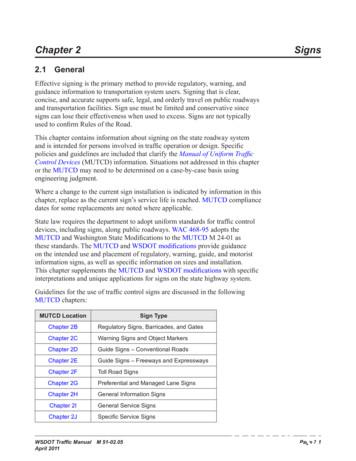
Signs Chapter 2 Page 2-2 WSDOT Traffic Manual M 51-02.05 April 2011 Chapter 2L Changeable Message Signs Chapter 2M Recreational and Cultural Interest Signs Chapter 2N Emergency Management Signs Part 6 Work Zone Signs Part 7 School Area Signs Part 8 Railroad and Light Rail Signs Part 9 Bicycle Facility Signs 2.2 Sign Design
Vital Signs They are called vital signs because they are important. They include: Temperature Pulse Respirations Blood pressure Vital signs and other physiologic measurements can be the bases for problem solving. Many facilities ha
Jun 16, 2021 · Vital Signs Vital Signs Including Pulse Oximetry upon initiation of Monoclonal Therapy infusion: Pre-infusion, every 15 min x1 hour; then every 30 min x2 after infusion has been completed Notify Provider of Vital Signs Changes in vital signs ( /-10%) during and immed
Vital Signs Integration – Welch Allyn CVSM MDET - Vital Signs Integration – Welch Allyn CVSM, v2.1, MDET002, 31/05/2021 Townsville Hospital and Health Service Page 3 GQEA-PUBLIC 8. To begin taking the patients vital signs, press Start on the screen next to Blood P
M2.06 Selection of Signs M2.07 Classification of Signs M2.08 Sign Materials M2.09 Storage and Handling of Signs M2.10 Installation of Signs M2.11 Sign Maintenance M2.12 Hidden Signs M2.13 Obsolete Signs M2.14 Temporary Signs M2.15 General Sign Support Information M2.16 Wood Posts M2.17 Steel Posts -- Small Signs M2.18 Timber Poles
Diamond Touch POS YES Vital/TSYS PC Charge Digital Dining (by MenuSoft) YES Vital/TSYS Digital Payment Tech YES Vital/TSYS Authorize.net. Dina Touch Pos YES PC Charge DINERWARE YES Vital/TSYS DirectTouch YES Vital/TSYS Vital/TSYS NETePay DataTran / Data Cap Dispensary Management YES Vital/


