Anatomy For Anaesthetists
Anatomy for Anaesthetists
This page intentionally left blank
A N AT O M Y F O RANAESTHETISTSHAROLD ELLISCBE, MA, DM, FRCS, FACS(Hon)Clinical Anatomist,Guy’s King’s and St Thomas’s School of Biomedical Sciences, LondonEmeritus Professor of Surgery,University of LondonSTANLEY FELDMANBSc, MB, FRCAEmeritus Professor of Anaesthetics,Charing Cross and Westminster Medical SchoolWILLIAM HARROP-GRIFFITHSMA, MB, BS, FRCAConsultant Anaesthetist,St Mary’s Hospital, Paddington, Londonwith a chapter on theAnatomy of Paincontributed byANDREW LAWSONFFARCSI, FANZCA, FRCA, MScConsultant in Anaesthesia and Pain Management,Royal Berkshire Hospital, ReadingEighth edition
1963, 1969, 1977, 1983, 1988, 1993, 1997, 2004by Blackwell Science Ltda Blackwell Publishing companyBlackwell Science, Inc., 350 Main Street, Malden,Massachusetts 02148-5020, USABlackwell Publishing Ltd, 9600 Garsington Road,Oxford OX4 2DQ, UKBlackwell Science Asia Pty Ltd, 550 Swanston Street,Carlton, Victoria 3053, AustraliaThe right of the Author to be identifiedas the Author of this Work has beenasserted in accordance with theCopyright, Designs and Patents Act 1988.All rights reserved. No part ofthis publication may be reproduced,stored in a retrieval system, ortransmitted, in any form or by anymeans, electronic, mechanical,photocopying, recording or otherwise,except as permitted by the UKCopyright, Designs and Patents Act1988, without the prior permissionof the publisher.First published 1963Second edition 1969Third edition 1977Reprinted 1979Fourth edition 1983Fifth edition 1988Reprinted 1990Sixth edition 1993Reprinted 1995Seventh edition 1997Reprinted 1998Italian first edition 1972Japanese fourth edition 1989German fifth edition 1992Library of Congress Cataloging-in-Publication DataEllis, Harold, 1926–Anatomy for anaesthetists / Harold Ellis, StanleyFeldman, William Harrop-Griffiths; with a chapteron the Anatomy of pain contributed by AndrewLawson.—8th ed.p. ; cm.Includes bibliographical references and index.ISBN 1-4051-0663-81. Human anatomy. 2. Anesthesiology.[DNLM: 1. Anatomy. 2. Anesthesia.QS 4 E47a 2003] I. Feldman, Stanley A.II. Harrop-Griffiths, William. III. Title.QM23.2.E42 2003611′.0024617—dc222003020753ISBN 1405 1066 38A catalogue record for this title is available from theBritish LibrarySet in 10/13.5pt Sabon by Graphicraft Limited,Hong KongPrinted and bound in Denmark, by Narayana Press,OdderCommissioning Editor: Stuart TaylerEditorial Assistant: Katrina ChandlerProduction Editor: Rebecca HuxleyProduction Controller: Kate CharmanFor further information on Blackwell Publishing,visit our website:http://www.blackwellpublishing.com
ContentsPart 1: The Respiratory Pathway 1The Mouth 3The Nose 7The Pharynx 16The Larynx 26The Trachea 42The Main Bronchi 48The Pleura 50The Lungs 53Part 2: The Heart 71The Pericardium 73The Heart 75Developmental Anatomy 86Part 3: The Vertebral Canal and its Contents 95The Vertebrae and Sacrum 97The Spinal Meninges 119The Spinal Cord 125Part 4: The Peripheral Nerves 137The Spinal Nerves 139The Cervical Plexus 146The Brachial Plexus 153The Thoracic Nerves 180The Lumbar Plexus 183The Sacral and Coccygeal Plexuses 192Part 5: The Autonomic Nervous System 213Introduction 215The Sympathetic System 218The Parasympathetic System 228Part 6: The Cranial Nerves 233Introduction 235The Olfactory Nerve (I) 238The Optic Nerve (II) 239v
viContentsThe Oculomotor Nerve (III) 242The Trochlear Nerve (IV) 244The Trigeminal Nerve (V) 245The Abducent Nerve (VI) 266The Facial Nerve (VII) 267The Auditory (Vestibulocochlear) Nerve (VIII) 272The Glossopharyngeal Nerve (IX) 273The Vagus Nerve (X) 276The Accessory Nerve (XI) 282The Hypoglossal Nerve (XII) 283Part 7: The Anatomy of Pain 285Introduction 287Classification of Pain 288Peripheral Receptors and Afferent fibres 288The Spinal Cord and Central Projections 290Modulation of Pain 294The Gate Control Theory of Pain 295The Sympathetic Nervous System and Pain 296Part 8: Zones of Anaesthetic Interest 297The Thoracic inlet 299The Diaphragm 305The Intercostal Spaces 311The Abdominal Wall 318The Antecubital Fossa 324The Great Veins of the Neck 330The Orbit and its Contents 336Index 349
AcknowledgementsThe first two editions of this textbook were prepared in collaboration with thatskilled medical artist Miss Margaret McLarty. The illustrations for the sixthedition were almost all drawn or redrafted by Rachel Chesterton; we thank herfor the excellent way in which they have been executed. Further illustrations forthe seventh and this edition were prepared by Jane Fallows with great skill.vii
IntroductionThe anaesthetist requires a particularly specialized knowledge of anatomy. Someregions of the body, for example the respiratory passages, the major veins andthe peripheral nerves, the anaesthetist must know with an intimacy of detail thatrivals or even exceeds that of the surgeon; other areas can be all but ignored.Although formal anatomy teaching is no longer part of the syllabus of the FRCAin the UK, its importance for the safe practice of anaesthesia is recognized bythe examiners, who always include questions on anatomy related to anaesthesiain this examination. The role of anatomy in anaesthetic teaching is often considered merely as a prerequisite for the safe practice of local anaesthetic blocks.However, it is also important in understanding the anatomy of the airway, thefunction of the lungs, the circulation, venous access, monitoring neuromuscularblock and many other aspects of practical anaesthesia. For this reason, this bookis not intended to be a textbook for regional anaesthetic techniques; there aremany excellent books in this field. It is an anatomy book written for anaesthetists,keeping in mind the special requirements of their daily practice.In this eighth edition, we have revised much of the text, we have taken theopportunity to expand and update the sections of special interest to anaesthetistsand we have included new and improved illustrations. William Harrop-Griffithsof St Mary’s Hospital, London, joins us as our new co-author. He brings withhim special expertise in modern anaesthetic technology and has greatly assistedus in updating the text and illustrations. Dr Andrew Lawson has fully updatedhis important section on the Anatomy of Pain and has given valuable advice onprocedures relevant to the practice of pain medicine.viii
Part 1The Respiratory Pathway
This page intentionally left blank
The MouthThe mouth is made up of the vestibule and the mouth cavity, the former communicating with the latter through the aperture of the mouth.The vestibule is formed by the lips and cheeks without and by the gums andteeth within. An important feature is the opening of the parotid duct on a smallpapilla opposite the 2nd upper molar tooth. Normally the walls of the vestibuleare kept together by the tone of the facial muscles; a characteristic feature of afacial (VII) nerve paralysis is that the cheek falls away from the teeth and gums,enabling food and drink to collect in, and dribble out of, the now patulousvestibule.The mouth cavity (Fig. 1) is bounded by the alveolar arch of the maxilla andthe mandible, and teeth in front, the hard and soft palate above, the anteriortwo-thirds of the tongue and the reflection of its mucosa forward onto themandible below, and the oropharyngeal isthmus behind.The mucosa of the floor of the mouth between the tongue and mandiblebears the median frenulum linguae, on either side of which are the orifices of theUvulaPalatopharyngealarchPalatine tonsilPalatoglossalarchFig. 1 View of the open mouth with the tongue depressed.3
4The Respiratory PathwayFrenulum linguaeSublingual foldOrifice ofsubmandibularductFig. 2 View of the open mouth with the tongue elevated.submandibular salivary glands (Fig. 2). Backwards and outwards from theseducts extend the sublingual folds that cover the sublingual glands on each side(Fig. 3); the majority of the ducts of these glands open as a series of tiny orificesalong the overlying fold, but some drain into the duct of the submandibular gland(Wharton’s duct).The palateThe hard palate is made up of the palatine processes of the maxillae and thehorizontal plates of the palatine bones. The mucous membrane covering thehard palate is peculiar in that the stratified squamous mucosa is closely connected to the underlying periosteum, so that the two dissect away at operationas a single sheet termed the mucoperiosteum. This is thin in the midline, butthicker more laterally due to the presence of numerous small palatine salivaryglands, an uncommon but well-recognized site for the development of mixedsalivary tumours.The soft palate hangs like a curtain suspended from the posterior edge of thehard palate. Its free border bears the uvula centrally and blends on either side withthe pharyngeal wall. The anterior aspect of this curtain faces the mouth cavityand is covered by a stratified squamous epithelium. The posterior aspect is part
The MouthSubmandibularduct and idSublingual glandLingual N.Anterior bellyof digastricLingual A.Fig. 3 Coronal section through the floor of the mouth.of the nasopharynx and is lined by a ciliated columnar epithelium under whichis a thick stratum of mucous and serous glands embedded in lymphoid tissue.The ‘skeleton’ of the soft palate is a tough fibrous sheet termed the palatineaponeurosis, which is attached to the posterior edge of the hard palate. Theaponeurosis is continuous on each side with the tendon of tensor palati and may,in fact, represent an expansion of this tendon.The muscles of the soft palate are five in number: the tensor palati, the levatorpalati, the palatoglossus, the palatopharyngeus and the musculus uvulae (seeFig. 13).The tensor palati (tensor veli palatini) arises from the scaphoid fossa at the rootof the medial pterygoid plate, from the lateral side of the Eustachian cartilageand the medial side of the spine of the sphenoid. Its fibres descend laterally to thesuperior constrictor and the medial pterygoid plate to end in a tendon that piercesthe pharynx, loops medially around the hook of the hamulus to be inserted intothe palatine aponeurosis. Its action is to tighten and flatten the soft palate.The levator palati (levator veli palatini) arises from the undersurface of thepetrous temporal bone and from the medial side of the Eustachian tube, entersthe upper surface of the soft palate and meets its fellow of the opposite side.It elevates the soft palate.The palatoglossus arises in the soft palate, descends in the palatoglossal foldand blends with the side of the tongue. It approximates the palatoglossal folds.5
6The Respiratory PathwayThe palatopharyngeus descends from the soft palate in the palatopharyngealfold to merge into the side wall of the pharynx: some fibres become inserted alongthe posterior border of the thyroid cartilage. It approximates the palatopharyngealfolds.The musculus uvulae takes origin from the palatine aponeurosis at the posteriornasal spine of the palatine bone and is inserted into the uvula. Injury to the cranialroot of the accessory nerve, which supplies this muscle via the vagus nerve, resultsin the uvula becoming drawn across and upwards towards the opposite side.The tensor palati is innervated by the mandibular branch of the trigeminalnerve via the otic ganglion (see p. 265). The other palatine muscles are suppliedby the pharyngeal plexus, which transmits cranial fibres of the accessory nervevia the vagus.The palatine muscles help to close off the nasopharynx from the mouth indeglutition and phonation. In this, they are aided by contraction of the upperpart of the superior constrictor, which produces a transverse ridge on the backand side walls of the pharynx at the level of the 2nd cervical vertebra termedthe ridge of Passavant.Partial clefts of palatePremaxillaVomerUnilateral completecleft palateFig. 4 Types of cleft palate deformity.Bilateral completecleft palate
The NoseParalysis of the palatine muscles results (just as surely as a severe degreeof cleft palate deformity) in a typical nasal speech and in regurgitation of foodthrough the nose.Cleft palateThe palate develops from a central premaxilla and a pair of lateral maxillaryprocesses: the former usually bears all four (occasionally only two) of theincisor teeth. All degrees of failure of fusion of these three processes may takeplace. There may be a complete cleft, which passes to one or both sides of thepremaxilla; in the latter case, the premaxilla prolapses forwards to produce amarked deformity. Partial clefts of the posterior palate may involve the uvulaonly (bifid uvula), involve the soft palate or encroach into the posterior partof the hard palate (Fig. 4).The NoseThe nose is divided anatomically into the external nose and the nasal cavity.The external nose is formed by an upper framework of bone (made up of thenasal bones, the nasal part of the frontal bones and the frontal processes of themaxillae), a series of cartilages in the lower part, and a small zone of fibro-fattytissue that forms the lateral margin of the nostril (the ala). The cartilage of thenasal septum comprises the central support of this framework.The cavity of the nose is subdivided by the nasal septum into two quite separatecompartments that open to the exterior by the nares and into the nasopharynx bythe posterior nasal apertures or choanae. Immediately within the nares is a smalldilatation, the vestibule, which is lined in its lower part by stiff, straight hairs.Each side of the nose presents a roof, a floor and a medial and lateral wall.The roof first slopes upwards and backwards to form the bridge of the nose(the nasal and frontal bones), then has a horizontal part (the cribriform plateof the ethmoid), and finally a downward-sloping segment (the body of thesphenoid).The floor is concave from side to side and slightly so from before backwards.It is formed by the palatine process of the maxilla and the horizontal plate of thepalatine bone.The medial wall (Fig. 5) is the nasal septum, formed by the septal cartilage,the perpendicular plate of the ethmoid and the vomer. Deviations of the septumare very common; in fact, they are present to some degree in about 75% of theadult population. Probably nearly all are traumatic in origin, and result from7
8The Respiratory PathwayFrontal sinusPerpendicular plateof ethmoidNasal boneCartilage of septumSphenoidalair sinusNasal vestibuleVomerPalatine processof maxillaHorizontal plateof palatine boneFig. 5 The septum of the nose.quite minor injuries in childhood or even at birth. The deformity does not usually manifest itself until the second dentition appears, when rapid growth in theregion produces deflections from what had been an unrecognized minor dislocation of the septal cartilage. Males are more commonly affected than females,a distribution which would favour this traumatic theory. Both nostrils may becomeblocked, either from a sigmoid deformity of the cartilage or from compensatoryhypertrophy of the conchae on the opposite side. The deviation is nearly alwaysconfined to the anterior part of the septum.The lateral wall (Fig. 6) has a bony framework made up principally of the nasalaspect of the ethmoidal labyrinth above, the nasal surface of the maxilla belowand in front, and the perpendicular plate of the palatine bone behind. This issupplemented by the three scroll-like conchae (or turbinate bones), each archingover a meatus. The upper and middle conchae are derived from the medial aspectof the ethmoid labyrinth; the inferior concha is a separate bone.Onto the lateral wall open the orifices of the paranasal sinuses (see p. 10) andthe nasolacrimal duct; the arrangement of these orifices is shown in Fig. 7.The sphenoid sinus opens into the spheno-ethmoidal recess, a depression betweenthe short superior concha and the anterior surface of the body of the sphenoid.The posterior ethmoidal cells drain into the superior meatus. The middle ethmoidal cells bulge into the middle meatus to form an elevation, termed the bullaethmoidalis, on which they open. Below the bulla is a cleft, the hiatus semilunaris,into which opens the ostium of the maxillary sinus. The hiatus semilunaris curves
The NoseCribriform plateof ethmoidSpheno-ethmoidalrecessSuperior conchaMiddle conchaInferior conchaVestibuleInferior meatusFig. 6 The lateral wall of the right nasal cavity.Hiatus semilunarisPosterior ethmoidal openingFrontal sinusSphenoid sinusOpening of sphenoid sinusinto spheno-ethmoidal recessFrontalsinus openingAnteriorethmoidalopeningMiddleethmoidal openingBulla ethmoidalisMaxillary ostiumEustachian orificePharyngeal recessOpening of nasolacrimal ductFig. 7 The lateral wall of the right nasal cavity; the conchae have been partially removed.forward in front of the bulla ethmoidalis as a passage termed the infundibulum,which drains the anterior ethmoidal air cells. In about 50% of cases the frontalsinus drains into the infundibulum via the frontonasal duct. In the remainder,this duct opens into the anterior extremity of the middle meatus.9
10The Respiratory PathwayThe nasolacrimal duct drains tears into the anterior end of the inferior meatusin solitary splendour.The paranasal sinusesThe paranasal air sinuses comprise the maxillary, sphenoid, frontal and ethmoidal sinuses. They are, in effect, the out-pouchings from the lateral wall ofthe nasal cavity into which they drain; they all differ considerably from subjectto subject in their size and extent, and they are rarely symmetrical. There aretraces of the maxillary and sphenoid sinuses in the newborn; the rest becomeevident about the age of 7 or 8 years in association with the eruption of thesecond dentition and lengthening of the face. They only become fully developedat adolescence.The maxillary sinus (the antrum of Highmore) is the largest of the sinuses.It is pyramid-shaped, and occupies the body of the maxilla (Fig. 8). The base ofthis pyramid is the lateral wall of the nasal cavity and its apex points laterallytowards the zygomatic process.Ethmoidalair cellsCrista galliSuperiorconchaOrbitAntralostiumMiddle andinferiorconchaeMaxillaryantrumNasalseptumHard palate(maxilla)Fig. 8 The maxillary sinus in coronal section.
The NoseThe floor of the sinus extends into the alveolar process of the maxilla, whichlies about 1.25 cm below the level of the floor of the nose. Bulges in the floorare produced by the roots of at least the 1st and 2nd molars; the number of suchprojections is variable and may include all the teeth derived from the maxillaryprocess, i.e. the canine, premolars and molars. The floor may actually be perforated by one or more of the roots.The roof is formed by the orbital plate of the maxilla, which bears the canalof the infra-orbital branch of the maxillary nerve. Medially, the antrum drainsinto the middle meatus; the ostium is situated high up on this wall and is thusinefficiently placed from the mechanical point of view. Drainage from this sinusis therefore dependent on the effectiveness of the cilia lining its wall. There maybe one or more accessory openings from the antrum into the middle meatus.The sphenoid sinuses lie side by side in the body of the sphenoid. Occasionally,they extend into the basisphenoid and the clinoid processes. They are seldomequal in size, and the septum between them is usually incomplete. They openinto the spheno-ethmoidal recess.The frontal sinuses occupy the frontal bone above the orbits and the rootof the nose. They are usually unequal, and their dividing septum may be incomplete. It is interesting that their extent is in no way related to the size of thesuperciliary ridges. They drain through the frontonasal duct into the middlemeatus.The ethmoidal sinuses or air cells are made up of some 8–10 loculi suspendedfrom the outer extremity of the cribriform plate of the ethmoid and boundedlaterally by its orbital plate. They thus occupy
block and many other aspects of practical anaesthesia. For this reason, this book is not intended to be a textbook for regional anaesthetic techniques; there are many excellent books in this field. It is an anatomy book written for anaesthetists, keeping in mind the special requirements of their daily practice.
Bruksanvisning för bilstereo . Bruksanvisning for bilstereo . Instrukcja obsługi samochodowego odtwarzacza stereo . Operating Instructions for Car Stereo . 610-104 . SV . Bruksanvisning i original
Clinical Anatomy RK Zargar, Sushil Kumar 8. Human Embryology Daksha Dixit 9. Manipal Manual of Anatomy Sampath Madhyastha 10. Exam-Oriented Anatomy Shoukat N Kazi 11. Anatomy and Physiology of Eye AK Khurana, Indu Khurana 12. Surface and Radiological Anatomy A. Halim 13. MCQ in Human Anatomy DK Chopade 14. Exam-Oriented Anatomy for Dental .
39 poddar Handbook of osteology Anatomy Textbook 10 40 Ross ,Pawlina Histology a text & atlas Anatomy Textbook 10 41 Halim A. Human anatomy Abdomen & lower limb Anatomy Referencebook 10 42 B.D. Chaurasia Human anatomy Head & Neck, Brain Anatomy Referencebook 10 43 Halim A. Human anatomy Head & Neck, Brain Anatomy Referencebook 10
10 tips och tricks för att lyckas med ert sap-projekt 20 SAPSANYTT 2/2015 De flesta projektledare känner säkert till Cobb’s paradox. Martin Cobb verkade som CIO för sekretariatet för Treasury Board of Canada 1995 då han ställde frågan
service i Norge och Finland drivs inom ramen för ett enskilt företag (NRK. 1 och Yleisradio), fin ns det i Sverige tre: Ett för tv (Sveriges Television , SVT ), ett för radio (Sveriges Radio , SR ) och ett för utbildnings program (Sveriges Utbildningsradio, UR, vilket till följd av sin begränsade storlek inte återfinns bland de 25 största
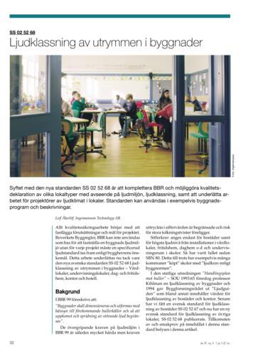
Hotell För hotell anges de tre klasserna A/B, C och D. Det betyder att den "normala" standarden C är acceptabel men att motiven för en högre standard är starka. Ljudklass C motsvarar de tidigare normkraven för hotell, ljudklass A/B motsvarar kraven för moderna hotell med hög standard och ljudklass D kan användas vid
LÄS NOGGRANT FÖLJANDE VILLKOR FÖR APPLE DEVELOPER PROGRAM LICENCE . Apple Developer Program License Agreement Syfte Du vill använda Apple-mjukvara (enligt definitionen nedan) för att utveckla en eller flera Applikationer (enligt definitionen nedan) för Apple-märkta produkter. . Applikationer som utvecklas för iOS-produkter, Apple .
Artificial Intelligence (AI) has the potential to make a significant difference to health and care. A broad range of techniques can be used to create Artificially Intelligent Systems (AIS) to carry out or augment health and care tasks that have until now been completed by humans, or have not been possible previously; these techniques include inductive logic programming, robotic process .


