CHAPTER 3 Cell Structure And Function
CHAPTER3Cell Structureand FunctionK E Y CO N C E P T S3.1 Cell TheoryCells are the basic unit of life.3.2 Cell OrganellesEukaryotic cells share many similarities.3.3 Cell MembraneThe cell membrane is a barrier that separates a cell from theexternal environment.3.4 Diffusion and OsmosisMaterials move across membranes because of concentrationdifferences.3.5 Active Transport, Endocytosis,and ExocytosisCells use energy to transport materials that cannot diffuseacross a membrane.BIOLOGYBIOLOGYView animated chapterconcepts. Cell Organelles Get Through a CellMembrane68Unit 2: CellsCL ASSZONE .COMRESOURCE CENTERKeep current with biology news. Get more information on Featured stories Prokaryotic and Eukaryotic News feedsCells Careers Diffusion and Osmosis
colored SEM; magnification 11,000 Why do these cellslook like fried eggs?ConnectingMacrophages (large tan cells) take inand digest foreign material, such asinvading bacteria (small red cells). Theyplay an important role in your immunesystem. Many macrophages travel the body,recognize foreign material, engulf it, andbreak it down using chemicals. They havean adaptable internal skeleton that helpsthem move and stretch out their “arms” tocapture invading particles.CONCEPTSTechnology The scanningelectron microscope (SEM)uses electrons to creategreatly magnified, threedimensional images ofsurface structures. Samplesmust be carefully prepared towithstand the vacuum and toprevent shriveling. Thismeans that any cell ororganism you see in an SEMimage is dead. In addition,images are generated in blackand white (left). The pictureabove is artificially colored tohighlight specific parts.Chapter 3: Cell Structure and Function69
3.1Cell TheoryKEY CONCEPTCells are the basic unit of life.MAIN IDEAS Early studies led to the developmentof the cell theory. Prokaryotic cells lack a nucleus and mostinternal structures of eukaryotic cells.VOCABULARYcell theory, p. 71cytoplasm, p. 72organelle, p. 72prokaryotic cell, p. 72eukaryotic cell, p. 72Connect You and all other organisms are made of cells. As you saw on theprevious page, a cell’s structure is closely related to its function. Today we knowthat cells are the smallest unit of living matter that can carry out all processesrequired for life. But before the 1600s, people had many other ideas about thebasis of life. Like many breakthroughs, the discovery of cells was aided by thedevelopment of new technology—in this case, the microscope.MAIN IDEAEarly studies led to the development of thecell theory.TAKING NOTESAs you read, make an outlineusing the headings as topics.Summarize details that furtherexplain those ideas.I. Main IdeaA. Supporting idea1. Detail2. DetailB. Supporting ideaAlmost all cells are too small to see without the aid of a microscope. Althoughglass lenses had been used to magnify images for hundreds of years, the earlylenses were not powerful enough to reveal individual cells. The invention ofthe compound microscope in the late 1500s was an early step toward thisdiscovery. The Dutch eyeglass maker Zacharias Janssen, who was probablyassisted by his father, Hans, usually gets credit for this invention.A compound microscope contains two or more lenses. Total magnification, theproduct of the magnifying power of each individual lens, is generally much morepowerful with a compound microscope than with a single lens.Discovery of CellsFIGURE 3.1 Hookefirst identified cellsusing this microscope.Its crude lensesseverely limited theamount of detailhe could see.70Unit 2: CellsIn 1665, the English scientist Robert Hooke used the three-lens compoundmicroscope shown in FIGURE 3.1 to examine thin slices of cork. Cork is thetough outer bark of a species of oak tree. He observed that cork is made oftiny, hollow compartments. The compartments reminded Hooke of smallrooms found in a monastery, so he gave them the same name: cells. Theplant cells he observed, f the nucleus. But many proteins are involved in turning geneson and off, and they need to access the DNA at certaintimes. The special structure of the nucleus helps it meetboth demands.The nucleus is composed of the cell’s DNA enclosed in adouble membrane called the nuclear envelope. Each memnucleusbrane in the nuclear envelope is similar to the membranesurrounding the entire cell. As FIGURE 3.7 shows, the nuclearenvelope is pierced with holes called pores that allow largemolecules to pass between the nucleus and cytoplasm.FIGURE 3.7 The nucleus storesand protects DNA. (colored SEM;magnification 90,000 )poresThe nucleus also contains the nucleolus. The nucleolus is adense region where tiny organelles essential for makingproteins are assembled. These organelles, called ribosomes, area combination of proteins and RNA molecules. They arediscussed on the next page, and a more complete descriptionof their structure and function is given in Chapter 8.Chapter 3: Cell Structure and Function75
Endoplasmic Reticulum and RibosomesFIGURE 3.8 The endoplasmicA large part of the cytoplasm of most eukaryotic cells is filled by theendoplasmic reticulum, shown in FIGURE 3.8. The endoplasmic reticulum (EHNduh-PLAZ-mihk rih-TIHK-yuh-luhm), or the ER, is an interconnectednetwork of thin folded membranes. The composition is verysimilar to that of the cell membrane and nuclear membranes.The ER membranes form a maze of enclosed spaces. The interiorendoplasmic reticulumof this maze is called the lumen. Numerous processes, includingthe production of proteins and lipids, occur both on the surfaceof the ER and inside the lumen. The ER must be large enough toribosomesaccommodate all these processes. How does it fit inside a cell?The ER membrane has many creases and folds. If you haveever gone camping, you probably slept in a sleeping bag thatcovered you from head to foot. The next morning, you stuffedit back into a tiny little sack. How does the entire sleeping bagfit inside such a small sack? The surface area of the sleeping bagdoes not change, but the folds allow it to take up less space.Likewise, the ER’s many folds enable it to fit within the cell.In some regions, the ER is studded with ribosomes (RY-buh-SOHMZ),tiny organelles that link amino acids together to form proteins. Ribosomesribosomerough ERare both the site of protein synthesis and active participants in the process.Ribosomes are themselves made of proteins and RNA. After assembly in thenucleolus, ribosomes pass through the nuclear pores into the cytoplasm,smooth ERwhere most protein synthesis occurs.Surfaces of the ER that are covered with ribosomes are called roughER because they look bumpy when viewed with an electron microscope.As a protein is being made on these ribosomes, it enters the lumen. Insidethe lumen, the protein may be modified by having sugar chains addedto it, which can help the protein fold or give it stability.Not all ribosomes are bound to the ER; some are suspended in the cytoplasm. In general, proteins made on the ER are either incorporated into thecell membrane or secreted. In contrast, proteins made on suspended ribosomes are typically used in chemical reactions occurring within the cytoplasm.FIGURE 3.9 The Golgi apparatus modifies, packages,Surfaces of the ER that do not contain ribosomes are called smooth ER.and transports proteins.SmoothER makes lipids and performs a variety of other specialized functions,(colored TEM; magnificationsuch as breaking down drugs and alcohol.about 10,000 )reticulum aids in the productionof proteins and lipids. (colored TEM;magnification about 20,000 )Golgi ApparatusGolgi apparatus76Unit 2: CellsFrom the ER, proteins generally move to the Golgi apparatus,shown in FIGURE 3.9. The Golgi apparatus (GOHL-jee) consists ofclosely layered stacks of membrane-enclosed spaces that process,sort, and deliver proteins. Its membranes contain enzymes thatmake additional changes to proteins. The Golgi apparatus alsopackages proteins. Some of the packaged proteins are storedwithin the Golgi apparatus for later use. Some are transported toother organelles within the cell. Still others are carried to themembrane and secreted outside the cell.
VesiclesCells need to separate reactants for various chemical reactionsuntil it is time for them to be used. Vesicles (VEHS-ih-kuhlz),shown in FIGURE 3.10, are a general name used to describe smallmembrane-bound sacs that divide some materials from the rest ofthe cytoplasm and transport these materials from place to placewithin the cell. Vesicles are generally short-lived and are formedand recycled as needed.After a protein has been made, part of the ER pinches off toform a vesicle surrounding the protein. Protected by the vesicle,the protein can be safely transported to the Golgi apparatus.There, any necessary modifications are made, and the protein ispackaged inside a new vesicle for storage, transport, or secretion.FIGURE 3.10 Vesicles isolateand transport specific molecules.(colored SEM; magnification 20,000 )vesiclesCompare and Contrast How are the nucleus and a vesicle similar anddifferent in structure and function?MAIN IDEAOther organelles have various functions.MitochondriaMitochondria (MY-tuh-KAHN-dree-uh) supply energy to the cell.Mitochondria (singular, mitochondrion) are bean shaped and havetwo membranes, as shown in FIGURE 3.11. The inner membrane hasmany folds that greatly increase its surface area. Within theseinner folds and compartments, a series of chemical reactions takesplace that converts molecules from the food you eat into usableenergy. You will learn more about this process in Chapter 4.Unlike most organelles, mitochondria have their own ribosomes and DNA. This fact suggests that mitochondria wereoriginally free-living prokaryotes that were taken in by larger cells.The relationship must have helped both organisms to survive.VacuoleA vacuole (VAK-yoo-OHL) is a fluid-filled sac used for the storageof materials needed by a cell. These materials may include water,food molecules, inorganic ions, and enzymes. Most animal cellscontain many small vacuoles. The central vacuole, shown inFIGURE 3.12, is a structure unique to plant cells. It is a single largevacuole that usually takes up most of the space inside a plant cell.It is filled with a watery fluid that strengthens the cell and helps tosupport the entire plant. When a plant wilts, its leaves shrivelbecause there is not enough water in each cell’s central vacuole tosupport the leaf ’s normal structure. The central vacuole may alsocontain other substances, including toxins that would harm predators, waste products that would harm the cell itself, and pigmentsthat give color to cells—such as those in the petals of a flower.FIGURE 3.11 Mitochondria generateenergy for the cell. (colored TEM;magnification 33,000 )outermembranemitochondrioninner membraneFIGURE 3.12 Vacuoles temporarilystore materials. (colored TEM;magnification 9000 )vacuoleChapter 3: Cell Structure and Function77
LysosomesFIGURE 3.13 Lysosomes digestand recycle foreign materialsor worn-out parts. (colored TEM;magnification 21,000 )Lysosomes (LY-suh-SOHMZ), shown in FIGURE 3.13, are membrane-boundorganelles that contain enzymes. They defend a cell from invading bacteriaand viruses. They also break down damaged or worn-out cell parts. Lysosomestend to be numerous in animal cells. Their presence in plant cells is stillquestioned by some scientists, but others assert that plant cells do have lysosomes, though fewer than are found in animal cells.Recall that all enzymes are proteins. Initially, lysosomal enzymes are madein the rough ER in an inactive form. Vesicles pinch off from the ER membrane, carry the enzymes, and then fuse with the Golgi apparatus. There, theenzymes are activated and packaged as lysosomes that pinch off from theGolgi membrane. The lysosomes can then engulf and digest targeted molecules. When a molecule is broken down, the products pass through thelysosomal membrane and into the cytoplasm, where they are used again.Lysosomes provide an example of the importance of membrane-boundstructures in the eukaryotic cell. Because lysosomal enzymes can destroy cellcomponents, they must be surrounded by a membrane that prevents themfrom destroying necessary structures. However, the cell also uses other methods to protect itself from these destructive enzymes. For example, the enzymesdo not work as well in the cytoplasm as they do inside the lysosome.lysosomeCentrosome and CentriolesFIGURE 3.14 Centrioles divideDNA during cell division.(colored TEM; magnification 35,000 )top viewcentriolesside viewThe centrosome is a small region of cytoplasm that produces microtubules. Inanimal cells, it contains two small structures called centrioles. Centrioles(SEHN-tree-OHLZ) are cylinder-shaped organelles made of short microtubulesarranged in a circle. The two centrioles are perpendicular to each other, asshown in FIGURE 3.14. Before an animal cell divides, the centrosome, includingthe centrioles, doubles and the two new centrosomes move to opposite ends ofthe cell. Microtubules grow from each centrosome, forming spindle fibers. Thesefibers attach to the DNA and appear to help divide it between the two cells.Centrioles were once thought to play a critical role in animal cell division.However, experiments have shown that animal cells can divide even if thecentrioles are removed, which makes their role more questionable. In addition, although centrioles are found in some algae, they are not found in plants.Centrioles also organize microtubules to form cilia and flagella. Cilia looklike little hairs; flagella look like a whip or a tail. Their motion forces liquidspast a cell. For single cells, this movement results in swimming. For cellsanchored in tissue, this motion sweeps liquid across the cell surface.Compare In what ways are lysosomes, vesicles, and the central vacuole similar?MAIN IDEAPlant cells have cell walls and chloroplasts.Plant cells have two features not shared by animal cells: cell walls and chloroplasts. Cell walls are structures that provide rigid support. Chloroplasts areorganelles that help a plant convert solar energy to chemical energy.78Unit 2: Cells
FIGURE 3.15 Cell walls shapeCell WallsIn plants, algae, fungi, and most bacteria, the cell membrane is surrounded bya strong cell wall, which is a rigid layer that gives protection, support, andshape to the cell. The cell walls of multiple cells, as shown in FIGURE 3.15, canadhere to each other to help support an entire organism. For instance, muchof the wood in a tree trunk consists of dead cells whose cell walls continue tosupport the entire tree.Cell wall composition varies and is related to the different needs of eachtype of organism. In plants and algae, the cell wall is made of cellulose, apolysaccharide. Because molecules cannot easily diffuse across cellulose, thecell walls of plants and algae have openings, or channels. Water and othermolecules small enough to fit through the channels can freely pass throughthe cell wall. In fungi, cell walls are made of chitin, and in bacteria, they aremade of peptidoglycan. The unique characteristics and functions of thesematerials will be discussed in Chapters 18 and 19.and support individual cells andentire organisms. (LM; magnification3000 )ChloroplastsFIGURE 3.16 Chloroplasts convertChloroplasts (KLAWR-uh-PLASTS) are organelles that carry out photosynthesis,a series of complex chemical reactions that convert solar energy into energyrich molecules the cell can use. Photosynthesis will be discussed more fully inChapter 4. Like mitochondria, chloroplasts are highly compartmentalized.They have both an outer membrane and an inner membrane. They also havestacks of disc-shaped sacs within the inner membrane, shown in FIGURE 3.16.These sacs, called thylakoids, contain chlorophyll, a light-absorbing moleculethat gives plants their green color and plays a key role in photosynthesis. Likemitochondria, chloroplasts also have their own r
KEY CONCEPTS 3 Cell Structure and Function 3.1 Cell Theory Cells are the basic unit of life. 3.2 Cell Organelles Eukaryotic cells share many similarities. 3.3 Cell Membrane The cell membrane is a barrier that separates a cell from the external environment. 3.4 Diffusion and Osmosis Mat
Part One: Heir of Ash Chapter 1 Chapter 2 Chapter 3 Chapter 4 Chapter 5 Chapter 6 Chapter 7 Chapter 8 Chapter 9 Chapter 10 Chapter 11 Chapter 12 Chapter 13 Chapter 14 Chapter 15 Chapter 16 Chapter 17 Chapter 18 Chapter 19 Chapter 20 Chapter 21 Chapter 22 Chapter 23 Chapter 24 Chapter 25 Chapter 26 Chapter 27 Chapter 28 Chapter 29 Chapter 30 .
TO KILL A MOCKINGBIRD. Contents Dedication Epigraph Part One Chapter 1 Chapter 2 Chapter 3 Chapter 4 Chapter 5 Chapter 6 Chapter 7 Chapter 8 Chapter 9 Chapter 10 Chapter 11 Part Two Chapter 12 Chapter 13 Chapter 14 Chapter 15 Chapter 16 Chapter 17 Chapter 18. Chapter 19 Chapter 20 Chapter 21 Chapter 22 Chapter 23 Chapter 24 Chapter 25 Chapter 26
UNIT-V:CELL STRUCTURE AND FUNCTION: 9. Cell- The Unit of Life: Cell- Cell theory and cell as the basic unit of life- overview of the cell. Prokaryotic and Eukoryotic cells, Ultra Structure of Plant cell (structure in detail and functions in brief), Cell membrane, Cell wall, Cell organelles: Endoplasmic reticulum, Mitochondria, Plastids,
DEDICATION PART ONE Chapter 1 Chapter 2 Chapter 3 Chapter 4 Chapter 5 Chapter 6 Chapter 7 Chapter 8 Chapter 9 Chapter 10 Chapter 11 PART TWO Chapter 12 Chapter 13 Chapter 14 Chapter 15 Chapter 16 Chapter 17 Chapter 18 Chapter 19 Chapter 20 Chapter 21 Chapter 22 Chapter 23 .
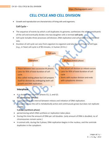
of the cell and eventually divides into two daughter cells is termed cell cycle. Cell cycle includes three processes cell division, DNA replication and cell growth in coordinated way. Duration of cell cycle can vary from organism to organism and also from cell type to cell type. (e.g., in Yeast cell cycle is of 90 minutes, in human 24 hrs.)
Many scientists contributed to the cell theory. The cell theory grew out of the work of many scientists and improvements in the . CELL STRUCTURE AND FUNCTION CHART PLANT CELL ANIMAL CELL . 1. Cell Wall . Quiz of the cell Know all organelles found in a prokaryotic cell
Class-XI-Biology Cell Cycle and Cell Division 1 Practice more on Cell Cycle and Cell Division www.embibe.com CBSE NCERT Solutions for Class 11 Biology Chapter 10 Back of Chapter Questions 1. What is the average cell cycle span for a mammalian cell? Solution: The average cell cycle span o
Jan 21, 2020 · pertaining to the cell theory, structure and functions, cell types and modifications, cell cycle and transport mechanisms. This module has seven (7) lessons: Lesson 1- Cell Theory Lesson 2- Cell Structure and Functions Lesson 3- Prokaryotic vs Eukaryotic Cells Lesson 4- Cell Types and Cell


