Regulation Of Cell Cycle Proteins By Chemokine Receptors .
Regulation of cell cycle proteins by chemokinereceptors: A novel pathway in humanimmunodeficiency virus neuropathogenesis?Renato Brandimarti,1,2 Muhammad Zafrullah Khan,1 Alessandro Fatatis,1 and Olimpia Meucci1QUERY SHEETQ1:Q2:Q3:Au: Pls. define EMSA.Au: Pls. define PTX and ERK.Au: Pls. complete reference.
Journal of NeuroVirology, 10(suppl. 1): 1–5, 2004 c 2004 Journal of NeuroVirologyISSN: 1355-0284 print / 1538-2443 onlineDOI: 10.1080/13550280490268287Regulation of cell cycle proteins by chemokinereceptors: A novel pathway in humanimmunodeficiency virus neuropathogenesis?Renato Brandimarti,1,2 Muhammad Zafrullah Khan,1 Alessandro Fatatis,1 and Olimpia Meucci151015201 Department2 Departmentof Pharmacology and Physiology, Drexel University College of Medicine, Philadelphia, Pennsylvania, USA;of Experimental Pathology, University of Bologna, ItalyIn order to test the hypothesis that alteration of cell cycle proteins are involvedin the neuronal damage caused by human immunodeficiency virus (HIV), theauthors have been studying the effect of chemokines on the CDK/Rb/E2F-1pathway—which is involved in neuronal apoptosis and differentiation.First, they have asked whether CXCR4, the specific receptor for thechemokine SDF-1 and X4-using gp120s, can regulate Rb and E2F-1 activity incultures of differentiated rat neurons. Although CCR3 and CCR5 are known tomediate infection of microglia by HIV-1, recent evidence indicate that CXCR4also play important roles in HIV-induced neuronal injury, and dual-tropic isolates that use CXCR4 to infect macrophages have recently been reported. Theauthors have focused on two specific brain areas in which CXCR4 is physiologically relevant, i.e., the cerebellum and the hippocampus. So far, the dataindicate that changes in the nuclear and cytosolic levels of Rb, which resultin the functional loss of this protein, are associated with apoptosis in theseneurons, and that SDF-1α and gp120IIIB affect this pathway. A summary of thefindings are presented. Journal of NeuroVirology (2004) 10(suppl. 1), 1–5.Keywords: E2F-1; gp120; HIV neuropathology; neuronal differentiation andapoptosis; Rb; SDF-1α2530The neurological manifestations of acquired immunodeficiency syndrome (AIDS) remain an importantcomplication of human immunodeficiency virus(HIV) infection, as they not only interfere withpatients’ daily activities, but also affect therapeuticdecisions and represent an independent risk factorfor mortality. Though in the last few years progressesAddress correspondence to Olimpia Meucci, MD, PhD, DrexelUniversity College of Medicine, Department of Pharmacology andPhysiology, 245 N. 15th Street, NCB no. 8804, Philadelphia, PA19102, USA. E-mail: Olimpia.Meucci@drexel.eduThis work was supported by grants from the NIH/NIDA (R01DA15014-01) and the American Foundation of AIDS Research(amfAR 02816-30-RG) to O.M. Some of the data presented at thismeeting are now in press as full-length paper. The authors wishto thank Dr. Peter Toogood (Pfizer Inc, Ann Arbor, MI) for kindlyproviding the PD0183812 compound, Dr. Todd Pappas (University of Texas Medical Branch, Galveston, TX) for generously sharing AMD3100, and Brian J. Musser for his valuable technicalassistance.Received 5 February 2003; accepted 9 March 2003.have been made regarding the understanding ofthe development of HIV infection in the centralnervous system (CNS), the basic mechanisms of HIVneuropathogenesis are still a matter of investigation(Bensalem and Berger, 2002; Mamidi et al, 2002).The combined effort of scientists interested in different aspects of neuroAIDS has provided evidencethat neuronal damage and loss may derive from acomplex series of events implicating both neuronaland non-neuronal cells, and that, in analogy to theimmune system, chemokine receptors act as HIVcoreceptors in the CNS (Berger et al, 1999). According to current models, CNS damage is caused by therelease of neurotoxic factors by immune-activatedand HIV-infected macrophages and microglia. Thesetoxins may include the HIV envelope proteins, gp120and gp41, other viral proteins (e.g., tat, nef), as well asvarious cellular products (i.e., cytokines, excitatoryamino acids, free radicals, and amines), which areable to induce neuronal injury, dendritic and synaptic damages, and neuronal and glial apoptoses (Bezzi35404550
2556065707580859095100105110Chemokine receptors and cell cycle proteins in the CNSR Brandimarti et alet al, 2001; Kaul et al, 2001; Nath, 2002). Astrocytesactivation and infection may also contribute to theneuronal damage, as their function is critical for neuronal survival and they can produce neurotoxins aswell (Lawrence and Major, 2002). One well-studiedviral protein with reported neurotoxic effects in vitroand in vivo is the HIV-1 envelope protein gp120,which normally mediates binding of HIV to targetcells.Recently, we have focused our attention on the effect of chemokine receptors on cell cycle proteinsinvolved in neuronal apoptosis and differentiation,namely the retinoblastoma gene product, Rb, andthe transcription factor, E2F-1 (Harbour and Dean,2000a), with the intent of evaluating their potentialinvolvement in HIV-induced neurodegeneration.In the mammalian CNS, neurons withdraw fromthe cell cycle immediately after their differentiation. However, they still express components of thecell cycle machinery. The cell cycle is tightly regulated by sequential activation of cyclin-dependent kinases (CDKs), which then regulate the activity of target substrates involved in specific transitional steps.For instance, the CDK4/6 and cyclin D1 complex isthought to control the G0 to G1 transition. One ofthe best characterized substrates of CDK4/6 is thetumor suppressor protein Rb. Rb is normally hypophosphorylated in quiescent cells and binds andinhibits members of the transcription factor family E2F, such as E2F-1. Phosphorylation of Rb induces release of E2F-1, which dimerizes with its transcription partner DP1/2 and activates genes requiredfor S phase transition. However, Rb may also associate with other transcription factors, which probably enable this protein to regulate gene expression inan E2F-independent manner. In addition to cell cycle progression, the Rb/E2F pathway has also beenshown to integrate with pathways that control differentiation and programmed cell death (Harbour andDean, 2000a, 2000b).Interestingly, overexpression of phosphorylated Rb(pRb) and E2F-1 have been reported in the brain(i.e., hippocampus, cortex, and basal ganglia) ofmonkeys with simian immunodeficiency virus (SIV)encephalitis (Jordan-Sciutto et al, 2000) and HIVdemented patients (Jordan-Sciutto et al, 2002), suggesting their involvement in HIV neuropathogenesis. Also, initial observations about the up-regulationof the cyclin D3 gene (which is upstream of Rbphosphorylation and E2F-1 deregulation) in HIV encephalopathy (HIVE) and SIV encephalopathy (SIVE)have recently been presented (H. Fox, this meeting). Some neurotrophins, such as nerve growth factor (NGF) and brain-derived growth factor (BDNF),may also alter the subcellular localization of Rb, E2F,and p53 in human neuroglia cultures (Jordan-Sciuttoet al, 2001).In order to study whether CXCR4 is able to affect the CDK/Rb/E2F1 pathway, we have analyzedthe changes in expression, localization, and activa-tion of Rb and E2F-1 induced by short-term (10 to30 min) and long-term (1 to 24 h) treatments withSDF-1α (and gp120IIIB ) in primary cultures of postmitotic neurons and in a human cell line (HOS) transfected with human recombinant CXCR4. Immunocytochemistry, immunoblotting, and gel retardationanalyses (EMSA) showed that SDF-1α affects thesubcellular localization of Rb and E2F-1 in a timedependent manner. Changes in the expression levelsof the two proteins were observed in cells treated withthe chemokine under different experimental conditions. Preliminary data indicate that gp120IIIB doesnot mimic the action of SDF-1α on Rb and E2F-1 expression and has opposite effects at the nuclear level.Initial evidence about the coupling of CXCR4 withthe cell cycle proteins were obtained in the HOS cells,which offer the advantage of homogenous CXCR4 expression in the absence of other chemokine receptors.In analogy to primary neurons (Meucci et al, 1998;Miller and Meucci, 1999), both SDF-1α (0.2 to 20 nM)and gp120IIIB (0.2 to 20 nM) activate CXCR4 in thesecells, and induce Ca2 responses sensitive to PTX andto anti-CXCR4 antibodies, as well as ERK phosphorylation. However, similarly to what we had foundin neurons treated with the chemokine fractalkineand/or gp120IIIB (Meucci et al, 2000), SDF-1α, but notgp120IIIB , was able to activate prosurvival pathways,such as Akt, in the HOS cells.Treatment with SDF-1α (20 nM) increased Rb levels in HOS cells as demonstrated by Western blotanalysis and immunocytochemistry. Despite its activity as a nuclear protein, Rb was also detectablein the cytosol of HOS cells. Although Rb stainingin the cytosol was quite low in control conditions,a significant increase in the protein levels was observed after a 3-h treatment with SDF-1α. Conversely,gp120IIIB (200 pM) induced an inhibition of the nuclear Rb content. The increase in Rb induced by SDF1α was blocked by the protein synthesis inhibitorcycloheximide (CHX; 1 µM). The effect of SDF-1αon Rb was also observed when cells were deprivedof serum (6 to 24 h), a condition that caused a dramatic decrease in Rb cytosolic levels. The additionof SDF-1α to serum-free medium prevented the cytosolic loss of Rb induced by the lack of trophicfactors.Experiments with different antibodies that recognize Rb phosphorylated at Ser795 and Ser780 (pRb),respectively, as well as in vitro kinase assays usinga glutathione S-transferase (GST) fusion protein containing the COOH-terminal region of Rb (amino acids769 to 921) indicate that SDF-1α treatment (20 nM,up to 60 min) does not directly induce Rb phosphorylation in these cells. Although a slight increase in theRb phosphorylated band was observed after SDF-1αtreatment, this effect was reverted by CHX, suggestingthat it was correlated to the overall increase in totalRb expression. Further studies with normal populations of dividing cells, such as glia, are currently inprogress to confirm these findings in other cell types.115Q1120125130Q2135140145150155160165170
Chemokine receptors and cell cycle proteins in the CNSR Brandimarti et al175180185190195200205210215220225230Initial results indicate that SDF-1α is able to affecttotal Rb expression in primary culture of mixed cortical glia as well.We next studied the effects of SDF-1α and gp120IIIBin differentiated neurons. Immunostaining and immunoblotting of hippocampal and cortical neuronswith anti-Rb antibodies showed that Rb is homogenously distributed to the nucleus and the cell body,whereas pRb staining is mostly localized to the cytoplasm in neurons maintained in the presence of glialtrophic factors, which support their survival. Conversely, when neurons are kept in a saline solutionin the absence of the glia support, an increase in thephosphorylated level of Rb is observed, particularlyin the nucleus. Preliminary experiments indicate thattreatment of neurons with SDF-1α (20 nM) reducedthe Rb phosphorylation induced by glia deprivation.On the contrary, treatment of neurons with gp120IIIB(200 pM) seemed to increase the level of phosphorylated Rb and to enhance the DNA binding activity ofE2F-1. These data suggest that changes in the expression and subcellular localization of Rb that translatein loss of Rb function are associated with apoptosisin differentiated neurons, and that SDF-1α is able toaffect the expression and the phosphorylation of Rb.Further experiments are in progress to support thisconclusion and correlate these observations with theaction of SDF-1α on the survival of hippocampal neurons and with gp120 neurotoxicity.Meanwhile, we have gathered interesting resultsin a different model of neuronal apoptosis that isknown to involve Rb deregulation (Padmanabhanet al, 1999), i.e., cerebellar granule neurons deprivedof depolarizing concentrations of potassium (Galliet al, 1995). When maintained in a serum-free lowpotassium medium (KCl 5 mM), granule neuronsundergo apoptosis, a process that involves severalchanges of cell cycle regulators (Park et al, 2000), including (i) accumulation of cyclin D1 and increasedCDK4 activity; (ii) phosphorylation and subsequentloss of the Rb protein; (iii) up-regulation of E2F-1.These alterations are implicated in apoptosis ofgranule neurons. This, along with the reported roleof SDF-1α in cerebellar development (Lu et al, 2002;Zhu et al, 2002), made this system very attractiveto investigate the effect of SDF-1α on neuronal cellcycle proteins.In line with previous reports, total Rb levels decreased when neurons were deprived of extracellular potassium, an effect observed after a relativelyshort incubation in K5 (1 h). The addition of SDF1α (20 nM) to the K5 medium inhibited Rb reductionwithin the first 2 to 6 h. A modest increase in Rb levels was also observed when neurons were exposed toSDF-1α in a medium containing 25 nM KCl, whereasSDF-1α was unable to affect Rb levels when added toneurons that were deprived of potassium 5 h earlier.There were no significant changes in the expressionlevels of CXCR4 between K5 and K25 neurons, as determined by immunocytochemistry. These data sug-3gest that the chemokine interferes with the earlieststeps of the death signal cascade triggered by K5.The ability of Rb to inhibit E2F-1 depends on itsphosphorylation, which also affects Rb localizationand degradation. Although several different sites ofphosphorylation have been identified on Rb, fourresidues COOH-terminal to the pocket domain (Ser975/807/811/780) are critical for its interaction withE2F proteins and are targets of CDK4/6. Accordingto previous reports on cerebellar granule neurons,phosphorylation of Rb by CDK4 is one of the earliestevents in the K5-induced apoptotic cascade. Thus, toevaluate whether the effect of SDF-1α on Rb may affect E2F-1 function, we probed the neuronal extractswith antibodies against Rb phosphorylated at Ser795or Ser780. As in the case of total Rb levels, we foundthat SDF-1α treatment prevents the phosphorylationof Rb caused by potassium deprivation, which is generally associated to the degradation of the protein andactivation of apoptotic genes by E2F-1. SDF-1α didnot alter expression of structural proteins, such asactin.In agreement with our findings on Rb levels andphosphorylation, cell survival experiments showedthat SDF-1α was able to reduce neuronal celldeath induced by potassium deprivation—an effect prevented by the CXCR4 antagonist AMD3100(100 ng/ml). The neuronal death induced by K5 after 20 to 22 h was reduced by 50% in the presence of SDF1-α (20 nM). A similar degree of protection (60%) was observed by incubating neurons withPD0183812 (0.5 µM), a specific and potent inhibitorof CDK4/6 (Fry et al, 2001), suggesting that SDF-1αmight inhibit Rb phosphorylation and its subsequentdegradation. As expected, PD0183812 was also ableto reduce phosphorylation and loss of Rb in theseneurons. Thus, in cerebellar granule neurons, SDF1α increases total Rb expression, prevents the phosphorylation of Rb induced by KCl deprivation (whichseems to precede Rb degradation in these neurons),and promotes neuronal survival. These findings indicate that Rb function is important in preventing neuronal death, and may be involved in the regulation ofneuronal survival by chemokines.One of the functions of Rb is to bind and inhibitthe activity of the transcription factor E2F-1 (Harbourand Dean, 2000a), which is implicated in apoptosisof the CNS. E2F-1 protein levels are a critical parameter to the apoptotic property of this transcription factor, and ectopic expression of E2F-1 in quiescent cells leads to entry into DNA synthesis andapoptosis. Thus, we studied the effect of SDF-1α onE2F-1 expression, localization, and DNA-binding activity by Western blot analysis and EMSA. Experiments in HOS cells have shown that when cells aretreated with SDF-1α in the presence of serum, a timedependent increase of E2F-1 in the cytosol (peak at3 h), associated with a slower decrease in the nucleus(peak at 24 h), is observed. The DNA-binding activity of E2F-1 in nuclear extracts of HOS cells treated235240245250255260265270275280285290
4Chemokine receptors and cell cycle proteins in the CNSR Brandimarti et alFigure 1 Simplified model for the involvement of cell cycle proteins in the regulation of survival and differentiation by neuronalchemokine receptors: SDF-1α may affect expression of survival genes by regulating Rb and E2F-1 function. Also, SDF-1α could affect theinteraction of Rb and/or E2F-1 with additional proteins involved in the regulation of cell survival (i.e., mdm2, Akt and MAP kinases).Thus, alterations of the CDK/Rb/E2F pathway—induced by activation of chemokine receptors under pathological conditions—may beimplicated in HIV-induced neuropathogenesis.295300305310315320325with SDF-1α for 3 h was reduced, and, in some experiments, a band of “protein-bound” E2F-1 appearsin SDF-1α–treated cells, suggesting that SDF-1α mayfavor the binding of the transcription factor to regulatory proteins. Additional experiments are requiredto characterize the nature of the E2F-1 complex anddetermine the DNA-binding activity in different experimental conditions (i.e., longer treatments, totalcell extracts).However, we found that protein levels of E2F-1 andits DNA-binding activity were higher than normalin preapoptotic cerebellar granule neurons. Both effects were inhibited by SDF-1α, suggesting that thechemokine may interfere with the apoptotic actionof E2F-1. This is in agreement with experiments withhippocampal neurons showing that the HIV envelopeprotein increases E2F-1 DNA-binding activity. This isa very interesting observation, considering that in thesame cultures (as well as in cortical neurons) gp120activates the p38 kinase, which is able to block Rbmediated inhibition of E2F-1. As the transcriptionalpotential of the E2F family members is dependentupon their nuclear localization, these data suggestthat SDF-1α could reduce E2F-1 proapoptotic activity by “retaining” it into the cytosol and/or favoringits binding to inhibitory proteins. To further supportthis possibility, we are currently testing the effects ofSDF-1α on additional proteins that are known to interact with Rb and E2F-1 and regulate their actions,such as mdm2 and p53. Although the data related top53 are still unclear, we have found that SDF-1α increases total mdm2 levels in HOS cells and granuleneurons. Mdm2 is thought to contribute to neuronalsurvival in several ways: by repressing p53 transcrip-tional activity and targeting it for degradation, by regulating E2F-1 accumulation and activity, as well asprotecting Rb from degradation. On the other hand,loss of Rb also leads to degradation of mdm2, whereasphosphorylation of mdm2 by Akt facilitates targetingof p53 for destruction. Our current working model,based on these and other observations, is reported inFigure 1.In conclusion, activation of the chemokine receptor CXCR4 modulates the activity of two importantregulators of cell survival and apoptosis, the tumorsuppressor protein Rb and the transcription factorE2F-1, in dividing cells as well as differentiated neurons. This suggests that chemokines may be directlyinvolved in the regulation of glia and neuronal survival and differentiation, and supports the potentialinvolvement of cell cycle deregulation in HAD.The ability of CXCR4 to regulate cell cycle progression and survival/apoptosis is not restricted tothe nervous system, but has long been recognized aspart of the important regulatory function of SDF-1in hematopoiesis (Furukawa, 1998). The chemokineis implicated in homing and survival of progenitor hematopietic cells as well as in their differentiation. Recent reports showed that SDF-1 suppressesapoptosis
Journal of NeuroVirology, 10(suppl. 1): 1–5, 2004 2004 Journal of NeuroVirologyc ISSN: 1355-0284 print / 1538-2443 online DOI: 10.1080/13550280490268287 Regulation of cell cycle proteins by chemokine
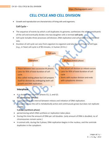
of the cell and eventually divides into two daughter cells is termed cell cycle. Cell cycle includes three processes cell division, DNA replication and cell growth in coordinated way. Duration of cell cycle can vary from organism to organism and also from cell type to cell type. (e.g., in Yeast cell cycle is of 90 minutes, in human 24 hrs.)
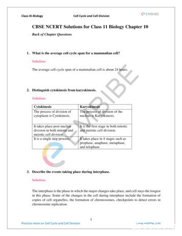
Class-XI-Biology Cell Cycle and Cell Division 1 Practice more on Cell Cycle and Cell Division www.embibe.com CBSE NCERT Solutions for Class 11 Biology Chapter 10 Back of Chapter Questions 1. What is the average cell cycle span for a mammalian cell? Solution: The average cell cycle span o
The Cell Cycle The cell cycle is the series of events in the growth and division of a cell. In the prokaryotic cell cycle, the cell grows, duplicates its DNA, and divides by pinching in the cell membrane. The eukaryotic cell cycle has four stages (the first three of which are referred to as interphase): In the G 1 phase, the cell grows.
Apr 11, 2017 · Chapter 13 –Moving Proteins into Membranes and Organelles 13.1 Targeting Proteins To and Across the ER Membrane 13.2 Insertion of Membrane Proteins into the ER 13.3 Protein Modifications, Folding, and Quality Control in the ER 13.4 Targeting of Proteins to Mitochondria and Chloroplasts 13.5 Targeting of Peroxisomal Proteins
Apr 12, 2012 · Concept 12.3: The cell cycle is regulated by a molecular control system In each experiment, cultured mammalian cells at two different phases of the cell cycle were induced to fuse. The frequency of cell division – Varies with the type of cell 1 These cell cycle differences – Result from regulation at the molecular level
The cell cycle includes all of the events in the life of an individual cell, from cell division to the period when a cell is not dividing while it carries out it’s regular functions. We can thus divide the cell cycle into: A. Interphase: Period of cell cycle when cell is not dividing 1. G1 Phase: Cellular organelles begin to duplicate. 2.
1 Cell Cycle and Mitosis THE CELL CYCLE The cell cycle, or cell-division cycle, is the series of events that take place in a eukaryotic cell between its formation and the moment it replicates itself. These events can be divided in two main parts: interphase (
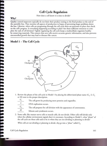
Model 1 - The Cell Cycle G1 s M Checkpoint G2 Checkpoint 1. Review the phases of the cell cycle in Model 1 by placing the abbreviated phase name (G,, S, G, or M) next to the proper description. The cell grows by producing more proteins and organelles. DNA replication occurs. The cell prepares for cell division with the appearance of cenrrosomes.


