Cell Model - SWIC
Cell doplasmicReticulum(NoteRibosomes)RibosomesLysosome
Cell Model
MITOSIS SLIDESINTERPHASEEARLYPROPHASELATEPROPHASE
MITOSIS SLIDESMETAPHASEANAPHASEANAPHASELATEPROPAS
MITOSIS SLIDESTELOPHASE
MITOSIS SLIDES
MITOSIS SLIDESANAPHASELATEPROPAS
MITOSIS SLIDES
MITOSIS MODELNucleus is intact.Centrioles are closetogetherInterphase
MITOSIS MODEL
MITOSIS MODELCentrioles aremoving apart.Nucleus (with chromatin)is still intact.Early Prophase
MITOSIS MODEL
MITOSIS MODELCentrioles are atopposite ends.Nucleus breaksapart, formingchromosomes.Late Prophase
MITOSIS MODEL
MITOSIS MODELSpindle FiberChromosomesare aligned in themiddle of the cellforming sisterchromatids.Metaphase
MITOSIS MODEL
MITOSIS MODELSister chromatidsseparate formingdaughterchromosomes.Anaphase
MITOSIS MODEL
MITOSIS MODELDaughterchromosomes becomefurther separated.Late Anaphase
MITOSIS MODEL
MITOSIS MODELChromosomes havecompleted theirseparation forming newnuclei. Cytokinesis causescleavage furrow.Telophase
MITOSIS MODEL
MITOSIS MODEL(Note cell boundarybetween cells.)Cytokinesis has beencompleted. We now have 2individual cells.Daughter Cells/ Interphase
MITOSIS MODEL
SIMPLE SQUAMOUS EPITHELIUMFunction: Diffusion and filtrationLocation: Lung alveoli, kidney glomerulus, capillary wallsSimpleSquamousEpithelium
Function:Location:
SIMPLE CUBOIDAL EPITHELIUMFunction: Secretion and some absorptionLocation: Any secretory gland, kidney tubules and other ducts
Function:Location:
SIMPLE COLUMNAR ion: Absorption or secretionLocation: Lining of small intestine, vas deferens, and other high-pressure ducts
Function:Location:
PSEUDOSTRATIFIED CILIATED COLUMNAR EPITHELIUMFunction: Protection, removal of foreign materialLocation: Nasal cavities, sinuses, pharynx, trachea, and bronchi of narEpithelium
Function:Location:
STRATIFIED SQUAMOUS EPITHELIUMFunction: Protection against abrasionLocation: Epidermis, oropharynx, anal canal
STRATIFIED SQUAMOUS EPITHELIUMFunction:Location:
AREOLAR CONNECTIVE ction: Attach one tissue/organ to another, separates musclesLocation: Under skin, surrounding vessels, glands, muscles and nerves
DENSE REGULAR CONNECTIVE TISSUEFibroblastNucleiCollagenFibersFunction: Connection of two structures togetherLocation: Tendons and ligaments
ADIPOSE TISSUEFunction: Energy storage, insulation under skin, cushioning around organsLocation: Under skin, around heart, kidneys, and e
Function: ,Location: , , ,
Function:Location: ,
Function: , ,Location: , , ,
ELASTIC icFibersFunction: Provide flexible supportLocation: Outer ear, epiglottis, larynx
Function:Location: , ,
HYALINE CARTILAGEFunction: SupportLocation: Costal cartilage, end of noseMatrixChondrocytesin Lacunae
Function:Location:
SKIN sclesArrectorPili (Subcutaneous)LayerPacinianCorpuscle
SKIN sumHair Bulb(containinghair papilla)PapillaStratumBasale
SKIN MODEL
SKIN MODEL
SKIN SLIDESEpidermisSebaceousGlandDermisHair Follicle(with HairRoot)AdiposeTissueHypodermis
SKIN SLIDESHair Follicle(with Hair Root)SweatGlandsSebaceousGlandSweatGlands
SKIN nlosum& Corneum.Usuallycannot le
SKIN SLIDESPacinianCorpuscleAdiposeTissue
SKIN SLIDES
SKIN SLIDES
SKIN SLIDES
SKIN SLIDES
SIMPLE SQUAMOUS EPITHELIUM Location: Simple Squamous Epithelium Function: Diffusion and filtration . SKIN SLIDES Adipose Tissue Pacinian Corpuscle . SKIN SLIDES . SKIN SLIDES . SKIN SLIDES . SKIN SLIDES . Aut
Completing this process correctly will ensure that you have a SWIC Student ID number for dual credit classes. Please follow instructions carefully. 1. To access the SWIC Web Application (also known as the New Student Information Form) please visit: www.swic.edu. Click on the yellow “App
Section B – ACT/New SAT Score or SWIC GPA Option Students earn points in this section from their ACT/New SAT scores. In lieu of the ACT/New SAT, students may use their SWIC GPA ONLY if they have at least 12 of the 37 general education credit hours for the MLT program posted on the transcript(s) received by SWIC no later than Feb. 1,
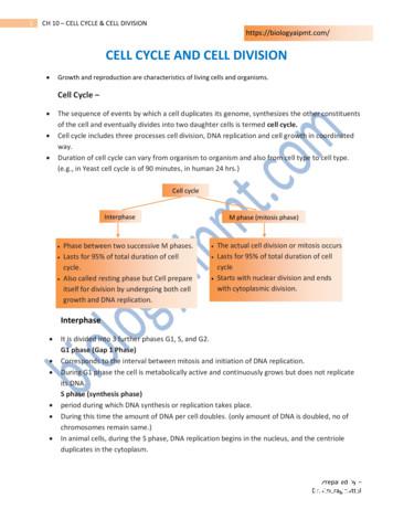
of the cell and eventually divides into two daughter cells is termed cell cycle. Cell cycle includes three processes cell division, DNA replication and cell growth in coordinated way. Duration of cell cycle can vary from organism to organism and also from cell type to cell type. (e.g., in Yeast cell cycle is of 90 minutes, in human 24 hrs.)
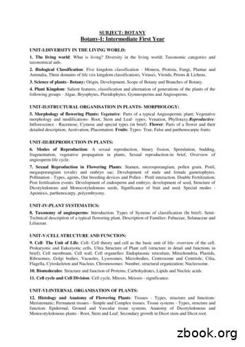
UNIT-V:CELL STRUCTURE AND FUNCTION: 9. Cell- The Unit of Life: Cell- Cell theory and cell as the basic unit of life- overview of the cell. Prokaryotic and Eukoryotic cells, Ultra Structure of Plant cell (structure in detail and functions in brief), Cell membrane, Cell wall, Cell organelles: Endoplasmic reticulum, Mitochondria, Plastids,
SWIC email accountsand . 3. for Financial Aid and Apply SWIC scholarships. 4. previous transcripts Send Enrollment Services to . 5. your photo ID Show proof of residency and Enrollment Services at . 6. the placement test Take see an academic advisorand . 7. for classes Register . 8. payment arrange
3D Cell Model Project You will be creating a 3-dimensional model to represent either a plant cell or an animal cell. 1. Choose either a plant or animal cell. Below are the organelles required to be included in your model: Plant Cell Animal Cell Cell Membrane Cell Membrane Nucleus Nucleus Cytoplasm Cytoplasm Mitochondria Mitochondria
The Cell Cycle The cell cycle is the series of events in the growth and division of a cell. In the prokaryotic cell cycle, the cell grows, duplicates its DNA, and divides by pinching in the cell membrane. The eukaryotic cell cycle has four stages (the first three of which are referred to as interphase): In the G 1 phase, the cell grows.
Many scientists contributed to the cell theory. The cell theory grew out of the work of many scientists and improvements in the . CELL STRUCTURE AND FUNCTION CHART PLANT CELL ANIMAL CELL . 1. Cell Wall . Quiz of the cell Know all organelles found in a prokaryotic cell


