Radiation, Cell Cycle, And Cancer - FAS
We have seen the enemy, and he is us!—Churchy LeFemme, aka Walt KellyRadiation, Cell Cycle, and CancerRichard J. Reynolds and Jay A. ScheckerWe live in remarkable times. The DNA within ourcells—the entire human genome—is steadfastlybeing mapped and deciphered. That work combinedwith new results from molecular and cellular biology are enabling researchers to reconstruct the inner workings of cells inunprecedented detail. We are beginning to build a holisticframework for understanding the human organism, one that integrates the distinct yet interrelated roles of DNA, genes, thecell, the body, and the environment. With it comes a better understanding of the cellular origins of many diseases, includingthe origins of cancer.The insights are timely. Cancer is one of the great scourges ofmodern civilization, for roughly one in five people inthe industrialized nations of North America, western Europe, and Asia will die of it. It is a disease of the cell that develops because of failures in the mechanisms that regulate cellgrowth. An individual cell multiplies without restraint until it and its progeny eventually overwhelm tissues and organs. Whatinitiates this process and how it progressesNumber 23 1995 Los Alamos Science51
Radiation, Cell Cycle, and Cancerhas been the subject of theoretical and experimental investigation almost since thestart of medical research. It has led to the identification of various cancer-causingsubstances, or carcinogens, in our diet and within our environment.Ionizing radiation* is one of those carcinogens, and its ability to induce cancer is notin doubt. The tragic experiences of the radium-dial painters during the early part ofthis century and the sobering epidemiological studies of the atomic-bomb survivorsof Hiroshima and Nagasaki bear witness to the fact that ionizing radiation can instigate a variety of cancer types. The bomb survivors, for example, display a small butstatistically significant increase in the level of several cancers, including leukemia,breast, thyroid, and skin cancer. Radiation and cancer definitely correlate.How does ionizing radiation cause cancer? How can a brief interaction with invisible particles smaller than an atom or the transient passage of massless electromagnetic waves cause a smoothly functioning, exceptionally well-organized cell to spiral chaotically out of control? Our cells for the most part are stable andpredictable entities, yet exposing them to levels of radiation well below the lethaldose can induce behavior that will eventually lead to the death of an entire organism. How does this happen?Answering these questions has proven to be extraordinarily difficult. Eventoday, the causes of cancer and the many ways the disease can progress are notcompletely understood. In the absence of a complete understanding, it has notbeen possible to determine the exact role that ionizing radiation plays in cancer induction. Nevertheless, a basic understanding does exist. Ionizing radiation candamage the DNA of chromosomes and potentially mutate the genes that reside onthose chromosomes. Because genes ultimately dictate cell function and behavior,ionizing radiation, through its capacity to induce genetic mutations, can bringabout a change in the basic nature of the cell. The cell becomes transformed,meaning that it is aberrant and is slowly evolving into a cancerous state.Although this picture is correct, it is somewhat superficial. It does not take intoaccount the rate of DNA damage or the particular type of damage that ionizing radiation induces, nor does it account for the powerful DNA repair mechanisms thathelp maintain the genome. It does not reveal that healthy cells have “defenses,” orcellular responses, that can limit excess proliferation and prevent cancer from developing. Augmenting the basic picture and elucidating what is known specifically about radiation and oncogenesis (the causes of tumor formation) is the main objective of this primer. In attaining that goal, we will spend a considerable amountof time building concepts and vocabulary, beginning with genes and gene expression. We will relate gene expression to cell function and then expand upon thenature of cell regulatory processes. We will learn that once a cell has becometransformed by some random, initial event, its progression towards cancer will bedriven by the abnormal behavior or removal of specific, critical proteins. We willlearn that within that set of “cancer-causing” genes, some are specifically correlated with DNA damage induced by ionizing radiation.*We will restrict our attention to ionizing raditation in this primer, that is, only nuclear emissions andx rays. Effects due to lower-energy electromagnetic radiations, such as ultraviolet radiation and emissions from power lines, will not be considered.52Los Alamos Science Number 23 1995
Radiation, Cell Cycle, and CancerGenes and Gene ExpressionWe are what we are because of our genes. This notion, along with the realizationthat DNA is the molecular carrier of heredity, are two of the seminal discoveriesof modern science. It has been discovered that a gene is composed of a specificDNA sequence, and gene sequences are distributed throughout our chromosomes(see “DNA, Genes, and Protein Synthesis”). Each chromosome is a single, longDNA molecule that is woven around a complex proteinstructure. Every person inherits a set of 23 chromosomes from each parent, and for every chromosomepassed to us by our mother, there is a correspondingchromosome contributed to us by our father. The 46chromosomes that compose the human genome can bearranged into 22 pairs of matching, or homologous,chromosomes, plus one pair of sex chromosomes—theX and Y chromosomes. Females possess an XX pair,whereas males possess an XY pair (Figure 1). Becauseeach chromosome in a homologous pair contains thesame set of genes, our cells have two copies, or alleles,of every gene. The DNA sequences of two alleles areusually very similar but not identical—each contains information from one of the two parents. What happenswhen a cell makes use of dissimilar gene copies?This question relates to gene expression, which was firstsystematically investigated by Gregor Johann Mendel(1822-1884), the “father” of modern genetics. Over thecourse of eight years, Mendel manipulated the breedingof several purebred strains of garden pea plants. Henoted the manifestation of certain characteristics of the plants, say flower color orpea texture, and how often those traits appeared in each successive generation.From his observations, he was able to deduce the statistical laws of inheritance,using as a hypothesis the existence of two inherited “units” for each trait.(Mendel’s units of heredity are what we now call genes. He used the word“Merkmale” to describe the units of heredity. The word gene was coined by theDutch botanist Wilhelm Ludwig Johannsen (1857-1927).) Mendel was also ableto deduce when certain traits would be observed, or expressed.Figure 1. The Human GenomeThe human chromosomes in this photograph were arranged to show the 22pairs of homologous chromosomes,plus the one pair of sex chromosomes(lower right). The original photo wastaken when the chromosomes had assumed their most condensed state.(For most of the life of a cell, a chromo-Take for example the trait flower color. Mendel found that a pea plant has a“gene” that dictates flower color, and that the gene has two “alleles,” one for violet flowers and one for white flowers. He also found that the violet allele had adominant mode of gene expression, that is, only one violet allele had to be presentfor the flowers to be violet. In contrast, the white allele had a recessive mode ofexpression, that is, both flower-color alleles had to be white for the flowers to bewhite.some is in a very loose, threadlikeform.) The chromosomes shown abovewere treated with a dye (Giemsa stain)that preferentially stains certain regionsand thereby produces the unique banding patterns that are used to identifyeach chromosome. Because the twosex chromosomes are different (X andMendel’s basic concepts about gene expression have been greatly expanded. Theterm “gene expression” is now used to describe the manifestation of traits at themolecular and cellular level. Expression begins with the processes of gene transcription and translation in which the DNA sequence that makes up a gene is usedas a template to synthesize a protein (see “DNA, Genes, and Protein Synthesis”).That protein then produces certain observable characteristics in the cell. Thus agene is said to be “expressed” when the protein that it specifies is actually synthesized and functioning in the cell.Y), or not homologous, the genomeshown is that of a male, namely thewell-known cytogeneticist T. C. Hsu ofthe University of Texas System CancerCenter. (Photo courtesy of T. C. Hsu.)continued on page 58Number 23 1995 Los Alamos Science53
Radiation, Cell Cycle, and CancerDNA, Genes, and Protein SynthesisThe cell is a marvelous ensemble of proteins, organic molecules, and organelles.Although it is on the order of ten microns or so in diameter, a cell is an incrediblechemical factory with the capability to synthesize more than 10,000 different proteins and enzymes and the ability to oversee thousands of simultaneous chemicalreactions. Figure A is a simple depiction of a mammalian cell in which we've selectively drawn only a few basic components (not to scale). The cell boundary isdefined by an outer, bilayer lipid membrane. The cell interior is filled with an aqueous colloidal fluid called the cytoplasm. Floating within the cytoplasm are thousands of proteins and large, macromolecular structures. We've indicated a ribosome, which is a protein complex required for the synthesis of proteins. We'vealso conspicuously highlighted the cell nucleus, which houses all of the nuclearDNA (our genome).Figure A. Basic Components of theCellCell membraneRibosomeCytoplasmIf the DNA molecules found in a human cell were laid end to end and stretchedout, the resulting line, though only 2 billionths of a meter wide, would be about twometers long, or about 200,000 times longer than the cell itself. Therefore, our DNAis packaged into dense constructs called chromosomes, each of which consists ofa single, linear DNA molecule containing millions of base pairs. The DNA is twisted and packed around proteins called histones, and that structure is itself twistedinto a secondary packing structure. There are at least four levels of twisting andpacking, but the degree of packing and the chromosome appearance can vary, depending upon both transcriptional activity (described below) and the stage of thecell's reproductive cycle.Human beings have a total of 46 chromosomes. Two of those chromosomes,called X and Y, determine the sex of the person. All males have an XY combination, whereas all females carry an XX combination. The other 44 chromosomescan be grouped into 22 pairs of “homologous" chromosomes. The individual members of each pair are very similar, but one is inherited from the mother and theother is inherited from the father (see Figure A and main article). For simplicity, wehave depicted only four chromosomes, representing two homologous irAs shown in Figure B, the double-stranded DNA that makes up a chromosomeconsists of two single-stranded molecules that are intertwined to form a doublehelix. The backbone of each single strand is a long chain consisting of repeatingsugar-phosphate subunits. The sugars appear as pentagon-shaped rings in FigureB. (DNA is an acronym for deoxyribose nucleic acid. Deoxyribose is the particulartype of sugar.) The sugar portion contains five carbon atoms, labeled 1' to 5', andthe backbone is constructed by linking, through a phosphodiester bond, the 5' carbon of one sugar to the 3' carbon of the next. Because of the asymmetry in thephosphodiester linkage, the phosphodiester backbone, as it is often called, can beassigned an orientation, either 5' to 3' or 3' to 5'. The two strands of the DNA double helix actually have opposite orientations. One strand can be said to move “up,”whereas the other moves “down.” Many proteins that interact with DNA are sensitive to this orientation and can distinguish one strand from the other.Attached to each sugar unit is one of four different nucleic acid bases: adenine (A),cytosine (C), guanine (G), and thymine (T). The bases can be further classified aspurines (A and G) or pyrimidines (C and T). In forming the double helix, the baseswill line up between the two DNA backbones, a base in one strand pairing with anopposing base in the complementary strand. The base pairs are chemically linkedby hydrogen bonds. In the standard Watson-Crick base pairing, each pair must be54Los Alamos Science Number 23 1995
Radiation, Cell Cycle, and Cancer5' endFigure B. DNA and ItsConstituents3' endOOO- PO3'5'5 -to-3 directionCGOO5'OO3'OP OOO- P OO3'5'ATOO5'OO3'OCarbon atomOxygen atomPhospherous atomCovalent bondHydrogen bondOP OOOO- PO3'5'PTAOSugarO5'OO3'OHydrogen atoms notinvolved in hydrogenbonding have beenomitted in this drawing.As a result, some carbonatoms and some nitrogenatoms appear to beunderbonded.5 -to-3 directionP OOOO- PO5'O3'CGPhosphodiesterbondOOOP OO3' end5' endcomprised of a purine coupled to a pyrimidine. Furthermore, the purine A can onlypair with the pyrimidine T, and the purine G can only pair with the pyrimidine C.Thus, the sequence of bases along one strand dictates a unique sequence ofbases along the second complementary strand. Together, the two strands incorporate a level of information redundancy into the double-stranded DNA molecule, because each strand can act as a template for synthesizing the other. Template-directed copying of each DNA strand is called replication.The information encoded within the DNA molecule enables the cell to synthesizeproteins. A gene, depicted schematically in Figure C, is that segment of DNA thatcodes for a single protein, and our genome contains roughly 50,000 to 100,000genes dispersed among the 46 chromosomes. Because the overwhelming majorityof cell processes are carried out by proteins, a cell goes to great lengths to ensurethat the integrity of the base sequence is maintained. This is the primary role ofDNA repair mechanisms (see “DNA Repair” on page 78).To translate the information encoded by DNA into a protein product, the cell mustgo through a multistep process. First, the coding region of a gene is read, or transcribed, into a copy of the DNA sequence. The copy takes the form of a moleculeof RNA, which is similar, with a few differences, to a single-strand of DNA. Aftersome processing, the RNA will leave the nucleus and enter the cell's cytoplasm.The information contained in the RNA will be translated by a ribosome, a largemacromolecule that guides the assembly of amino acids into the protein product.DNA BasesPyrimidinesCTPurinesAGGene segments range from thousands to millions of base pairs in length. Therefore, Figure C depicts the DNA as a solid bar containing different subregions. Wehave indicated the coding region, which contains the actual sequence used forprotein synthesis, and two regulatory DNA sequences that are used to control theNumber 23 1995 Los Alamos Science55
Radiation, Cell Cycle, and CancerRegulatory factorsTranscription complexTranscription factorRNA polymerase IIDownstream3'5'Gene coding region3'ATG(Start codon)Regulatory sequencesTAA(Stop codon)5'DoublestrandedDNAPromoter regionFigure C. Gene Structure and Transcriptionrate and frequency of transcription. The double helix is actually a fairly open structure that permits access to the chemical groups of the DNA bases. Proteins calledregulatory factors will recognize these groups and selectively bind to specific DNAsequences. By physically distorting the helix (bending and folding the DNAstrands) or by promoting protein-protein interactions, the regulatory factors can either facilitate or inhibit transcription. The regulatory regions may be far removedfrom the coding region and may even be located "downstream."Transcription of the DNA sequence into an RNA copy is initiated at the promoter region, which also contains a specific DNA sequence (TATA) that is recognized by atranscription factor. This factor is a protein that binds to the DNA and initiates theself-assembly of a transcription complex consisting of perhaps 10 or more proteins,including RNA polymerase II (RNA Pol II). The RNA Pol II complex will transcribethe DNA coding sequence. Thus, the initial step in creating a protein is tied to thepresence (or sometimes the absence) of transcription factors and regulatory proteins. That is one way the cell has of regulating the expression of a gene.As depicted in Figure D, RNA Pol II instigates the unwinding of the DNA doublehelix, which enables it to "read" and transcribe one of the two DNA strands. Because of the Watson-Crick base-pairing rules, the RNA molecule that is producedcontains all of the information that was originally encoded in the DNA strand. AsRNA Pol II moves along, the relaxed strands of previously transcribed DNA sections rewind. After the gene has been completely transcribed, RNA Pol II will leavethe DNA and some processing of the RNA molecule occurs. The resulting RNAstrand (now called messenger RNA, or mRNA) leaves the cell nucleus and will beused as the template for protein synthesis.Figure D. Transcription of DNA to RNARNA polymerase II3'5'3'3'A5'T5'GACRNA transcript56DNA templateRNA precursorsLos Alamos Science Number 23 1995
Radiation, Cell Cycle, and CancerAminoacyl-tRNA synthetaseAlaAttachment sitetRNAAminoacyl-tRNACGGAlaAmino acidAnticodonC GGAmino-acid sequence(protein)GGCCodon for alanine3'C G GC G A AG G GC G CaC G CCCC C G5'mRNARibosomeFigure E. Translation of mRNA to ProteinThe process of converting the mRNA template into an actual protein is called translation, as shown in Figure E. Translation takes place at the ribosome. The sequence of RNA bases contained in the mRNA transcript is interpreted as a seriesof "words," or codons, consisting of three consecutive RNA bases. With some exceptions, each codon corresponds to a specific amino acid. These are the smallmolecules from which proteins are constructed. For example, the DNA base sequence GCC codes for the amino acid alanine. The exceptions are three stopcodons (TAA, TAG, TGA) that are used as punctuation and indicate the terminationof an amino-acid sequence.A molecule called transfer RNA (tRNA) is the actual link between a codon and anamino acid. One end of the tRNA has an "anticodon" that pairs according toWatson-Crick rules with a codon in the mRNA template. The other end of thetRNA is bound to an amino acid that corresponds to that codon in the mRNA template. The top of Figure E shows the reaction that places the correct amino acidonto the corresponding tRNA. That reaction is catalyzed by a family of specificenzymes called the aminoacyl-tRNA synthetases. The ribosome facilitates the pairing of the anticodon region of a tRNA molecule to the mRNA codon and catalyzesthe transfer of the amino acid to the growing protein chain. The ribosome stepsalong the mRNA molecule, adding an amino acid to the chain at each step, until itreaches a stop codon. At that point, the protein product is finished, and the ribosome detaches. Numerous ribosomes will often attach to the same mRNA, so thatmany copies of the same protein are produced for each DNA transcription event.It is clear, then, that DNA plays a critical role in protein synthesis. A single genecan get transcribed many times, and each time it is transcribed, many identicalproteins are produced. If a gene coding for a major regulatory protein becomesmutated, then that single mutation can mean the difference between a normal anda dysfunctional cell. Number 23 1995 Los Alamos Science57
Radiation, Cell Cycle, and Cancercontinued from page 53But a question remains. We have approximately 50,000 to 100,000 genes. Doesevery cell make use of all the genes that are encoded in its genome? The answeris no, and the reason has to do with a much more fundamental concept of gene expression—the notion of regulation. A gene embodies not only DNA sequencesthat code directly for protein construction but also regulatory sequences that control various aspects of the transcription process. Regulatory sequences include thepromoter region, where transcription is initiated, and regions that control the rateand frequency of transcription. Those regulatory regions are recognized by regulatory factors, which are a class of proteins that bind to certain DNA sequencesand either directly or indirectly (by attracting other proteins) inhibit or enhancetranscription.Therefore, the mere presence of a gene within our genome does not guarantee thatit will be expressed. Instead, the production of a protein from the gene is dependent upon a very complicated relationship between DNA, regulatory proteins, andprotein synthesis. In fact, the cell has at least six levels of control on gene expression, beginning with regulation of the promoter region and ending with the breakdown and removal of the protein product. Once produced, however, many proteins must first be activated by other proteins, form a complex, or both, beforebeing able to play a part in cell processes. Protein activation and participation inprotein complexes are but two examples of how a gene product can be regulated.The expression of a particular gene and the behavior of its protein product cantherefore change due to a number of factors. Abnormal behavior can certainly bethe direct result of a DNA mutation, but it may be expressed through a type ofdomino effect that links the action of one protein to the function of another.Cell DifferentiationCancer is a disease of cells, and human beings have lots of them. We are composed of approximately 1013 to 1014 individual cells, most of which are not identical. Instead, they have differentiated into roughly 350 types. Differentiationmeans that a cell has become specialized in function and structure and has compromised its independence and some of its capabilities in favor of being a cooperative member of a tissue and organism. Our cells, for the most part, are immobile,and therefore, the specialized cells in any given tissue depend heavily on other tissues to provide nutrients and basic resources, to remove waste, to create environmental stability, and to provide protection.This interdependence is distinct from a single-cell organism. A free living cell isself-sufficient and behaves in a manner that best aids its own survival. Certainly,one survival mechanism is proliferative advantage. For example, the rod-shapedbacteria Escherichia coli can divide and produce two new bacteria every 30 minutes. In principle, then, E. coli has the reproductive capacity to produce well over200 trillion progeny in just 24 hours!*Clearly, the differentiated cells of a multicellular organism cannot exhibit this typeof proliferative behavior, nor can they be insensitive to the needs of other cells.Instead, everything about a differentiated cell, including when it reproduces, itsshape and size, and the chemicals and proteins it synthesizes, is essentially determined by the needs of the tissue and the organism of which that cell is a part. Ex*This number is a theoretical extrapolation. The actual number of bacteria that would be generated islimited by the availability of resources and by the necessity to remove heat and waste by-products.58Los Alamos Science Number 23 1995
Radiation, Cell Cycle, and Cancererting its influence through a multitude of intricate, intercellular controls, the bodyensures that the behavior of individual cells is directed towards sustaining theoverall health of the organism. This paradigm of specialization is obviously a successful one that imparts survivability to all the cells of the body and to the entireorganism.How do cells differentiate? The process is only beginning to be understood. It isnot simply that each specialized cell has a different set of genes, because all thecells of the body are genetic clones and possess the same genome. Nor does differentiation result from a change in the information content of the DNA or theamount of DNA present. Rather, specialization comes about because only a particular set of genes are expressed. Those expressed genes determine if the cellwill be a nerve cell or a skin cell, if it is mobile within tissue (such as amacrophage), or if it grows slowly or rapidly. In one sense, the genome is analogous to a library that contains books on all subjects. When we wish to specializeour area of interest, we select from the library only those books that are appropriate for our needs and leave the other books undisturbed.An epigenetic change within the genome—one that modifies gene expression without changing the information content of the DNA—appears to be the mode bywhich cells differentiate. Many differentiated cells pass their traits on to theirprogeny; that is, a liver cell begets a liver cell, which implies that the epigeneticchanges to the genome are conferred to daughter cells. The transmission is believed to happen through a chemical modification of DNA sequences known asmethylation. The methylation patterns are maintained during DNA replication, butthe way in which they are originally established and the way they become modified is not fully understood.However, not all cells are descendants of fully differentiated cells. Instead, thespecialized cells of many tissues and organs originate from a class of relatively undifferentiated cells called stem cells. The successive progeny of stem cells displayincreasing degrees of specialization, and that process may continue for several cellgenerations. The stem cells are highly unusual. Their specific role in the tissue isto renew lost or damaged cells, but at the same time, they must maintain their ownpopulation. As illustrated in Figure 2, stem cells have the peculiar property ofgenerating dissimilar progeny. One of the cells that is produced remains a stemcell, whereas the other cell begins to specialize in response to external signals.Those signals evidently help trigger the epigenetic changes. But each type of stemcell is slightly different, and the pathway of specialization that their progeny follow is also distinct. Thus, basal cells are ultimately the source of epithelial cells(those cells that make up the skin layers and the lining of the intestinal and respiratory tracts), whereas the hemopoietic stem cells are the precursors of about tenor more different cell types that make up the blood and the immune system.As the progenitor of many tissue cells, the stem cells perform a function that highly differentiated cells have relinquished, namely repeated cell division. The terminally differentiated cells of the skin or the hemopoietic system are so specializedthat they rarely divide. Stem cells renew those nondividing cells, and therefore,the role of the stem cells within the scheme of the organism is that of proliferation. Unlike E. coli, however, the reproductive potential of a stem cell is strictlyregulated (as it is for all other types of cells). Cells will only multiply as a consequence of having received numerous extracellular signals. Growth factors, or mitogens, are positive regulators that stimulate proliferation. Other signals will inhibit growth and are considered to be negative regulators. Normally, cellularNumber 23 1995 Los Alamos Science59
Radiation, Cell Cycle, and CancerHemopoietic stem cellExternal differentiationsignalsFigure 2. Cell Differentiation andStem CellsThe hemopoietic stem cells are responsible for generating about a dozen different types of cells, including the vari-Partiallydifferentiatedstem cellous kinds of blood cells and the celltypes that make up the immune system.The figure illustrates the generation ofa highly specialized cell, theHemopoietic stem cellmacrophage (a cell that lives within tissue and is descended from a whiteExternal differentiationsignalsblood cell), from a relatively undifferentiated stem cell. The process of differentiation takes place over several cellgenerations, and it occurs because ofthe expression of different genes.External differentiationsignalsWhen a stem cell divides into two cells,one of them will remain a stem cell.The other, in response to external signals, begins to specialize. This daugh-Expressedgeneter expresses new genes (colored seg-Protein productof expressed genements on the chromosomes) that aretranscribed and translated into proteins(colored shapes in the cytoplasm). Thenew proteins modify the cell’s functionand appearance. The process of specialization continues through severalmore generations until finally a celltype reaches a terminal stage of differentiation. If a macrophage divides,both of its progeny will remain asmacrophages. Because stem cells andtheir partially differentiated progeny divide frequently, cancers often emergefrom those cell types.Macrophage(phagocyticwhite blood cell)Teminally differentiatedprogenyMacrophage(phagocyticwhite blood cell)proliferation is controlled by the cell’s interpretation and response to these reciprocal types of regulation.One of the major differences between a normal cell and a cancer cell is that thelatter responds in an unbalanced manner to regulatory signals and proliferates atinappropriate times. A precancerous, or neoplastic cell, might undergo changes inthe way it responds to regulatory signals, and it might divide independently of theneeds of the tissue. If the modified behavior results in a proliferative advantagefor a cell line, the uncontrolled growth can ultimately lead to the disruption of tissue function. Because stem cells are the most rapidly and frequently dividing cellsin our body, they are in one sense “primed” to express proliferative advantages.Most
The human chromosomes in this pho-tograph were arranged to show the 22 pairs of homologous chromosomes, plus the one pair of sex chromosomes (lower right). The original photo was taken when the chromosomes had as-sumed their most condensed state. (For most of the life of a cell, a chromo-some is in a very loose, threadlike form.)
Part 6: Modeling the cell cycle in a normal cell Part 7: Modeling the cell cycle in a cancer cell Living Environment Major Understandings: Gene mutations in a cell can result in uncontrolled cell division, called cancer. Exposure of cells to certain chemicals and radiation increases mutations and thus increases the chance of cancer.
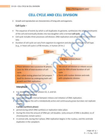
of the cell and eventually divides into two daughter cells is termed cell cycle. Cell cycle includes three processes cell division, DNA replication and cell growth in coordinated way. Duration of cell cycle can vary from organism to organism and also from cell type to cell type. (e.g., in Yeast cell cycle is of 90 minutes, in human 24 hrs.)
Ovarian cancer is the seventh most common cancer among women. There are three types of ovarian cancer: epithelial ovarian cancer, germ cell cancer, and stromal cell cancer. Equally rare, stromal cell cancer starts in the cells that produce female hormones and hold the ovarian tissues together. Familial breast-ovarian cancer
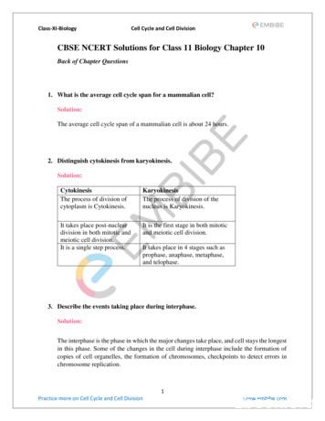
Class-XI-Biology Cell Cycle and Cell Division 1 Practice more on Cell Cycle and Cell Division www.embibe.com CBSE NCERT Solutions for Class 11 Biology Chapter 10 Back of Chapter Questions 1. What is the average cell cycle span for a mammalian cell? Solution: The average cell cycle span o
Medical X-rays or radiation therapy for cancer. Ultraviolet radiation from the sun. These are just a few examples of radiation, its sources, and uses. Radiation is part of our lives. Natural radiation is all around us and manmade radiation ben-efits our daily lives in many ways. Yet radiation is complex and often not well understood.
The Cell Cycle The cell cycle is the series of events in the growth and division of a cell. In the prokaryotic cell cycle, the cell grows, duplicates its DNA, and divides by pinching in the cell membrane. The eukaryotic cell cycle has four stages (the first three of which are referred to as interphase): In the G 1 phase, the cell grows.
Non-small cell lung cancer is the most common type of lung cancer and accounts for 84% of cases. There are different types of non-small cell lung cancer, including: Adenocarcinoma - a cancer that forms in the outer parts of the lung. Squamous cell carcinoma - a cancer that forms from a cell lining the airway.
Non-Ionizing Radiation Non-ionizing radiation includes both low frequency radiation and moderately high frequency radiation, including radio waves, microwaves and infrared radiation, visible light, and lower frequency ultraviolet radiation. Non-ionizing radiation has enough energy to move around the atoms in a molecule or cause them to vibrate .


