Developmental Cell Article - Indstate.edu
Developmental CellArticleTwo Forkhead Transcription Factors Regulatethe Division of Cardiac Progenitor Cellsby a Polo-Dependent PathwayShaad M. Ahmad,1 Terese R. Tansey,1 Brian W. Busser,1 Michael T. Nolte,5 Neal Jeffries,2 Stephen S. Gisselbrecht,4Nasser M. Rusan,3 and Alan M. Michelson1,*1Laboratoryof Developmental Systems Biology, Genetics and Developmental Biology Centerof Biostatistics Research3Laboratory of Molecular Machines and Tissue Architecture, Cell Biology and Physiology CenterNational Heart Lung and Blood Institute, National Institutes of Health, Bethesda, MD 20892, USA4Division of Genetics, Department of Medicine, Brigham and Women’s Hospital and Harvard Medical School, Boston, MA 02115, USA5Department of Biological Sciences, University of Notre Dame, Notre Dame, IN 46556, USA*Correspondence: j.devcel.2012.05.0112OfficeSUMMARYThe development of a complex organ requires thespecification of appropriate numbers of each of itsconstituent cell types, as well as their proper differentiation and correct positioning relative to each other.During Drosophila cardiogenesis, all three of theseprocesses are controlled by jumeau (jumu) andCheckpoint suppressor homologue (CHES-1-like),two genes encoding forkhead transcription factorsthat we discovered utilizing an integrated genetic,genomic, and computational strategy for identifyinggenes expressed in the developing Drosophila heart.Both jumu and CHES-1-like are required duringasymmetric cell division for the derivation of twodistinct cardiac cell types from their mutual precursorand in symmetric cell divisions that produce yet a thirdtype of heart cell. jumu and CHES-1-like control thedivision of cardiac progenitors by regulating theactivity of Polo, a kinase involved in multiple stepsof mitosis. This pathway demonstrates how transcription factors integrate diverse developmentalprocesses during organogenesis.INTRODUCTIONThe remarkable cellular diversity present within metazoanorgans illustrates several important themes in developmentalbiology, including a requirement for the specification of appropriate numbers of distinct cell types, the proper differentiationof these cells and their correct positioning within the organ(Rosenthal and Harvey, 2010). Taken together, the existence ofmultiple organ-specific cell types implies that numerous biological processes must work in unison during development, andraises an intriguing question: how is the requisite integration ofthese diverse developmental pathways achieved?The formation of the Drosophila embryonic heart providesa particularly amenable system for addressing this question(Bodmer and Frasch, 2010; Bryantsev and Cripps, 2009). Anorgan that pumps hemolymph throughout the body cavity, theDrosophila heart is composed of two groups of cells arrangedin a metamerically repeated and stereotyped pattern (Figures1A–1C): an inner group of Myocyte enhancer factor 2 (Mef2)expressing contractile cardial cells (CCs) that form a lineartube, surrounded by a sheath of pericardin (prc) and Zn fingerhomeodomain 1 (zfh1)-expressing nephrocytic pericardial cells(PCs). Neither the CCs nor the PCs constitute a uniform population, as revealed both by their distinct cell lineages and by thecomplexity of their individual gene expression programs. Fromanterior to posterior, and named for the transcription factorsthey express, there are two Seven-up-CCs (Svp-CCs), twoTinman-Ladybird-CCs (Tin-Lb-CCs), and two CCs expressingonly Tin (the posterior-most Tin-CCs) in each hemisegment. Alarger number of PCs surround the cardial cells: two Svp-PCsand two Odd-skipped-PCs (Odd-PCs) are positioned laterally,two Even-skipped-PCs (Eve-PCs) are situated dorsolaterally,and a row of Tin-PCs and Tin-Lb-PCs runs immediately ventralto the CCs (Azpiazu and Frasch, 1993; Bodmer, 1993; Jaglaet al., 1997; Ward and Skeath, 2000).A stereotyped series of asymmetric and symmetric cardiacprogenitor cell divisions gives rise to these eight differentiatedcell types (Alvarez et al., 2003; Han and Bodmer, 2003). Thedifferential expression of multiple genes, and both the distinctlineage and intricate but invariant positioning of the individualheart cell types, argue for a high degree of functional precisionand regulatory complexity in the generation of the heart. Thishypothesis is borne out by classical genetic studies, whichshowed that the development of the Drosophila heart fromthe dorsal-most region of the mesoderm, a tin-expressingdomain referred to as the cardiac mesoderm (CM), is dependent on contributions from multiple signals and transcriptionfactors that are conserved between flies and vertebrates (summarized in Figure 1D; reviewed by Bodmer and Frasch, 2010;Bryantsev and Cripps, 2009; Chien et al., 2008). Thus, the identification of genes that regulate cardiac development, anddetailed investigations of their expression and function inDrosophila, are likely to provide considerable insight into therelated mechanisms controlling cardiogenesis in vertebrates,including human.Developmental Cell 23, 97–111, July 17, 2012 ª2012 Elsevier Inc. 97
Developmental CellFkh Factors Regulate Cardiogenesis via Polo KinaseFigure 1. Strategy for Gene Expression Profiling of the Drosophila Embryonic Heart(A) Staining for expression of Mef2 protein reveals the cardial cells (CCs, arrow) in the heart of a stage 16 embryo.(B) Staining for expression of Pericardin (Prc) protein reveals the pericardial cells (PCs, arrow) in the heart of a stage 16 embryo.(C) Schematic diagram showing the stereotyped positions of the eight different cell types composing the Drosophila embryonic heart. An individual hemisegmentis indicated by the dashed red box.(D) Regulatory network responsible for the development of the cardiac mesoderm and heart (Bodmer and Frasch, 2010; Bryantsev and Cripps, 2009).(E) Genetic perturbations used for gene expression profiling, along with the expected changes in cardiac mesoderm gene levels relative to wild-type mesoderm.‘‘tinD-positive’’ represents dorsal mesodermal cells isolated from wild-type embryos using targeted expression of GFP driven by the tinD enhancer (Yin et al.,1997).(F) Detection curves showing the number of genes from the training set detected as a function of q-value cutoff. The predictive value of individual genotype/wildtype comparisons (various colors; see legend) are compared to randomly generated rankings (thin black lines) and to composite rankings derived from a uniform(gray) or a weighted (violet) combination of all data sets.98 Developmental Cell 23, 97–111, July 17, 2012 ª2012 Elsevier Inc.
Developmental CellFkh Factors Regulate Cardiogenesis via Polo KinaseHere, we describe an integrated strategy that we developedand applied to identify 70 genes expressed in the DrosophilaCM or heart. We further show that one gene discovered withthis approach, jumeau (jumu), plus its homolog, Checkpointsuppressor homologue (CHES-1-like)—both of which encodeFkh transcription factors—mediate both asymmetric andsymmetric cardiac progenitor cell divisions by regulatinga Polo-kinase dependent pathway.RESULTSA Genomic Screen for Genes Expressed in the CardiacMesoderm or HeartAs an initial step to identify regulators and effectors of heartdevelopment in Drosophila, we screened for genes expressedin the CM or differentiated heart using an integrated genetic,genomic and computational strategy that we previously appliedto study somatic muscle gene expression (Estrada et al., 2006).Two essential aspects of our approach are the use of specificgenetic backgrounds to selectively perturb CM gene expression based on prior knowledge of cardiogenic pathways(Figures 1D and 1E), and the availability of a training set of 40genes already known to be expressed in the CM. Previousstudies revealed that activation of the fibroblast growth factorreceptor (FGFR)- and epidermal growth factor receptor(EGFR)-driven receptor tyrosine kinase (RTK)/Ras, Wingless(Wg), or Decapentaplegic (Dpp) pathways produces extra CMcells compared with wild-type, thereby elevating levels of CMgene expression (Azpiazu and Frasch, 1993; Bodmer, 1993;Carmena et al., 1998; Frasch, 1995; Gisselbrecht et al., 1996;Grigorian et al., 2011; Michelson et al., 1998; Staehling-Hampton et al., 1994). In contrast, activation of Notch results in fewerCM cells and thus reduced levels of CM gene expression (Hartenstein et al., 1992; Mandal et al., 2004). Moreover, becausethe CM arises from the tin-expressing dorsal mesoderm, CMgenes will be highly enriched in cells purified from this subregion of the whole embryo.To generate a compendium of gene expression profiles associated with CM development, we used flow cytometry to purifythe entire mesoderm from both wild-type stage 11 embryosand similar embryos from nine informative genetic backgrounds,as well as tin-expressing dorsal mesoderm from equivalentlystaged wild-type embryos (Figure 1E). We then used a statisticalmeta-analysis method (Estrada et al., 2006) fitted to the trainingset of 40 known CM genes to rank all Drosophila genes by theirlikelihood of being expressed in the CM based on their collectivebehavior in this expression profiling compendium. Any gene that(1) is upregulated with activation of the RTK/Ras, Wg or Dpppathways, upregulated with Dl loss-of-function, downregulatedwith Notch activation, and downregulated with wg loss-offunction, and (2) is enriched in tin-expressing mesoderm relativeto the entire mesoderm, has a high probability of being ex-pressed in the CM (Table S1A available online and ExperimentalProcedures).To validate the predictions of this meta-analysis, we usedlarge-scale whole-embryo in situ hybridizations to assess thein vivo expression patterns of highly ranked CM gene candidates. Of 136 randomly selected genes that provided informative in situ hybridization results among the top-ranked 400candidates and that did not include any of the training setgenes, 70 were expressed in the CM and/or heart. Thus, themeta-analysis predicted cardiac genes with an accuracy of51.4% (Table S1B). Further analyses revealed that 37 genesare expressed in both the CM and the mature heart, 22 genesare expressed in the CM but not in the heart, and 11genes are expressed in the heart but not in the CM (TableS1B and Figure S1).To gain insight into the biological processes in which thesegenes are involved, the 110 training set plus newly validatedCM and heart genes were queried for the relative enrichment ofGene Ontology (GO) terms. Overrepresented terms (Table S1C)include categories associated with mesoderm development,cardiac differentiation, cell fate specification, transcriptionalregulation, migration, tube morphogenesis, and the RTK/Raspathway. Another enriched category was nervous system development, which likely reflects the pleiotropic effects of manydevelopmental regulators, and the fact that many of the identifiedgenes are also expressed in the nervous system (data notshown). Among the unexpected overrepresented categorieswere cytokinesis and cell division, the relevance of whichbecame apparent upon a detailed analysis of the cardiogenicfunctions of jumu, CHES-1-like, and polo.The Fkh Genes jumu and CHES-1-like Are Involved inDrosophila Heart DevelopmentPrevious studies have shown a striking conservation of transcription factors involved in both Drosophila and vertebratecardiogenesis. Genes encoding transcription factors were alsooverrepresented among the 110 CM- and heart-expressedgenes. One such gene is jumu, which encodes a Fkh subclassN transcription factor (Lee and Frasch, 2004) that is continuouslyexpressed in the CM and differentiating heart from embryonicstages 11 to 13 (Figures 2A and S1Y–S1Y000 ). In addition, weexamined the expression pattern of the only other DrosophilaFkh subclass N gene, CHES-1-like, and found that it is also expressed in the CM during stages 11 and 12 (Figures 2B andS1Z–S1Z000 ).Given the presence of these two Fkh transcription factors inthe embryonic CM, and the fact that this class of proteins isinvolved in mammalian cardiogenesis, we next used a wholeembryo RNA interference (RNAi) assay to assess whether jumuand CHES-1-like play a role in Drosophila cardiac development.RNAi directed against either jumu or CHES-1-like resulted inincorrect numbers and an uneven distribution of both CCs and(G) Weight factors that reflect the relative contribution of each condition (isolated whole mesoderm for nine genotypes plus purified wild-type tinD-positive cells)to the detection rate of the genes from the training set.(H) All genes were ranked according to their degree of CM-like expression patterns across the entire set of conditions, using their weighted T-scores. The ranks ofthe training set genes (blue) are plotted as thin vertical lines, revealing the extent to which optimization concentrates the training set at the top of the rank list. Thep-value is from the Wilcoxon-Mann-Whitney U test.See also Figure S1 and Table S1.Developmental Cell 23, 97–111, July 17, 2012 ª2012 Elsevier Inc. 99
Developmental CellFkh Factors Regulate Cardiogenesis via Polo KinaseFigure 2. jumu and CHES-1-like EmbryonicExpression and Loss-of-Function CardiacPhenotypes(A and B) jumu (A) and CHES-1-like (B) mRNAs areexpressed in the cardiac mesoderm at embryonicstage 11 (arrows).(C–E) Whole embryo RNAi results for dsRNA corresponding to lacZ (C), jumu (D), and CHES-1-like(E) in live embryos in which CCs express a nuclearlocalized form of GFP under control of a Handenhancer (Han and Olson, 2005), and PCs expressboth Hand-GFPnuc and a nuclear form of DsRedunder control of a heart enhancer from the Himgene (Him-DsREDnuc; A.M.M. and S. Michaud,unpublished data). Arrows indicate incorrectnumbers and uneven distribution of CCs and PCs.(F–M) Mef-2 antibody staining of CCs in wild-typeembryos (F), in embryos homozygous for hypomorphic jumu mutations (G and H), a jumu nulldeficiency (I), a CHES-1-like null mutation (J), inembryos with CM-targeted RNAi against jumu (K)and CHES-1-like (L), and in embryos homozygousfor both the jumu and CHES-1-like null mutations(M). Localized increases in CC number (blackarrows), localized reductions in CC number (whitearrows), incorrectly positioned CCs (black arrowheads), CC nuclei larger than normal (white arrowhead), and hemisegments missing all CCs (twinwhite arrows) are shown.See also Figure S2.PCs (Figures 2C–2E), indicating that both of these Fkh factorsare essential for normal heart development.Loss of Either jumu or CHES-1-like Function Results inLocalized Changes in Cardial Cell Number, Giant Nuclei,and Incorrectly Positioned Heart CellsWe undertook a more detailed analysis of the cardiogenic effectsof jumu and CHES-1-like by examining the phenotypes associated with loss-of-function mutations in these genes. Stainingwith antibodies against the nuclear protein Mef2 (which is expressed in CCs of the heart, as well as in somatic myoblasts)revealed that the uniform and symmetrically aligned distributionof CCs seen in wild-type embryos (Figure 2F) is markedly disrupted in embryos homozygous for jumu hypomorphic mutations(jumu06439 and jumuDf2.12, Figures 2G and 2H), a jumu null deficiency (Df(3R)Exel6157, Figure 2I) that deletes both jumu andanother gene not involved in heart development (Cheah et al.,2000; Strödicke et al., 2000; see also Table S2), and a nullmutation that we generated in CHES-1-like (Df(1)CHES-1-like1,Figures 2J and S2; see Experimental Procedures). Each mutantexhibited different hemisegments having localized increasesor decreases in CC number, occasional enlarged CC nuclei, orCCs that were misaligned with other CCs within a hemisegmentor with their counterparts across the dorsal midline. Similarphenotypes were also observed wheneither jumu or CHES-1-like activity wasknocked down by CM-targeted RNAidirected by the Hand-GAL4 and tinDGAL4 drivers (Figures 2K and 2L), indicating that the requirement of these Fkhgenes for correct heart development is autonomous to thecardiac mesoderm. Embryos doubly homozygous for both thejumu null deficiency and the CHES-1-like null mutation exhibiteda more severe phenotype, often missing entire hemisegments ofCCs (Figure 2M). Taken together, these results suggest a role forabnormal cell division as the origin of the jumu and CHES-1-likemutant heart phenotypes, which is consistent with the knowninvolvement of jumu in nervous system development (Cheahet al., 2000).jumu and CHES-1-like Are Required for BothAsymmetric and Symmetric Divisions of CardiacProgenitor CellsTwo asymmetric progenitor cell divisions generate all theSvp-expressing heart cells, with each division producing oneSvp-CC and one Svp-PC per hemisegment (Figure 3A, yellowand red cells, respectively) (Gajewski et al., 2000; Ward andSkeath, 2000). In contrast, a pair of symmetric cell divisions givesrise to the four Tin-CCs in each hemisegment, the two Tin-LbCCs and the two posterior-most Tin-CCs (Figure 3A, green cells)(Han and Bodmer, 2003). These lineage relationships are shownin Figure 3G.We took advantage of this ability to distinguish the products ofasymmetric and symmetric cardiac progenitor cell divisions to100 Developmental Cell 23, 97–111, July 17, 2012 ª2012 Elsevier Inc.
Developmental CellFkh Factors Regulate Cardiogenesis via Polo KinaseFigure 3. Cell Division Defects Underlyingthe Cardiac Phenotypes of jumu andCHES-1-like Mutants(A) Heart from an otherwise wild-type embryobearing the svp-lacZ enhancer trap showing TinCCs (green), Svp-CCs (yellow), and Svp-PCs (red).(B–F) Hearts from embryos that are homozygousfor either the jumu null deficiency (B–E) or theCHES-1-like null mutation (F), demonstrating celldivision defects that underlie the cardiac phenotypes shown in Figure 2. Mutant hemisegmentsare indicated by dashed ovals, with the Romannumerals corresponding to those in the schematicdiagram in (G). Insets in (C) (different perspectivesfrom a three-dimensional reconstruction) showthat each of the posterior Svp-CCs in the highlighted segment demonstrating mutant phenotypeIII consist of two nuclei that failed to dissociate.See also Movie S1. Similarly, insets in (E) (differentperspectives from a three-dimensional reconstruction) show that the highlighted hemisegmentcontains four Tin-CCs, with one of the Tin-CCs(arrowhead) consisting of two nuclei that failed todissociate during additional symmetric cell division (mutant phenotypes IV and V).(G) Schematic showing cell lineage relationships ina wild-type heart and the different cell divisiondefects responsible for the jumu and CHES-1-likecardiac phenotypes.See also Figure S3 and Table S2.determine whether cell division defects are responsible forthe heart phenotypes seen in jumu and CHES-1-like mutants.Indeed, one source of the localized increase in CC number inembryos lacking jumu or CHES-1-like function is an abnormalasymmetric cell division that causes a Svp progenitor cell to yieldtwo Svp-CCs instead of one Svp-CC and one Svp-PC (phenotype I in Figures 3B, 3F, and 3G). Conversely, in some casesa Svp progenitor produces two Svp-PCs instead of a Svp-CCand a Svp-PC, resulting in a localized reduction in CC numberin jumu mutants (phenotype II in Figures 3B and 3G).Occasional karyokinesis defects also occurred during theasymmetric division of Svp progenitor cells in both jumuand CHES-1-like mutants (phenotype III in Figures 3C, 3F,and 3G). This finding is more clearly illustrated in a three-dimensional reconstruction of microscopic images corresponding tothe two highlighted opposing hemisegments in Figure 3C (alsosee Movie S1). Note that the posterior-most Svp-CC nuclei ineach hemisegment are arrested in the process of dividing, witheach appearing to possess two nuclei that are unable tocompletely dissociate. The karyokinesis defects did not changethe number of Svp-CCs, but there was a reduction in the numberof associated Svp-PCs. In addition, depending on when thekaryokinesis arrest occurred, some of the Svp-CC nuclei appeared larger than normal. Mutations in jumu and CHES-1-likealso caused karyokinesis defects in the symmetrically dividingTin-CCs, which resulted in a localized reduction in the numberof these cells (phenotype IV in Figures 3C, 3D, 3F, and 3G).We also observed localized increases in the number ofTin-CCs in jumu and CHES-1-like mutant embryos (phenotype Vin Figures 3D, 3F, and 3G). Additional cell division is the likelysource of these extra Tin-CCs because in mutant embryossome hemisegments had wild-type numbers of Svp-CCs andTin-CCs but one or more Tin-CCs were arrested in the processof undergoing extra cell division (Figure 3E).Finally, a small fraction of hemisegments in both jumu null andCHES-1-like null mutant hearts exhibit two phenotypes thatcannot be explained by any of the previously considered mechanisms: (1) hemisegments containing only one Svp-CC and oneSvp-PC (Figures S3A and S3B), and (2) hemisegments with a totalof six Svp-expressing cells (Figures S3C and S3D). Defects in theearlier round of cell divisions that give rise to the Svp progenitorscan explain both of these phenotypes. In the first case, thismechanism would produce only one Svp progenitor cell in ahemisegment—which, in turn, could give rise to only two Svp heartcells—and in the second case, it would generate three Svpprogenitor cells that subsequently divide to yield six Svp cardiaccells. A quantitative summary of the jumu and CHES-1-like mutantphenotypes, the statistical significance of each class, and themechanisms by which they arise are found in Tables S2A and S2B.Figures 3D and 3F illustrate one possible reason for incorrectlypositioned CCs in jumu and CHES-1-like mutants. When onehemisegment contains as many as eight CCs, and its counterpart across the dorsal midline has as few as five such cells,keeping the hemisegments aligned requires one of the rows ofCCs to bulge out (Figure 2G). Alternatively, some of the excessCCs may be displaced from their normal linear arrangement(Figures 3D and 3F). This latter model is supported by the observation that, in jumu and CHES-1-like mutants, segments in whichopposing hemisegments have unequal numbers of CCs exhibitsignificantly more incorrectly positioned cells than do segmentswith hemisegments containing the same number of CCs (TablesS2C and S2D).In summary, all of the heart phenotypes observed in jumuand CHES-1-like mutants can be accounted for by defects inDevelopmental Cell 23, 97–111, July 17, 2012 ª2012 Elsevier Inc. 101
Developmental CellFkh Factors Regulate Cardiogenesis via Polo Kinasedifferent aspects of the asymmetric or symmetric division ofcardiac progenitor cells.Asymmetric Cell Division Defects in jumu and CHES-1like Mutants Are a Consequence of Defective NumbProtein Localization in Svp Cardiac Progenitor CellsMembrane-associated Numb protein localizes on one side ofasymmetrically dividing neural precursor cells and segregatesto only one of the two daughter cells where it antagonizes theactivity of Notch, leading to differences in progeny cell fates(Rhyu et al., 1994; Spana and Doe, 1996). Although Numbexpression has not previously been examined in Svp cardiacprogenitor cells, the identification of supernumerary Svp-PCsin numb mutants was used in a prior study to infer that numbplays a similar role in the Svp progenitors, with the daughtercell that inherits most of Numb protein assumed to adopt aSvp-CC fate (Ward and Skeath, 2000). We pursued this hypothesis in more detail by both genetic interaction and Numb proteinlocalization experiments.If the Svp progenitor cell division defects in jumu and CHES-1like mutants is a consequence of the wild-type functions of thesegenes being mediated via Numb localization during asymmetriccell division, then strong pairwise genetic interactions shouldoccur between numb and each of jumu and CHES-1-like alleles.To examine this possibility, the heart phenotypes of singlemutant heterozygotes of these three genes were quantitatedand compared with those of embryos that are doubly heterozygous either for mutations in both jumu and numb, or for mutations in both CHES-1-like and numb (Figures 4A and 4B andTables S2A and S2B). Double heterozygotes for both jumu andthe numb null mutations exhibit asymmetric cell division defectsin Svp-expressing cells that are significantly more severe (p 0.0018) than the additive effects of each of the two single heterozygotes. In contrast, defects in the symmetric cell divisions thatyield the Tin-CCs in the double heterozygotes are not significantly different (p 0.7198) from the additive effects of the singlejumu and numb heterozygotes. A similar synergistic geneticinteraction between CHES-1-like and numb occurs for asymmetric (p 0.0124) but not for symmetric (p 0.5863) cardiaccell divisions. Together, these results are consistent with jumuand CHES-1-like acting through numb to regulate the asymmetric cell division of Svp cardiac progenitor cells.To directly test whether Numb mislocalization is associatedwith jumu and CHES-1-like mutant cardiac cell fate phenotypes,we first stained wild-type embryos carrying the svp-lacZenhancer trap for expression of both Numb and b-galactosidase.Numb protein is asymmetrically localized in a crescent at onepole of normal Svp progenitor cells (Figure 4C). Thus, only oneof the two daughter cells should inherit most of this proteinand adopt a Svp-CC fate.In contrast, in embryos homozygous for single or double nullmutations of jumu and CHES-1-like, Numb protein is found ina more diffuse halo surrounding most of the nuclei in all dividingSvp progenitor cells (Figures 4D–4F). This finding implies that,after cell division, both progeny cells inherit roughly equalamounts of Numb protein, resulting in an inability to distinguishone cell from the other and with both taking on the same fate.Of note, similar Numb localization defects are also detected insome dividing Svp progenitor cells from embryos doubly hetero-zygous for mutations in the Fkh genes and numb, but not in numbheterozygotes (Figures 4G–4J).Proper asymmetric localization of Numb protein in the Svpprogenitor cells during asymmetric cell division requires itsphysical interaction and colocalization with phosphorylatedPartner of Numb (Pon) protein (Lu et al., 1998; Wang et al.,2007). Intriguingly, synergistic genetic interactions are alsoobserved between jumu and pon, and between CHES-1-likeand pon, during asymmetric, but not during symmetric, cell divisions of the Svp progenitor cells (Figures 4K and 4L and TablesS2A and S2B). These findings suggest that the utilization ofnumb by jumu and CHES-1-like during asymmetric cell divisionalso involves pon function.Loss of polo Function Phenocopies the Cardiac Defectsof jumu and CHES-1-like MutantsThe requirement of both jumu and CHES-1-like for the properlocalization of Numb during the asymmetric cell division ofcardiac progenitors led us to consider that other regulators ofmitosis might be involved in the effects of these Fkh transcription factors. One plausible candidate is polo, which encodesa kinase that phosphorylates Pon, the protein that serves asan adaptor for Numb during its asymmetric cellular localization(Wang et al., 2007), and that, as noted previously, exhibits synergistic genetic interactions with both jumu and CHES-1-likeduring asymmetric division of Svp cardiac progenitors. Intriguingly, Polo kinase not only regulates asymmetric cell divisionbut also has been implicated in multiple steps of mitosis,meiosis, and cytokinesis (Archambault and Glover, 2009),observations that correlate with the other jumu and CHES-1like mutant cardiac phenotypes. Furthermore, a polo orthologplays a role in cardiac myocyte proliferation during zebrafishheart regeneration (Jopling et al., 2010). Of additional significance, polo ranked very highly in our statistical meta-analysisfor identifying genes expressed in the CM (rank position 66;Table S1A). Moreover, in situ hybridization revealed that thepolo transcript is indeed transiently detected in the CM duringembryonic stages 11 to 12 when cardiac progenitor cells divide(Figure 5A).To test the hypothesis that jumu and CHES-1-like function inheart development by a polo-mediated pathway, we initiallyexamined the cardiac expression of Mef2 in polo mutants.Embryos homozygous for either of two strong polo hypomorphicmutations, polo9 and polo10 (Donaldson et al., 2001), exhibitlocalized increases or decreases in Mef2-positive CC number,larger than normal CC nuclei, and incorrectly positioned CCs(Figures 5B–5D), all of which phenocopy jumu and CHES-1-likemutants. Furthermore, the same five classes of cell divisiondefects as previously described for jumu and CHES-1-likemutants occur with polo loss-of-function (Figures 5E–5I andTable S2A). These observations suggest that jumu and CHES1-like act through a polo-mediated pathway to regulate thedivision and fates of cardiac progenitor cells, a possibility thatwe examined with the following series of additional experiments.Synergistic Genetic Interactions between jumu,CHES-1-like, and poloIf jumu, CHES-1-like, and polo function together during cardiogenesis, they might exhibit strong genetic interactions. To102 Developmental Cell 23, 97–111, July 17, 2012 ª2012 Elsevier Inc.
Developmental CellFkh Factors Regulate Cardiogenesis via Polo KinaseFigure 4. Asymmetric Cell Division Defects in jumu and CHES-1-like Mutants Are a Consequence of Defective Numb Protein Localization inSvp Cardiac Cell Progenitors(A and B) Fraction of hemisegments exhibiting asymmetric and symmetric cell division defects for single and double heterozygotes of mutations in jumu and numb(A) and CHES-1-like and numb (B). The black dashed line indicates the expected results in the double heterozygotes if the phenotypes were purely additive.(C) A dividing Svp-expressing cardiac progenitor cell from a wild-type embryo carrying the svp-lacZ enhancer trap, showing that Numb protein (green, arrow) isloca
Developmental Cell Article Two Forkhead Transcription Factors Regulate the Division of Cardiac Progenitor Cells by a Polo-Dependent Pathway Shaad M. Ahmad,1 Terese R. Tansey,1 Brian W. Busser,1 Michael T. Nolte,5 Neal Jeffries,2 Stephen S. Gisselbrecht,4 Nasser M. Rusan,3 and Alan M. Michelson1,* 1Laboratory of Developmental Systems Biology, Genetics and Developmental Biology Center
Amendments to the Louisiana Constitution of 1974 Article I Article II Article III Article IV Article V Article VI Article VII Article VIII Article IX Article X Article XI Article XII Article XIII Article XIV Article I: Declaration of Rights Election Ballot # Author Bill/Act # Amendment Sec. Votes for % For Votes Against %
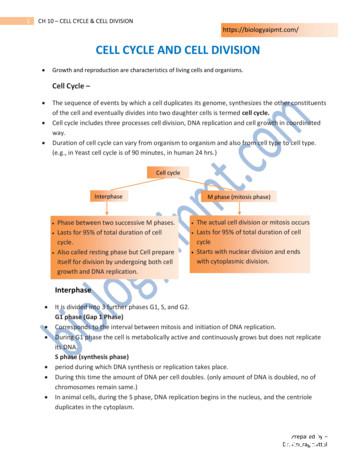
of the cell and eventually divides into two daughter cells is termed cell cycle. Cell cycle includes three processes cell division, DNA replication and cell growth in coordinated way. Duration of cell cycle can vary from organism to organism and also from cell type to cell type. (e.g., in Yeast cell cycle is of 90 minutes, in human 24 hrs.)
UNIT-V:CELL STRUCTURE AND FUNCTION: 9. Cell- The Unit of Life: Cell- Cell theory and cell as the basic unit of life- overview of the cell. Prokaryotic and Eukoryotic cells, Ultra Structure of Plant cell (structure in detail and functions in brief), Cell membrane, Cell wall, Cell organelles: Endoplasmic reticulum, Mitochondria, Plastids,
The Cell Cycle The cell cycle is the series of events in the growth and division of a cell. In the prokaryotic cell cycle, the cell grows, duplicates its DNA, and divides by pinching in the cell membrane. The eukaryotic cell cycle has four stages (the first three of which are referred to as interphase): In the G 1 phase, the cell grows.
Many scientists contributed to the cell theory. The cell theory grew out of the work of many scientists and improvements in the . CELL STRUCTURE AND FUNCTION CHART PLANT CELL ANIMAL CELL . 1. Cell Wall . Quiz of the cell Know all organelles found in a prokaryotic cell
Stent Type Stent Design Free Cell Area (mm2) Wallstent Closed cell 1.08 Xact Closed cell 2.74 Neuroguard Closed cell 3.5 Nexstent Closed cell 4.7 Precise Open cell 5.89 Protégé Open cell 20.71 Acculink Open cell 11.48 Stent Free Cell Area Neuroguard IEP Carotid Stent
Stent Type Stent Design Free Cell Area (mm2) Wallstent Closed cell 1.08 Xact Closed cell 2.74 Neuroguard Closed cell 3.5 Nexstent Closed cell 4.7 Precise Open cell 5.89 Protégé Open cell 20.71 Acculink Open cell 11.48 Neuroguard IEP Carotid Stent Stent Free Cell Area
and Career-Readiness Standards for English Language Arts. The second section includes the MS CCRS for ELA for kindergarten through second grade. The third section includes the MS CCRS for ELA for grades 3-5. The fourth section includes the MS CCRS for ELA, including Literacy in Social Studies, Science, and Technical Subjects. The final section .


