Ch 4 - Functional Anatomy Of Prokaryotic And Eukaryotic Cells
1/22/2017Prokaryotic cellsFunctional Anatomy of Prokaryoticand Eukaryotic CellsChapter 4BIO 220 DNA circular (usually) and not enclosed withina nucleus DNA not associated with histones (HU, IHF, HNS) Generally lack membrane-enclosed organelles Cell wall contains peptidoglycan Divide by binary fissionBinary fissionFig. 6.12Fig. 10.11
1/22/2017Size, shape, and arrangement ofbacterial cells Shape coccus (cocci)Fig. 4.1Size, shape, and arrangement ofbacterial cells Shape bacillus (bacilli)Fig. 4.2Size, shape, and arrangement ofbacterial cells Shape spiral Vibrio – curved rods– Vibrio cholerae Spirilla – corkscrew– Campylobacter jejuni Spirochetes – axialfilamentsBacterial cell shape is dependent on Genetics– Most bacteria are monomorphic – keepsame shape– Some are pleomorphic – can change shape May be due to genetics, environment, orlack of a cell wall–Rhizobium, Corynebacterium– Borrelia burgdorferiFig. 4.42
1/22/2017Prokaryotic cell structure Structures external to cell wallStructures external to cell wall Cell wall Structures internal to cell wallGlycocalyx “Sugar coat” of cell The glycocalyx is a viscous, gelatinous layerlocated outside the cell wall that is composedof polysaccharides and/or polypeptides If it appears organized and firmly adheres to theoutside of the cell wall it is called a capsule If instead it is unorganized in appearance andmore loosely attached to the cell wall it is calleda slime layerFig. 4.63
1/22/2017Glycocalyx functions Allows certain bacteria to resist phagocyticengulfment– Blocks the ability of phagocytes to recognizeantigenic cell wall components (LPS, peptidoglycan)– i.e. Streptococcus pneumoniae, Bacillus anthracisGlycocalyx functions Helps protect cells against dehydration Helps trap nutrients in bacterial cells Allows some bacteria to adhere toenvironmental surfaces– Biofilm formation– i.e. Streptococcus mutans, Vibrio choleraeFlagella Used for motility Flagellar arrangements include atrichous,peritrichous, polar (monotrichous,lophotrichous, amphitrichous)Parts of a flagellum Filament– Composed of globular protein (flagellin)– H antigens help distinguish between serovars(variations within a species) of gram (-) bacteria Hook– Attaches filament to cell Basal body– Anchors flagellum to cell wall and plasmamembraneFig. 4.74
1/22/2017Prokaryotic flagella Move like a propeller (rotates from basalbody), whereas eukaryotic flagella move like awhip Not covered by membrane Motility patterns– Runs – bacterium moves in one direction for aperiod of time (moves 10-20 times its length)– Tumbles – periodic, abrupt, random changes indirectionFig. 4.8Runs and tumblesTaxis Movement of a bacterium toward or awayfrom a particular stimulus is called taxis Chemotaxis PhototaxisFig. 4.9a5
1/22/2017Axial filamentsFimbriae Spirochetes use axialfilaments for motility Axial filaments aresimilar to flagella,except that theywrap around the cellbeneath an outersheath Spirochetes move ina spiral fashion Some gram-negative bacteria have hair-likeappendages (pilin) Fimbriae can occur on cell poles or along cellsurface, may be few to several hundred Fimbriae adhere to each other and to surfacesin and out of the body Neisseria gonorrhoeae,Escherichia coliFig. 4.11Pili Longer than fimbriae and only a few per cell Involved in motility– Twitching (grappling hook) and gliding motilityCell wall Involved in DNA transfer– Conjugation6
1/22/2017Cell wall functions Prevent osmotic lysis of bacterial cells Helps maintain shape of bacterium Point of anchorage for flagella (when present)Fig. 4.6Cell wall compositionPeptidoglycan (murein)Peptidoglycan –composed of repeatingdisaccharide subunitsconnected bypolypeptidesForms a lattice that surrounds and protects the cell.Fig. 4.12Fig. 4.13a7
1/22/2017Cell wall: gram–positive bacteriaGram–positive cell wall Thick peptidoglycan layer (many layers) Contain teichoic acids (alcohol phosphate)– Lipoteichoic acid – spans the peptidoglycan layerand is linked to the plasma membrane– Wall teichoic acid – linked to peptidoglycan– May regulate transport of cations in/out of cells,role in cell growth, prevent cell wall breakdown,provide much of the cell wall’s antigenic specificity Granular layer is between the cell wall andplasma membrane of cellFig. 4.13bCell wall: gram–negative bacteria Thin peptidoglycan layer and an outermembraneOuter membrane (OM) Lipopolysaccharides (LPS), lipoproteins,phospholipids LPS has three parts:– Lipid A – embedded in outer layer of OM (endotoxin)– Core polysaccharide – structural role– O polysaccharide – antigenic Lipoproteins – anchor peptidoglycan to OM Porins permit the passage of molecules such asnucleotides, disaccharides, peptides, amino acids,B12 and iron Presence of OM helps bacteria evade complementand phagocytosisFig. 4.13c8
1/22/2017Gram staining Why do we see differences in how gram ( )and gram (-) bacteria appear with the gramstaining protocol? Gram stain protocol– Primary stain – crystal violet– Mordant – Grams iodine– Decolorizer– Counterstain – safraninFig. 4.13c9
1/22/2017Mycoplasma (pneumoniae)Acid-fast cell walls Used to identify the bacteria of the genus . . . Cell walls contain a high concentration of ahydrophobic waxy lipid (mycolic acid)surrounding a thin layer of peptidoglycanFig. 11.24Plasma membraneStructures internal to the cell wallFig. 4.1410
1/22/2017Plasma membrane functions Selective permeability Breakdown of nutrients and the production ofenergyPM transport Passive transport– Simple diffusion– Facilitated diffusion– Osmosis Active transport– Group translocationFig. 4.15Simple diffusionFig. 4.16Facilitated diffusionFig. 4.1711
1/22/2017OsmosisFig. 4.18Fig. 4.17Active transportCytoplasm Requires the use of energy Thick, aqueous, semitransparent, and elastic In group translocation, as a substance istransported across the wall of a prokaryoticcell, the substance is chemically altered whicheffectively traps it within the cell Mostly water, but also contains proteins,CHOs, lipids, inorganic ions Cytoskeleton present, but made of differentproteins from those found in eukaryotic cells(MreB and ParM, crescentin, FtsZ)12
1/22/2017CytoplasmNucleoid Lacks a nuclearenvelope Contains bacterialchromosome Plasmids – small,circular dsDNA thatreplicatesindependently ofbacterial chromosome Nucleoid Ribosomes InclusionsFig. 4.6InclusionsRibosomes What is the function of ribosomes? Ribosomal subunits Metachromatic granules (volutin)– Reserve of inorganic phosphate for ATP synthesis– Corynebacterium diphtheriae Polysaccharide granules Lipid inclusions– Mycobacterium, Bacillus, Spirillum Sulfur granules– AcidithiobacillusFig. 4.1913
1/22/2017Inclusions Carboxysomes– Ribulose 1,5-diphosphate carboxylase (CO2fixation) Gas vacuoles Magnetosomes– Fe3O4– Decompose H2O2– MagnetospirillumEndospores Members of Clostridium and Bacillus can formendospores, which are essentially dehydrated,stripped-down, “resting” bacterial cells When released into the environment,endospores can survive extremes in heat,dehydration, toxic chemical and radiationexposureFig. 4.20Sporulation (sporogenesis)Endospores Contain a large amount of dipicolinic acid(DPA), which may serve to protect bacterialDNA Endospore core contains DNA, a little RNA,ribosomes, enzymes, small molecules Germination is the process by which anendospore returns to its vegetative state Germination is triggered by high heat ormolecules called germinants (alanine &inosine)Fig. 4.2114
1/22/2017Endospores – Why should I care? Endospores are resistant to processes thatnormally kill vegetative cells Home canning – botulism– Clostridium botulinumFig. 10.1Eukaryotic cells DNA organized into chromosomes containedwithin a nucleus DNA is associated with histones Do have membrane-enclosed organelles Cell walls lack peptidoglycan Cell division by mitosis/meiosis andcytokinesisFig. 4.2215
1/22/2017Cell wall and glycocalyxCilia and Flagella When present, eukaryotic cell walls tend to besimpler than prokaryotic cell walls and have adifferent composition Algae & plants – cellulose Fungi – cellulose or chitin Yeasts – polysaccharides (mannan and glucan) Protozoa have an outer protein covering calleda pellicle GlycocalyxFig. 4.23PhagocytosisPlasma membrane Similar to prokaryotes Eukaryotic membranes contain carbohydratesand sterols Plasma membrane transport– No group translocationFig. 16.816
1/22/2017Cytoplasm Everything inside the plasma membrane withthe exception of the nucleus Cytosol vs. cytoplasm Cytoskeleton Cytoplasmic streamingRibosomes Sites of protein production 80S (60S & 40S) vs 70S May be free or attached to ER PolyribosomesNucleusFig. 4.24Endoplasmic reticulumFig. 4.2517
1/22/2017Golgi complex & LysosomesVacuoles (Plants) Derived from Golgi complexMay serve as temporary storage organellesHelp bring food into the cellStorage of wastesMay accumulate waterFig. 4.26MitochondriaFig. 4.27Chloroplasts (Algae green plants)Fig. 4.2818
1/22/2017Peroxisomes and centrosomesFig. 4.22Endosymbiotic theory Origin of eukaryotic cells Ancestral eukaryotic cell developed a“nucleus” when PM folded around DNA This cell likely ingested aerobic bacteria Support– Mitochondria/chloroplasts resemble bacteria– These organelles contain circular DNA– Organelles reproduce independently– Ribosomes resemble those of prokaryotes– Antibiotic action19
- May regulate transport of cations in/out of cells, role in cell growth, prevent cell wall breakdown, provide much of the cell wall's antigenic specificity Granular layer is between the cell wall and plasma membrane of cell Gram-positive cell wall Fig. 4.13b Cell wall: gram-negative bacteria Thin peptidoglycan layer and an outer
Clinical Anatomy RK Zargar, Sushil Kumar 8. Human Embryology Daksha Dixit 9. Manipal Manual of Anatomy Sampath Madhyastha 10. Exam-Oriented Anatomy Shoukat N Kazi 11. Anatomy and Physiology of Eye AK Khurana, Indu Khurana 12. Surface and Radiological Anatomy A. Halim 13. MCQ in Human Anatomy DK Chopade 14. Exam-Oriented Anatomy for Dental .
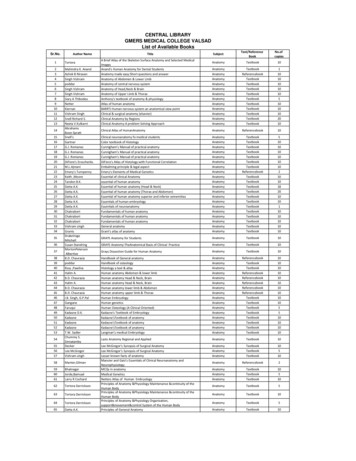
39 poddar Handbook of osteology Anatomy Textbook 10 40 Ross ,Pawlina Histology a text & atlas Anatomy Textbook 10 41 Halim A. Human anatomy Abdomen & lower limb Anatomy Referencebook 10 42 B.D. Chaurasia Human anatomy Head & Neck, Brain Anatomy Referencebook 10 43 Halim A. Human anatomy Head & Neck, Brain Anatomy Referencebook 10
Descriptive anatomy, anatomy limited to the verbal description of the parts of an organism, usually applied only to human anatomy. Gross anatomy/Macroscopic anatomy, anatomy dealing with the study of structures so far as it can be seen with the naked eye. Microscopic
HUMAN ANATOMY AND PHYSIOLOGY Anatomy: Anatomy is a branch of science in which deals with the internal organ structure is called Anatomy. The word “Anatomy” comes from the Greek word “ana” meaning “up” and “tome” meaning “a cutting”. Father of Anatomy is referred as “Andreas Vesalius”. Ph
Pearson Benjamin Cummings Anatomy and Physiology Integrated Anatomy – Gross anatomy, or macroscopic anatomy, examines large, visible structures Surface anatomy: exterior features Regional anatomy: body areas Systemic anatomy: groups of organs working
Anatomy titles: Atlas of Anatomy (Gilroy) Anatomy for Dental Medicine (Baker) Anatomy: An Essential Textbook (Gilroy) Anatomy: Internal Organs (Schuenke) Anatomy: Head, Neck, and Neuroanatomy (Schuenke) General Anatomy and Musculoskeletal System (Schuenke) Fo
Functional Anatomy of the Upper Extremity CHAPTER 6 Functional Anatomy of the Lower Extremity CHAPTER 7 Functional Anatomy of the Trunk Functional Anatomy SECTION II Hamill_ch05_137-186.qxd 11/2/07 3:55 PM Page 137. Hamill_ch05_137-186.qxd 11/2/07 3:55 PM Page 138. CHAPTER 5
abdomen and pelvis volume 5 8 cbs anatomy 1 25 chaurasia, b.d. bd chaurasia's human anatomy: lower limb abdomen and pelvis volume 6 8 cbs anatomy 1 26 chaurasia, b.d. bd chaurasia's human anatomy: lower limb abdomen and pelvis volume 7 8 cbs anatomy 1 27 chaurasia, b.d. bd chaurasia's human anatomy: lower limb abdomen and pelvis volume 8 8 cbs .


