Respiratory System - Anatomy
Respiratory SystemAnatomyOverviewOf all the substances that cells and thewhole body must have to survive, O2 is byfar the most crucial A person can live a few weeks withoutfood, a few days without water, but only afew minutes without O2 Constant removal of carbon dioxide fromthe body is just as important for survivalas a constant supply of O2 Functions The organs of the respiratory systemperform several functions:– Gas exchange via diffusion Deliveryof O2 to body cellsof CO2 produced by body cells Elimination– Regulation of blood pH– Filter, warm & humidify the air we breathe– Contain receptors for the sense of smell– Production of vocal sounds– Excretion of heat & water1
Respiration EnsuresO2 is supplied to body cellsis removed from the body cells Respiration CO2 homeostatic mechanism– Helps maintain a constant environment body cells to function effectivelyRespiratory Organs Organsof the respiratory system– Nose & nasal cavities– Pharynx– Larynx– Trachea– Bronchi– Lungs– Alveoli Basic structure is that of a tube with many branches endingin millions of extremely tiny, very thinthin-walled sacs calledalveoliConsists of passageways that filter incoming air & carry itinto the lungs2
Respiratory Tract Divisions Assist in thedescription ofsymptoms associatedwith commonrespiratory problemssuch as a coldNose Upper respiratory tract-Nose-Pharynx-Larynx PharynxLarynxLower respiratory tract in the thorax– trachea, bronchial tree & lungsRespiratory Tract Nose, pharynx, larynx, trachea, bronchi &bronchioles are hollow tubes– Form air passageways– Constitute conducting portion of respiratorysystem Air sacs & alveoli– Respiratory portion of the respiratory system– Gas exchange occurs in the alveoli (largesurface area)– Alveoli sacs are delicate elastic membraneswith extensive capillary network of thepulmonary circulation3
Anatomy of theRespiratory SystemUpper Respiratory TractNose Air enters the respiratorytract through the externalnares or nostrilsFlows into the right & leftnasal cavities, (lined byrespiratory mucosa)A partition called the nasalseptum separates thesetwo cavitiesAir may also enter via themouth - the nasal cavities& mouth meet at theregion at the back of themouth pharynxSurface is moist frommucus & warm fromblood flowNerve endingsresponsible for thesense of smell(olfactory receptors)are located in thenasal mucosaThree conchaeprotrude into thenasal cavityThese increase surfacearea over which airmust flow as it passesthrough the nasalcavity4
NoseThe structure of the conchae increases the surface area overwhich inhaled air travels ensuring that it is thoroughlywarmed & filteredNose Blood vessels in the nasal mucosa cool hot air &warm cold airAir entering the nose is generally contaminatedwith one or more common irritants such asinsects, dust, pollen & bacteriaAir is purified removing almost all contaminantsbefore inspired air reaches the lungsMucus secreted by mucosa adds moisture to dryair while trapping fine dust particles & micromicroorganismsCiliated cells of the mucosa move contaminatedmucus into the throat where it is swallowed ClinicalExample:– Because the mucosa lines the nose,sinus infections often develop fromcolds in which the nasal mucosa isinflamed– When the nasal cavity is blocked, the airin the sinuses is absorbed– Sometimes a sinus headache is incurred& localised over the inflamed area5
Paranasal SinusesFour paranasal sinusessinuses- the frontal,maxillary, sphenoidal & ethmoidal – draininto the nasal cavities Paranasal sinuses are lined with mucousmembrane that assists in the productionof mucus for the respiratory tract Hollow spaces help to lighten the skull &serve as resonant chambers for theproduction of sound Paranasal SinusesYou can see how a sinus headache would be quite uncomfortable asthe pressure within the cavity would build up & be unable to escape(see slide 15)Pharynx Extendsfrom the nasal cavities tothe larynx Behind the nasal cavities & abovethe soft palate is the nasopharynx Dorsally is the oropharynx digestive & respiratory passagewaysmeet Inferior to oropharynx lies thelaryngopharynx immediately beforethe larynx6
Three regions of the pharynxPharynx Twoauditory tubes, the Eustachiantubes open from the middle ear intothe lateral walls of the nasopharynx– Equalise air pressure between thenasopharynx & the middle earPharynx Pharyngeal tonsils lie on posterior wall ofnasopharynx– Traps airborne infectious agents– Swollen tonsils are referred to as adenoids which mayobstruct the passage of air Palatine tonsils lie on the lateral aspects of thepharynx behind the mouth– Function same as pharyngeal tonsil– Tonsillitis inflammation of the palatine tonsils obstructs nasopharynx, forcing mouth breathing air is not properly moistened, warmed or filtered beforereaching the lungs7
Question:How might the position of the tonsils assist in performing their immunefunction of trapping and destroying pathogens?Pharynx Thepharynx is a passageway forboth the digestive & respiratorysystems Distally, the pharynx branches intotwo tubes– Oesophagus stomach– Larynx Larynx lungsLarynxCartilaginous structure connecting thepharynx & trachea at the level of thecervical vertebrae Connective tissue containing nine pieces ofcartilage arranged in boxbox-like formation Largest cartilage is the thyroid cartilage,AKA "Adam's apple" – Thyroid cartilage is visible in the ventral aspectof the throat and is more pronounced in adultmales than adult females8
The cricoid cartilage resembles a signet ring– Connects larynx & trachea The epiglottis, a leafleaf-shaped "lid" at the entry tothe larynx– Seals off the respiratory tract when food passes into theoesophagus Opening to the larynx is called the glottis– During swallowing the larynx is pulled upward, theepiglottis closes to route food/fluid to the stomach– If anything other than air enters the larynx, a coughreflex is triggered to expel the substance & prevent itgoing to the lungsThe larynxQuestion:What is the advantageof having cartilaginousrings in the airways?Transvere sectionthrough the tracheaLarynxLarynx is a passageway for air & produces soundTwo folds of tissue project from the lateral wallsof the larynx vocal cords Exhalation vocal cords vibrate produce sounds that can be modified into wordsby muscles of the neck, lips, tongue, & cheeks Length of vocal cords determines pitchfemales & children have shorter vocal cords voices of a higher pitch – Read page 842 Jenkins, Kemnitz & TortoraStructures of voice production9
Trachea Larynx opens into a rigid tube tracheaTrachea is 12 to 15cms long in the midline ofthe neckSupported & held open by a stack of CC-shapedrings of cartilage open at the dorsal aspectThe area between adjacent cartilages & the tipsof cartilage contains connective tissue & smoothmuscleThe trachea is an open passageway for incoming& outgoing airCiliated cells filter air before it enters the bronchiTracheaBy pushing against your throat about aninch above the sternum, you can feel theshape of the trachea Only if you use considerable force can yousqueeze it closed Air has no other way to get to the lungs, &complete tracheal obstruction can squeezethe trachea shut & cause death in amatter of minutes – Eg. choking on food, tumour or infectioncausing inflammation of the lymph nodes ofthe neckBronchi The trachea branches into two primary bronchi– Same structure as the trachea– Right bronchus is slightly larger & more vertical than theleft Bronchi become smaller & smaller secondarybronchi then tertiary bronchiAs they extend further into the lungs diameter isreduced to about one millimetreBronchi are now called bronchiolesThe amount of cartilage reduces as the tubesbecome smaller & smaller disappearing in thedistal bronchioles10
Note the branching structure of the bronchi as the tubes becomesmaller & smallerBronchioles Bronchioles are composed of smooth musclesupported by connective tissueSubdivide until they form the smallest airpassageways terminal bronchiolesTerminal bronchioles extend into the alveoliAlveoli resemble a single grape & are effective in gasexchange as they are thinthin-walled & in contact with ablood capillaryMembrane inside each alveoli is covered in surfactantwhich reduces surface tension, keeping them fromcollapsing as air moves in & out during respirationBranching & rebranching of the bronchi & bronchioleswithin the lungs is called the bronchial treeBronchiClinical Examples: Inflammation of the bronchial tree is commonlyknown as bronchitis Asthma also affects the bronchial tree– Asthma is accompanied by periodic attacks of wheezing& difficult breathing– Caused by spasms of the smooth muscles (as there is nocartilage to hold them open)– Often triggered by allergens in the environment– Read page 844844-845 Jenkins, Kemnitz & TortoraBronchi11
Lungs Paired organs occupying most of the space of thethoracic cavityConsist of millions of small, cupcup-shaped outpockets (sacs) called alveoliRespiratory membranes of alveoli are a thinbarrier in which gases can pass by diffusion 300 million alveoli in an average adultLungs are separated from one another by amedian dividing wallCalled the mediastinum– contains the heart, thymus, oesophagus, large bloodvessels embedded in connective tissueLungs Lungsare conical shaped withelastic, spongy texture due to thenature of the alveoli Right lung is subdivided into threelobes Left lung is subdivided into two lobes Each lobe is divided into smallerlobules, each lobule is serviced by alarge bronchiole12
Microscopic Anatomy of theLungs The walls of the alveoli are one cellsthick The surface area of the alveoli ishuge 70m2 As the capillary network is soclosely associated with the cell wallrespiratory gases are easily diffusedacross the surfaceRespiratory MembraneRespiratory membrane separates the airin the alveoli from the blood insurrounding capillaries Consists of four cell layers – Alveolar wall of Type I & Type II alveolar cells– Epithelial basement membrane– Capillary basement membrane– Capillary endothelium– Read page 849849-850 Jenkins, Kemnitz & TortoraAlveoliHistology of AlveoliEpithelial spiratory/alveoli.jpg13
PleuraTwoTwo-layered membrane surrounding eachlung Inner layer visceral pleura – covers the surface of each lung– reaches into the fissures between the lobes ofthe lung– encloses the mediastinum Outer layer parietal pleura– lines the inner surface of the thoracic cavityWho remembers Fred Dagg & his song ‘If it weren't for yourgumboots’? The pleurisy mentioned in the song is an inflammation ofthe pleurae. It is a very painful condition as it reduces the ability ofthe pleural surfaces to move over each other causing rubbing/frictionwith each breath.PleuraVisceral & parietal pleura are continuouswith one another where the primarybronchus, blood vessels & nerves entereach lung Two layers of the pleura form a collapsedsac Area within the sac pleural cavity – Fluid in the cavity keeps the twotwo-pleural membranes inclose contact with each other & allows them to glidesmoothly over each other– Fluid adheres the two layers of the pleura to one another14
Respiratory Mucosa Membranelining most of the airdistribution tubes in the respiratorysystem respiratory mucosa Respiratory mucosa is covered withmucus & lines the tubes of therespiratory tree Protective mucus is an important airpurification mechanismRespiratory Mucosa 125ml of respiratory mucus is produced dailyForms a continuous blanket that covers the liningof the air distribution tubes in the respiratory treeMucus moves upward to the pharynx on millionsof hairlike cilia that cover the epithelial cells inthe respiratory mucosaCigarette smoke paralyses cilia accumulationsof mucus & the typical smoker’smoker’s cough, which isan effort to clear the secretions– Read Cari’Cari’s story in chapter 22 of Jenkins, Kemnitz &Tortora for more on the effects of smoking on therespiratory tractRespiratory Mucosa This image is showsthe respiratory mucosa The cilia lining theepithelium are clearlyseen Mucus producinggoblet cells are labmanual2002/labsection2/Respiratory03 files/image002.jpg15
respiratory problems such as a cold Upper respiratory tract-Nose-Pharynx-Larynx Nose Pharynx Larynx Lower respiratory tract in the thorax - trachea, bronchial tree & lungs Respiratory Tract Nose, pharynx, larynx, trachea, bronchi & bronchioles are hollow tubes - Form air passageways - Constitute conducting portion of respiratory system
Human Anatomy and Physiology II Laboratory The Respiratory System This lab involves two exercises in the lab manual entitled “Anatomy of the Respiratory System” and "Respiratory System Physiology". In this lab you will look at lung histology, gross anatomy, and physiology. Complete the review sheets from the exercise and take the online quiz on
Clinical Anatomy RK Zargar, Sushil Kumar 8. Human Embryology Daksha Dixit 9. Manipal Manual of Anatomy Sampath Madhyastha 10. Exam-Oriented Anatomy Shoukat N Kazi 11. Anatomy and Physiology of Eye AK Khurana, Indu Khurana 12. Surface and Radiological Anatomy A. Halim 13. MCQ in Human Anatomy DK Chopade 14. Exam-Oriented Anatomy for Dental .
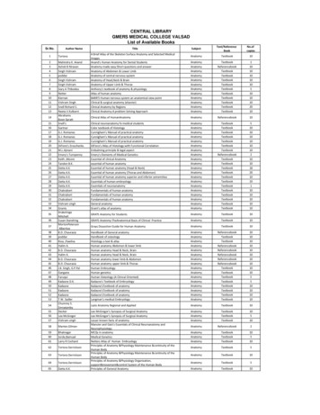
39 poddar Handbook of osteology Anatomy Textbook 10 40 Ross ,Pawlina Histology a text & atlas Anatomy Textbook 10 41 Halim A. Human anatomy Abdomen & lower limb Anatomy Referencebook 10 42 B.D. Chaurasia Human anatomy Head & Neck, Brain Anatomy Referencebook 10 43 Halim A. Human anatomy Head & Neck, Brain Anatomy Referencebook 10
Anatomy titles: Atlas of Anatomy (Gilroy) Anatomy for Dental Medicine (Baker) Anatomy: An Essential Textbook (Gilroy) Anatomy: Internal Organs (Schuenke) Anatomy: Head, Neck, and Neuroanatomy (Schuenke) General Anatomy and Musculoskeletal System (Schuenke) Fo
HASPI Medical Anatomy & Physiology 14a Lab Activity The Respiratory System A healthy respiratory system is crucial to an individual’s overall health, and respiratory distress is often one of the first indicators of a life-threatening illness. The function of the respiratory system is to exchange gases between the external air and the body.
Station 1: The Respiratory System Respiratory System Anatomy - Using the "Respiratory System" chart, identify the labeled organs or parts of the organ A-S in Table 1 below. If there are any that you cannot identify, use a textbook or online resource. A smaller version of the charts are included for later review. Table 1: The Respiratory .
Descriptive anatomy, anatomy limited to the verbal description of the parts of an organism, usually applied only to human anatomy. Gross anatomy/Macroscopic anatomy, anatomy dealing with the study of structures so far as it can be seen with the naked eye. Microscopic
Text and illustrations 22 Walker Books Ltd. Trademarks Alex Rider Boy with Torch Logo 22 Stormbreaker Productions Ltd. MISSION 3: DESIGN YOUR OWN GADGET Circle a word from each column to make a name for your secret agent gadget, then write the name in the space below. A _ Draw your gadget here. Use the blueprints of Alex’s past gadgets on the next page for inspiration. Text and .


