Appendix G: The Pantone “Our Color Wheel” Compared To
Appendix G - 1Appendix G:The Pantone “Our color Wheel” compared to the ChromaticityDiagram (2016) 1There is considerable interest in the conversion of Pantone identified color numbers to other numbers within the CIEand ISO Standards. Unfortunately, most of these Standards are not based on any theoretical foundation and haveevolved since the late 1920's based on empirical relationships agreed to by committees. As a general rule, theseStandards have all assumed that Grassman’s Law of linearity in the visual realm. Unfortunately, this fundamentalassumption is not appropriate and has never been confirmed. The visual system of all biological neural systems relyupon logarithmic summing and differencing.A particular goal has been to define precisely the border between colors occurring in the local language andvernacular. An example is the border between yellow and orange.Because of the logarithmic summations used in the neural circuits of the eye and the positions of perceived yellowand orange relative to the photoreceptors of the eye, defining the transition wavelength between these two colors isparticularly acute.The perceived response is particularly sensitive to stimulus intensity in the spectral region from560 to about 580 nanometers.This Appendix relies upon the Chromaticity Diagram (2016) developed within this work. It has previously beendescribed as The New Chromaticity Diagram, or the New Chromaticity Diagram of Research. It is in fact afoundation document that is theoretically supportable and in turn supports a wide variety of less well foundedHering, Munsell, and various RGB and CMYK representations of the human visual spectrum. In general, it does notsupport any CIE Standards related to human vision; but it does provide a method for understanding how theseempirical representations came about.G.1 The Pantone color wheel versus the Chromaticity Diagram (2016)This section will concentrate on the development of the Pantone Color Space (known as Our Color Wheel) and theChromaticity Diagram (2016) developed in the work, “Processes in Biological Vision” (PBV). In the developmentof the comparison, additional comparisons will be represented with citations to further details in PBV.It will be asserted that there can be no precise mathematical equation(s) between these two color spaces because ofthe crudeness of the definition of the Pantone color space.To quote Pantone’s website,“In 1963, Pantone revolutionized the printing industry with the colorful PANTONE MATCHINGSYSTEM , an innovative tool allowing for the faithful selection, articulation and reproduction of consistent,accurate color anywhere in the world. The tool organizes color standards through a numbering system andchip format, which have since become iconic to the Pantone brand.”Elsewhere on that web page, they assert the proprietary nature of their numbering system and chip format.Pantone was acquired by X-Rite, Inc in 2007, and X-Rite was acquired by Danaher in 2012.It is likely that Pantone originally employed several pigments well known to artists in the preparation of their colorsamples, such as the list on page 31 of Hope & Walch2, an encyclopedia of color information.G.1.1 “Our Color Wheel” of pdfs/BA0646OurColorWheel.pdf provides what Pantone calls “Ourcolor wheel.” The wheel is conceptual and based on what they identify as the primary colors of red, blue and1January 27, 20192Hope, A. & Walch, M. (1990) The Color Compendium. NY: Van Nostrand Reinhold
2 Processes in Animal Visionyellow. They then define secondary colors as green, orange and violet. Each secondary color is made of equal partsof the adjacent primaries. They then define tertiary colors as made of equal parts of the primary on one side and thesecondary on the other side. The result is a wheel of only 12 discrete colors and no specific way to subdivide them.The equivalent Munsell Color Space can be subdivided into at least 120 discrete colors, hue, and an unlimitednumber of saturation levels. It can also accommodate a large number of lightness levels.“The Pantone Book of Color3,” authorized and printed by Pantone presents 1024 color swatches with fanciful namesand a ink formula proprietary to Pantone. No numerical codes are associated with the color swatches in this book.The first swatch in the book, labeled “Winter White” exhibits a distinct yellow caste. The color representation onthe cover does not include a central neutral (white) region.Recently, the multiple volumes of the Pantone Book of Color include thousands of annotated color samples tosupport various methods of printing on packaging, textile & plastics materials.The Pantone “Our color wheel” is neither a CMYK system used by printers in process color applications nor a RGBsystem as used in active sources (monitors, projection systems, etc.) It is a hybrid most closely related to theMunsell Color Space. However, it is not a direct overlay of the Munsell Color Space. They define a set of “Colorsin Common” which do not conform to any other system. They do adopt the Munsell Color Space concepts oflightness (value), and saturation (hue) but then they deviate and introduce tints and shades. “A shade is the hue plusblack, and a tint is the hue plus white. There are only five defined saturation steps between white and black. Bycombining the concepts of saturation and lightness, they obscure these independent parameters. They do not speakin terms of saturation as it is used in Munsell Color Space; zero is neutral (colorless) and the saturation can go up to(theoretical) high levels (15, 36, etc.). Simultaneously, the lightness can go from very high to very low withoutaffecting the saturation and hue (in the first order). The Munsell Color Space illustrates second order limits on thehuman color space due to the signal processing inherent in the neural system.The equivalent Munsell Color Space can be subdivided into at least 120 discrete colors, hue, and an unlimitednumber of saturation levels. It can also accommodate a large number of lightness levels (at least 14 on a logarithmicscale).G.1.2 The Chromaticity Diagram (2016)The Chromaticity Diagram (2016) has been presented in many forms. The basic form is shown in Figure G.1.2-1 Itis developed theoretically in Part 1a of Chapter 17 beginning with Section 17.3.3 on page 238 . It is developed morefully for applications and compared with other color spaces in Part 1b of Chapter 17 beginning with a variety ofdefinitions in Section 17.3.4 . Confirmation of the null axes at 494 and 572 nm was obtained by Wright in 1929. Seepage 17 of Part 1b.The parameters, O–, P– & Q– represent the signals propagated through the neural system to the brain andrepresented by O LnS - LnUV, P LnS - LnM and Q LnL - LnM. There is a caveat with respect to the equationrelated to Q that will not be introduced here. See Section 17.3.3 in Part 1a above.The Chromaticity Diagram (2016) is compatible with the axes of Hering Color Space, of Munsell Color Space, ofRGB Color Space, and CMYK Color Space. It also provides specific wavelengths for the individual color spaces inthese representations.3Eiseman,L. & Herbert, L. (1990) The Pantone Book of Color. Monachie NJ: Pantone, Inc.
Appendix G - 3Figure G.1.2-1 The Chromaticity Diagram (2016). The basic form is shown with thenulls at O 0 at 395 nm, P 0 at 494 nm, and Q 0 at 572 nanometers representingthe subtractions of the logarithms of the stimulus intensity within the neural systembetween the UV - S, S - M, and M - L photoreceptor channels.
4 Processes in Animal VisionG.1.3 The archaic CIE representations up through 1975When initially defining the photometric performance of the human visual system, the CIE was unable to demonstratethat performance in a consistent, and mutually acceptable and collegial manner. As a result, they collected the crudedata available in the 1920's and early 1930's and used it to describe a “Standard Observer,” that should never beinterpreted as exhibiting the average performance of the visual system of actual human observers.The x(λ), y(λ) & z(λ) functions defined by the CIE do not even remotely resemble the actual spectral sensitivities ofthe chromophores of vision. Similarly defining the luminance in terms of y(λ) where this function is defined asidentical to the adopted visibility function, V(λ), only complicates the problem. Finally, it is appropriate to point outthe function, P(λ), used to define the power density is only appropriate if the sensory neurons of the visual modalityare energy sensitive. In fact, they are fundamentally quantum counters. Using P(λ) in vision modeling discriminatesagainst the short wavelength region of the spectrum since the photons in this area contain more energy/photon thanin the long wavelength region.G.1.3.1 The archaic chromaticity function as an exampleThe CIE chromaticity diagram of 1934, modified in1951 are basically mathematical models developed in the 1920'sand early 1930's when the technology available was quite limited. The various laboratories contributing to the finalCIE 1934 all assumed the human neural system was based on linear summations and differences. This was a fatalerror. They also used gelatin filters and low temperature light sources (around 2700 Kelvin). The gelatin filterstypically had a spectral width of 20–25 nanometers wide, with very poorly defined skirts, and smeared out colors tothis degree of precision. The spectral width current 5 nanometer interference filters, with very steep skirts, givetotally different results. The low color temperature light sources were deficient in the blue region (althoughcompensation was attempted in those early days). The investigators did not recognize the presence of ultra-violetphotoreceptors in the human eye and theis led to major disparities among the experimenters based on the differentialadaptation of the various receptors. This resulted in hilarious arguments among the principles as so welldocumented.The 1961 CIE UCS (uniform color space, which included the Lab & Luv subsets) failed badly. It was based entirelyon a set of piece wise linear equations that did not represent reality very well. Between 1961 and 1975, theequations representing the UCS, Lab & Luv color spaces were modified several times. The 1961 CIE UCS and itsderivatives are no longer supported by the CIEThe 1976 CIE UCS dropped the Lab and Luv subsets. It remained based on a set of arbitrary piece-wise linearequations but at least they more closely matched the real world, as well as the theoretical world. See the lower rightframe of figure G.1.1-2 below. The CIE 1976 UCS is relatively equiangular when overlaid on the ChromaticityDiagram (2016). However, as shown, it does not extend to the limits of the human visual space. See Section17.3.5.4 (page 48) for more specific information.G.1.3.2 The archaic visibility function as an exampleThe so-called Photopic Visibility Function V( λ) of the CIE originally dated 1924, has been obsolete at least sincethe early 1950's when better instrumentation led to a new understanding of the function. Wright, one of the majorparticipants in the development of the Visibility Function during the 1920's, provided a very interesting descriptionof the development of the function at a symposium in 1969. Wright’s words as they appear in Boynton4, arereproduced in Section 17.2.3.6.5,“The CIE Colorimetry Committee recently in their wisdom have been looking at the old 1931 observer andhave been smoothing, and interpolating, the data to obtain more consistent calculations with computers. Thishas also involved some extrapolation and, in smoothing, they have added some additional decimal places.When I look at the revised table of the x (bar), y(bar), z(bar) functions, I am rather surprised to say the least.You see, I know how inaccurate the actual measurements really were. (Laughter from audience) Guild didnot take any observations below 400 nm and neither did I, and neither did Gibson and Tyndall on the V(8)curve, and yet at a wavelength of 362 nm, for example, we find a value y(bar) of 0.000004929604! This, inspite of the fact that at 400 nm the value of y(bar) may be in error by a factor of 10 (Laughter).”In 1951, Judd proposed a new photopic visibility function with significantly higher sensitivity than the 1924 standard4Boynton, R. (1979) Human Color Vision. NY: Holt, Rinehart and Winston
Appendix G - 5in the spectral region of 400 to 450 nm. The CIE chose not to accept the Judd modification cuting the wide use ofphotometry based on the 1924 standard.Wyszecki & Stiles (1982) discuss the state of the competing visibility standards available at that time (pages 392409). The situation was so confused that their 2nd Edition of 1982 omitted a section 5.7.3 of the 1st Edition (1967)although it was called out on page 396. In the 1980 time period, the title Visibility Function was replaced by theLuminous Efficiency Function rather than the more descriptive, spectral sensitivity function of human vision.The recent explosive growth in the application of narrow spectral band light sources based on semiconductorphysics, i.e., light emitting diodes and semiconductor-based light emitting lasers, has placed new pressure on the CIEto provide theoretically-based, or at least a theory of vision compatible, visibility function.----Figure G.1.3-1 compares the archaic CIE Visibility Function and recent measurements using 5 nm interferencefilters with virtually vertical skirts. The information in this figure is considerable and very important.The left frame displays the CIE 1931 Photopic Luminosity fct, the CIE 1934 Scotopic Luminosity fct., and the 1958TCI–58 Chromaticity Model of the CIE’s successor organization, the Technical committee on Illumination (TCI) ofthe International Standards Organization (ISO). All data is presented on a relative intensity scale. The responsedrawn (with smoothing) through the statistical data from many observers under a variety of measurement conditions(red line) exhibits a significantly different spectral peak wavelength than the widely published CIE 1931 PhotopicLuminosity fct. (previously known as the Visibility fct., V( λ); 532 nm versus the old value of 555 nm based on theStandard (unreal) Observer. This is the peak attributed to and measured for the M–channel (green) photoreceptor. Italso shows peaks corresponding to the S–channel (blue) photoreceptor at 437 nm and the L–channel (red)photoreceptor at 625 nm. It also illustrates the logarithmic summation of the response of the photoreceptor,particularly in the blue-green region where the depth of the notch between them is much less than expected under thelinear summation assumption.The right frame displays data collected using 5 nm interference filters and careful attention to the differentialadaptation of the eye before the start of measurements (personal communication). This author helped develop theprotocol used in the experiments of Babucke and helped track down internal reflections in the instrumentation thatinitially distorted the data. The curve is the absolute intensity of the response of one individual in radiometric unitsfor a fully dark adapted eye of KM. The absolute intensity threshold is remarkably constant across the humanspectrum until it nears the long wavelength limit assymptote near 0.675 microns (675 milli-microns). In the shortwavelength region, the threshold stimulus remained high down to at least 0.415 microns indicating the participationof the UV photoreceptor with a theoretical peak at 0.342 microns. A sharp cutoff at 0.400 microns could beexpected due to the lens of the eye.Figure G.1.3-1 Comparison of Visibility Functions, the CIE 1931 function and a recentvisibility functions using modern instrumentation. See text. Right frame fromBabucke, 2008.In the absence of the lens (known as the aphakic condition) the response of the human retina would be expected toshow a sensitivity down to about 0.320 microns as illustrated by Stark & Griswold and by Tan in Section 17.2.3.1.
6 Processes in Animal VisionFigure G.1.3-2 reproduces their data.Figure G.1.3-2 Comparison of aphakic vision and the theoretical model. The datacurves were normalized with respect to each other by Griswold & Stark. Thetheoretical spectra are normalized with respect to each other but separately from thedata. The four chromophore absorption curves are shown normalized separately asa reference. The composite theoretical curves are for a quality factor of 4.8 and areonly illustrative. See text for discussion. Data points from Griswold & Stark, 1992.While the experimental data in the figure appears to be quite good, it must be noted that at the scale of the figure, thedifferences between Griswold & Stark and Tan are generally greater than 2:1. Some points differ by 3:1. Thetheoretical curves proposed by this work can easily be drawn within the envelope of these combined works asshown.Thornton confirmed the theoretical spectral responses at the bottom of the figure in 1999 (Section 5.5.10.4.3 andSection 5.5.10.4.4). Besides the reference absorption curves at the bottom of the figure, two composite spectralsensitivity curves are shown. The upper line (Red) is for photopic vision and the lower (Green) is for pure scotopicvision; This loss of the long wavelength performance at low stimulus levels is due the long wavelengthphotoreceptor responding to the square of the stimulus intensity.The data in these sections confirm the tetrachromatic character of the human retina with spectral peaks conformingto the four photoreceptor types; Rhodonine(11) at 625 nm, Rhodonine(9) at 532 nm, Rhodonine(7) at 437 nm andRhodonine(5) at 342 nm. (Section 5.5.8 through Section 5.5.10) There is no data in any of the representationssupporting a broadband photoreceptor historically associated with a “Rod.” The term “Rod” is archaic and neverexisted in a biological eye. It was used in ancient time, up through the 20th Century, in the absence of an adequatetheoretical model and sufficiently sophisticated laboratory instrumentation.-----
Appendix G - 7Further complicating the laboratory confirmation problem is the significant difference between measurements usinga filtered photometric response (with the intensity measured on a photometer) and measurements made on an equalenergy basis (with intensity measured with a radiometer). The typical photometer uses a filter that is anapproximation to the archaic CIE Visibility Function. Only measurements based on a single photometer or based onradiometry are reproducible.G.1.4 Comparison of the Pantone color wheel and the Chromaticity Diagram (2016)Figure G.1.4-1 compares the Pantone, “Our color wheel,” to the New Chromaticity diagram with two separateoverlays; a composite showing the Munsell Color Space and the capability of process color and kinescopes toreproduce the visual color range, and an overlay of the CIE UCS 1976 color space.The frame on the lower left of this figure can be found in Section 17.3.5.2. The frame on the lower right of thisfigure can be found in Section 17.3.5.4.Pantone chose to assume the primary colors of human vision are red, blue and yellow. Young (1802) was one of thefirst philosophers to considered the choice between red, blue and yellow primaries and red, blue and green primariesin 1802. At the time, Young vacillated between the two sets. After initially assuming the first choice, he changedhis mind and ever after assumed the red, blue and green triad. As in the current Pantone case (beginning in 1963),the choice was made in the absence of any technical knowledge or viable theory. It is now clear that thephotoreceptors of the eye exhibit peak sensitivities at 342, 437, 532 and between 610 & 625 nanometers(corresponding to the ultraviolet, blue, green and red spectral channels). The caveat relating to the long wavelengthpeak is discussed in Section 17.3.3 of Part 1a of Chapter 17. The 437, 532 and 625 nm peaks correspond to thepeaks in the individual photoreceptors of the visual spectrum measured in the laboratory. All four peaks have beenmeasured in the laboratory on aphakic (lens-less) human eyes.The fundamental assumption by Young, and perpetuated by the CIE since the 1930's is false. That false assumptionis that the human color space is equilateral with the three primaries, red, blue and green are at the apices of such aequilateral triangle. In fact, the color channel differences represented by the O, P and Q channels of color signalinformation are treated as statistically independent by the brain. Mathematically, this means the channels are treatedas orthogonal and can be represented by the three axes of a rectangular volume. As shown in Section,17.3.3, theresulting color space is a right parallelepiped with the red, blue and green signaling channels at the corners of a righttriangle and not an equilateral triangle, as previously long assumed, when presented in two dimensions.To accommodate the limited ultraviolet sensitivity of the human eye, the representation of the O– channel is rotatedinto the plane of the 2-dimensional Chromaticity Diagram (2016) for convenience. This means the diagram exhibitsa discontinuity along the 437 nm axis. P– values larger than 10 are not shown correctly and straight lines cannot bedrawn between point in the main presentation and points at wavelengths shorter than 437 nm.Until a variant of the Chromaticity Diagram (2016) is adopted by the CIE based on a right triangle for the threecorners representing the red, blue, & green primaries,, any researcher should use great caution when invoking the1931 through 1976 Diagrams as a foundation for, as a tool in or as corroboration of his work (Section 17.3.3).Pantone does not describe the wavelength of any primary color. Thus it cannot be precisely converted to any othernotation.
8 Processes in Animal VisionFigure G.1.4-1 “Our color wheel” from Pantone (top) compared to the ChromaticityDiagram (2016) overlaid with the Munsell Color Space (bottom, left) and the CIE 1976UCS (bottom, right). Also shown on the left is the capability of “process color” andkinescopes to reproduce the visual spectrum. By inverting the Pantone wheel, it canbe shown (bottom, center) as an approximate overlay on the Chromaticity Diagrambut it does not accommodate the cyan color at 494 nm or differentiate between redand magenta adequately. See text. Top figure from Pantone, 2019.The CIE chromaticity diagram of 1934, modified in1951 are basically mathematical models developed in the 1920'sand early 1930's when the technology available was quite limited. The various laboratories contributing to the finalCIE 1934 all assumed the human neural system was based on linear summations and differences. This was a fatalerror. They also used gelatin filters and low temperature light sources (around 2700 Kelvin). The gelatin filterswere typically 20 nanometers wide and smeared out colors to this degree of precision. Current 5 nanometerinterference filters give totally different results. The low color temperature light sources were deficient in the blueregion (although compensation was attempted in those early days). The investigators did not recognize the presenceof ultra-violet photoreceptors in the human eye and theis led to major disparities among the experimenters based onthe differential adaptation of the various receptors. This resulted in hilarious arguments among the principles as so
Appendix G - 9well documented.The 1961 CIE UCS (uniform color space, which included the Lab & Luv subsets) failed badly. It was based entirelyon a set of piece wise linear equations that did not represent reality very well.Since the 1961 CIE Lab Standard, a variant of one of the subspaces, has survived, the a*,b* space. However, itremains empirically based and fails to represent the human color space adequately.The 1976 CIE UCS dropped the Lab and Luv subsets. It remained based on a set of arbitrary piece-wise linearequations but at least they more closely matched the real world.In the last few years, there have been many articles written describing a set of cone-fundamentals associated psychophysically with human vision. The psychology community supports a far different set of spectral responses of thethree chromophores of trichromatic vision than does the application oriented printing and display devicecommunities. Two questions arise. First; What is the physiological relevance of the spectral color space to thechromophores of biological vision? The answer to this question would make many of the earlier papers defining theprimary colors, the most vivid colors and many other designations (some of which are mentioned in Section 2.1 andSection 17.3.8 of this work) obsolete and archaic. Second; How precisely can the colors in a representation of thatcolor space be made to agree with the absolute scale of the Chromaticity Diagram (2016)?G.2 The a*, b* color spaceThe a*, b* color space of the 1976 CIELAB appears quite similar to the McLeod-Boynton color space of 19xxx.See Section 17.3.5.5.2. They both introduce an alternate set of coordinates useful for low saturation colors near theneutral (white) point.To support the above discussion, Figure G.2.1-1 reproduces a rendition of the a*,b* color space found inAbildgaard5. The location of the colors varies significantly from that presented by Malacara in 20026. Neitherrepresentation appears to be realistic. The Abildgaard colors seem to be too saturated for the scale locations shown.Are the scales shown the same as described by Malacara for his a*, b* color space? Malacara labeled his colorlocations as approximate. Figure 5.20 in the first edition of Malacara when inverted does not resemble the 2016Diagram but Malacara did include wavelength for the termini of his axes. The wavelengths explain why his figuredoes not contain a neutral (white point corresponding to LN). His axes when plotted on the Chromaticity Diagram(2016) is skewed far to the left (short wavelength axis). Malacara (2002) dedicated17 pages to consideration of the1960 Luv & 1960 Lab and 1976 L*a*b* color spaces. He also provided a figure 5.10 showing the distortions in CIEx,y color space compared to that of the Munsell Color Space. His figures 5.18, 5.19 and 5.21 address the highlydistorted form of the a*, b* color space. The curves in these figures all trail off toward infinity in the magenta area(as suggested by the CIE overlay on the Chromaticity Diagram 2016 in section G.2.1 of this Appendix. Figures 5.18& 5.19 provide scales for the a*, b* color space that may be the scales used in the Abildgaard representation.Malacara completely rewrote the material on a*, b* Color Space in sections 6.6 to 6.8 in his 2nd Edition7.When inverted, the Abildgaard representation resembles the Chromaticity Diagram (2016) but there are significantdifferences. Blue is not the complement to yellow in the human (and biological) color space. Similarly, the cyan atthe –a* terminus is not complementary to magenta. Cyan is complementary to a Reddish color. This should beobvious from the location of the CMY inks of the CMYK color map where the C, M & Y components are equallyspaced ( 120 apart) along a circle centered on the neutral point. This figure does not accommodate the sensitivity ofthe human eye to wavelengths shorter than 400 nm.5Abildgaard, M. yk kontra ISO12647 og Pantone v2013.xlsx6Malacara, D. (2002) Color Vision and Colorimetry: Theory and Applications. Bellingham, Wa: SPIE Press7Malacara, D. (2011) Color Vision and Colorimetry: Theory and Applications, 2nd Ed. Bellingham, Wa: SPIE Press
10 Processes in Animal VisionFigure G.2.1-1 The a*, b* Color Space associated with the CIEDE 2000 Standard. Thecenter is not defined in terms of the CIE Visibility Function of 1931. There is a typoat the termination of the –a* axis. No explanation was given for why the maximumscales are given as 110. The colors in this representation vary considerably fromthose of Malacara in 2002. From Michael Abildgaard, 2013.Figure G.2.1-2 reproduces the Malacar figure. Without complete definition, he gave the extremes of the a* axis asat 498 nm & magenta. He gave the b* axis as extending from 475 to 574 nm. These values correspond to thedefined a*, and b* axes at their intersection of the spectral locus in the CIE Chromaticity Diagram of 1931 (2 field).The unstated parameter is the color temperature used to define the white point of the CIE Chromaticity Diagram of1931. It was most likely D50 or 5000 Kelvin. The colors in the figure appear to show low saturation, suggestingthey are examples from near the intersection of the two axes (low saturation in the language of Munsell ColorSpace). Malacara did not provide scales for the a*, b* axes.
Appendix G - 11Figure G.2.1-2 Alternate coloration of the a*, b* color space ala Malarca. No distinctyellow appears in the figure. The coloration in this figure appears soft. No saturatedred is used. See text. From Malacara, 2002In his presentation at the 2004 meeting of IS&T/SID, Fairchild gives hue values of 24 for red, 90 for yellow, 162 for green and 246 for blue without further details, including the color at 0 ( a*).There is little commonality in coloration of the a*, b* color space between abildgaard, Fairchild and Malacara.Differences of 15 are the norm. Apparently all colors were unrelated to the chromophores of human vision exceptthe underlying framework assumes the values of the psychology community rather than the values of this work; 437,532 & 625 nm.
12 Processes in Animal Vision
Appendix G - 13G.2.1 The character of the a*, b* color spaceThe character of the a*, b* Color Space has been developed in detail in Section 17.5.3.4. The space is deceptivelydistorted as shown by the ellipses overlaid on the a*, b* space in that section. The a*, b* space is far fro
Pantone was acquired by X-Rite, Inc in 2007, and X-Rite was acquired by Danaher in 2012. It is likely that Pantone originally employed several pigments well known to artists in the preparation of their color samples, such as the list on page 31 of Hope & Walch2, an encyclopedia of color information. G.1.1 “Our Color Wheel” of Pantone
Pantone 1235 Pantone 185 Pantone Process Blue Pantone 360 Pantone Cool Grey 10. 02 Kube Drinking Water Filter Installation Instructions Visit: kubewater.co.uk Pantone 1235 Pantone 185Pantone Process BluePantone 360Pantone Cool Grey 10 Thank you We are thrilled you're only moments away from amazing filtered
pantone 1555 u/c pantone 1565 u/c pantone 1575 u/c pantone 1585 u/c pantone 1595 u/c pantone 1605 u/c pantone 1615 u/c pantone 162 u/c pantone 163 u/c pantone 164 u/c
Yellow Pantone : Process Yellow C* Ocean Blue Pantone : 307C* Deep Blue Pantone : 286C* Bright Blue Pantone : 2935C* Blue Pantone : 281C* Aqua Green Pantone : Green C* Green Pantone : 3425C* Hot Green Pantone : 375C* Leaf Green Pantone : 355C* Intense Red Pantone : 186C* Burgundy Pantone : 7421C* Red
especially when teamed with the robust colors of fall '08. Blue Iris PANTONE 18-3943 Royal Lilac PANTONE 18-3531 Shady Glade PANTONE 18-5624 Caribbean Sea PANTONE 18-4525 Aurora Red PANTONE 18-1550 Shitake PANTONE 18-1015 Withered Rose PANTONE 18-1435 Twilight Blue PANTONE 19-3938 Burnt Orange PANTONE 16-1448 Ochre PANTONE 14-1036 www .
PANTONE Blue 072 C PANTONE Process Yellow C PANTONE Process Magenta C PANTONE Process Cyan C E50 V50 B50 40.0 37.9 22.1 Y30 W50 59.5 40.5 M50 E50 Y30 R50 54.5 39.5 5.6 0.4 E50 W50 B50 G50 45.9 37.5 14.5 2.1 PANTONE 100 C PANTONE 101 C PANTONE 102 C PANTONE Yellow C W50 Y30 Y50 89.3 10.1 0.6 W50 Y30 72.8 27.2 Y30 W50 85.4 14.6 Y30 Y50 98.7 1.3
Pantone Process Yellow C Yellow Orange Pantone 165 C Orange Green Pantone 375 C Green Silver Pantone 877 C Silver Browns Pantone 470 C Brown Pantone 1545 C Brown The New U Theme Colors Pantone 139 C Gold Pantone 342 C Green Pantone 1795 C Red.
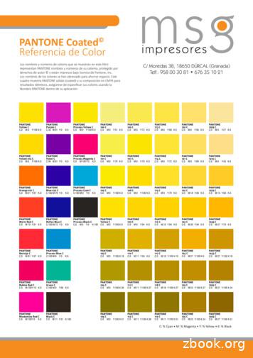
This chart shows PANTONE solid color simulations. For identical results, be sure to specify your colors using the exact PANTONE name within your application. PANTONE Coated Color Reference PANTONE Yellow C C:0 M:0 Y:100 K:0 PANTONE Yellow 012 C C:0 M:0 Y:100 K:0 PANTONE Orange 021 C C:0 M:51 Y:87 K:0 PANTONE Warm Red C
Pantone, Inc., 590 Commerce Blvd., Carlstadt, NJ 07072-3098. Tel: 201.935.5500 Web site: www.pantone.com. PANTONE Colors displayed here may not match PANTONE-identified standards. Consult current PANTONE for fashion and home color system publications for accurate color. PANTONE and other Pantone, Inc. trademarks are the property of Pantone, Inc.


