Breast MRI Quality Control
D.M.Reeve: AAPM 2010 Breast MRI Quality Control Breast MRI Quality Control Donna M. Reeve, MS, DABR, DABMP Department of Imaging Physics Educational Objectives Discuss the importance of breast MRI quality control (QC). Provide an overview of the new ACR Breast MRI Accreditation program (BMRAP) including quality control requirements. Describe MRI system and coil designs currently available for breast MR imaging Discuss breast coil quality control procedures
D.M.Reeve: AAPM 2010 Breast MRI Quality Control Importance of breast MR QC Quality control of MRI systems used for diagnostic breast MR imaging and biopsy guidance Is important to ensure production of high quality images by evaluating whether MRI scanner and coils used for breast imaging are performing consistently over time. Should be part of a comprehensive MRI quality control program. May be required to satisfy accreditation program requirements ACR Breast MRI Accreditation Program ACR Breast Magnetic Resonance Imaging Accreditation Program (BMRAP) launched in May 2010 under breast imaging accreditation programs (mammography, stereotactic breast biopsy, and breast ultrasound). Separate from the ACR MR Accreditation Program (MRAP) Provides accreditation for MR systems used for breast imaging:* imaging: Dedicated breast MRI systems or Whole body MRI systems with detachable table-top breast coil dedicated tables with integrated breast coils
D.M.Reeve: AAPM 2010 Breast MRI Quality Control Breast MRI RF Coils www.auroramri.com Philips MammoTrak SENSE 16 Channel www.sentinellemedical.com Invivo 3T Precision Breast Array 8 Channel Guidance documents www.acr.org
D.M.Reeve: AAPM 2010 Breast MRI Quality Control Accreditation fees www.acr.org Breast MRI Accreditation Program Requirements, 5/10/2010 Personnel Qualifications – Radiologist Initial qualifications: Certification in Radiology or Diagnostic Radiology (ABR, American Osteopathic Board of Radiology, Royal College of Physicians and Surgeons of Canada or Le College des Medecins du Quebec) AND Supervision, interpretation and reporting of 150 breast MRI exams in last 36 months or 100 breast MRI exams in a supervised situation. OR Not Board Certified Completion of an ACGME or AOA approved diagnostic radiology residency program AND Interpretation and reporting of 100 breast MRI exams in the last 36 months in a supervised situation.
D.M.Reeve: AAPM 2010 Breast MRI Quality Control Personnel Qualifications – Radiologist AND 15 hours of Cat 1 CME in MRI (including clinical applications of MRI in breast imaging, MRI artifacts, safety and instrumentation in the last 36 months. Continuing Experience: Upon renewal, 75 breast MRI examinations in prior 24 months. Continuing Education: 5 hours of Category 1 CME in breast MRI in the prior 36 months. Personnel Qualifications – Technologist Initial qualifications: 1. 2. 3. Registered in MRI (ARRT, ARMRIT, or CAMRT) OR Registered in radiography by ARRT and/or unlimited state license, and 6 months supervised clinical MRI scanning experience. OR Associate’s or Bachelor’s degree in allied health field and certification in another clinical imaging field and 6 months supervised clinical MRI scanning experience. AND Licensure in state in which he/she practices (if required for MRI techs) Supervised experience in breast MRI AND Supervised experience in the IV administration of MR contrast (if performed by the technologist)
D.M.Reeve: AAPM 2010 Breast MRI Quality Control Personnel Qualifications – Technologist Continuing Experience: Upon renewal, 50 breast MRI examinations in prior 24 months. Continuing Education: All: 24 hours of CE every 2 years CE includes credits pertinent to the technologist’s ACR accredited clinical practice Registered technologists: CE in compliance with requirements of certifying organization State licensed technologists, all others: CE relevant to imaging and the radiologic sciences, patient care Personnel Qualifications – Medical Physicist/MR Scientist Initial qualifications Medical Physicist: 1. 2. 3. Board Certification in Radiological Physics or Diagnostic Radiological Physics (ABR), in MRI Physics (ABMP), or in Diagnostic Radiology Physics ro MRI Physics (CCPM) Not board certified: graduate degree in relevant fields and formal course work in biological sciences and 3 years documented experience in a clinical MRI environment Grandfathered: Surveys of at least 3 MRI units between January 1, 2007 and January 1, 2010. MR Scientist: Graduate degree in a physical science involving nuclear MR or MRI 3 years experience in a clinical MRI environment.
D.M.Reeve: AAPM 2010 Breast MRI Quality Control Personnel Qualifications – Medical Physicist/MR Scientist Continuing Experience: Upon renewal, 2 MRI unit surveys in prior 24 months. Continuing Education: Upon renewal, 15 CEU/CME (half must be Category 1) in the prior 36 months (must include credits pertinent to the accredited modality). Personnel Qualifications – Medical Physicist/MR Scientist Must be familiar with MRI safety, FDA guidance for MR diagnostic devices, other regulations pertaining to the performance of the equipment being monitored. Be knowledgeable about MR physics, MRI technology, including function, clinical uses, performance specifications of MRI equipment, equipment, calibration processes and limitations of the performance testing hardware, procedures, and algorithms. Working understanding of clinical protocols and optimization. Maintain proficiency in CE programs to ensure familiarity with current concepts, equipment, and procedures. www.acr.org Breast MRI Accreditation Program Requirements, 5/10/2010
D.M.Reeve: AAPM 2010 Breast MRI Quality Control ACR Breast MRI Accreditation Program Annual and acceptance testing requirements Technologist QC requirements MRI Safety policies and practices Periodic maintenance and documentation Æ same as for MRI Accreditation Program BMRAP Clinical Images Facilities must submit clinical images and corresponding data for each magnet performing breast MRI* examinations at their site. Dedicated bilateral breast coil capable of simultaneous bilateral imaging. Facilities performing breast MRI must have the capacity to perform mammographic correlation, directed breast ultrasound and MRI-guided intervention, or create a referral arrangement with a cooperating BMRAP accredited facility that could provide these services. 45 days to acquire clinical exams No phantom image submission is required at this time.
D.M.Reeve: AAPM 2010 Breast MRI Quality Control BMRAP Clinical Images Submit 2 bilateral breast MRI cases from different patients 1. Known, enhancing, biopsy-proven carcinoma 2. BI-RADS category 1 (negative) or 2 (benign findings) Cases may not be older than 2 months BMRAP Clinical Images Exams must include these 4 sequences: www.acr.org Breast MRI Accreditation Program Requirements, 5/10/2010
D.M.Reeve: AAPM 2010 Breast MRI Quality Control BMRAP Clinical Images www.acr.org Breast MRI Accreditation Program Requirements, 5/10/2010 BMRAP Quality Control Program QC program identical to MRAP. Acceptance, annual, post-upgrade/repair testing, including annual testing of all RF coils Daily/weekly QC: Choice of phantom and action criteria is up to facility. Decision made by “qualified medical physicist/MR scientist in cooperation with the system vendor”. Large ACR phantom in head coil Dedicated breast MR systems may use small ACR phantom in breast coil. Other vendorvendor-supplied phantom
D.M.Reeve: AAPM 2010 Breast MRI Quality Control ACR MR Accreditation Phantoms Manufacturer: J.M Specialty Parts 1050 “large phantom” 780 “small phantom” Breast Coil QC
D.M.Reeve: AAPM 2010 Breast MRI Quality Control Breast MRI RF Coils Multiple configurations to allow imaging in unilateral or bilateral modes. Multi-channel: 8, 16, 32 independent channels Smaller elements increase SNR Multiple elements provide anatomical coverage - fixed or adjustable positions Allows use of parallel imaging to improve imaging speed or “trade” speed for increased resolution or SNR. RF receive-only multiple transmit RF (Philips Achieva 3.0 TX) Breast RF Coil Quality Control To establish baseline coil performance in order to monitor coil performance over time. Coil inspection Signal-to-noise ratio (SNR) Signal uniformity Phased array coils: compare SNR for individual channels Artifact evaluation (including ghosting) Using QC protocol Using clinical protocol
D.M.Reeve: AAPM 2010 Breast MRI Quality Control Breast RF Coil Quality Control Coil testing: Important to test coils: after installation of new scanner or new coils annually during physics testing whenever artifacts or coil problems occur Manufacturers provide a coil manual for each coil includes description of clinical use of the coil may include detailed description of coil test procedure may include pass/fail limits may only say “establish baseline and monitor over time” Breast RF Coil Quality Control Coil inspection Inspect coil, cables, cable insulation, ports and connectors for damage Could present a safety issue or result in low SNR or image artifacts. www.invivocorp.com
D.M.Reeve: AAPM 2010 Breast MRI Quality Control Breast RF Coil Quality Control Artifact evaluation Evaluate images acquired using coil QC protocol To troubleshoot artifacts observed on patient images may acquire images of QC phantom using clinical protocol. Breast RF Coil Quality Control Coil testing: Test all available coil configurations(bilateral, unilateral). Note any error messages that occur during scanning. In our experience the vendor is more likely to respond to coil QC failure when manufacturer’s QC procedure is followed. Uniformity: Follow procedure in 2004 ACR MRI QC Manual (min, max signal intensity within small ROI)
D.M.Reeve: AAPM 2010 Breast MRI Quality Control Breast RF Coil Quality Control Consistent scan/measurement methods: Identical phantom and positioning within coil Homogeneous phantom (sphere, cylinder, custom) ACR or other phantom Identical scan parameters: Pulse sequence, timing parameters, slice thickness and position, matrix, FOV, receive bandwidth, etc Record center frequency, transmit gain/attenuation, receiver gains gains Identical measurement methods, ROI positions SNR, signal uniformity, ghosting, stability tests Evaluation of channel performance Helpful to acquire screen capture of ROI positions and photograph photograph phantom setup SNR Methods Signal Signal measured in ROI within magnitude image Noise 1. Noise measured in background area of signal image (in air) free of artifacts. 2. Noise measured in “pure noise” noise” image acquired with no RF excitation. 3. NEMA approach: Noise measured in subtraction image: 2 signal images acquired with identical protocol and prescan parameters (center frequency, transmit gain or attenuation, receiver gains). SNR 0.655S SNR 0.655S σ bkg σ noise SNRNEMA S 2 σ sub 0.655 factor: noise distribution in magnitude image is Rician (not Gaussian) Determination of Signal-to-Noise Radio (SNR) in Diagnostic MRI, NEMA Standards MS 1-2008. www.nema.org
D.M.Reeve: AAPM 2010 Breast MRI Quality Control Examples of Breast Coil QC procedures Breast RF Coil Quality Control GE HD 8 channel phased array (1.5T, 3T) Automated QC procedure requires careful positioning/landmarking positioning/landmarking Custom phantom NEMA subtraction SNR of individual elements Pass/fail results
D.M.Reeve: AAPM 2010 Breast MRI Quality Control Breast RF Coil Quality Control Images acquired with individual coil elements Coronal Sagittal composite image Noise image Breast RF Coil Quality Control
D.M.Reeve: AAPM 2010 Breast MRI Quality Control Breast RF Coil Quality Control Breast RF Coil Quality Control
D.M.Reeve: AAPM 2010 Breast MRI Quality Control Breast RF Coil Quality Control Breast RF Coil Quality Control www.sentinellemedical.com Left Lateral Coil Array Medial Array Right Lateral Coil Array Bilateral imaging mode Unilateral biopsy mode
D.M.Reeve: AAPM 2010 Breast MRI Quality Control Bilateral Imaging mode Consistent positioning of QC phantoms and coils Unilateral Interventional mode
D.M.Reeve: AAPM 2010 Breast MRI Quality Control Breast RF Coil Quality Control Unilateral biopsy mode Bilateral imaging mode Breast RF Coil Quality Control Signal ROI: SE T1, unilateral and bilateral imaging modes Noise image (no RF excitation)
D.M.Reeve: AAPM 2010 Breast MRI Quality Control Breast RF Coil Quality Control Small ACR phantoms Bilateral mode Images courtesy of R. Price, PhD SE, Philips Achieva 1.5T, 16 channel breast array Breast RF Coil Quality Control Small ACR phantom Unilateral mode Images courtesy of R. Price, PhD SE, Philips Achieva 1.5T, 16 channel breast array
D.M.Reeve: AAPM 2010 Breast MRI Quality Control Acknowledgements Thanks to Ron Price, PhD and Raja Muthupillai, PhD for sharing their breast quality control program experiences and images. References 1. ACR Magnetic Resonance Imaging (MRI) Quality Control Manual, 2004. (under revision) 2. ACR Technical Standards for Diagnostic Medical Physics Performance Monitoring of MRI Equipment, revision 2009. www.acr.org 3. Breast MRI Accreditation Program Requirements, 5/10/2010. www.acr.org 4. Determination of Signal-to-Noise Radio (SNR) in Diagnostic Magnetic Resonance Imaging, NEMA Standards MS 1-2008. www.nema.org
D.M.Reeve: AAPM 2010 Breast MRI Quality Control
1. ACR Magnetic Resonance Imaging (MRI) Quality Control Manual, 2004. (under revision) 2. ACR Technical Standards for Diagnostic Medical Physics Performance Monitoring of MRI Equipment, revision 2009. www.acr.org 3. Breast MRI Accreditation Program Requirements, 5/10/2010. www.acr.org 4. Determination of Signal-to-Noise Radio (SNR) in Diagnostic
clinical practice the role of breast mri in clinical practice 514 reprinted from australian Family physician Vol. 38, No. 7, July 2009 Breast mri Breast MRI is performed using a standard MRI machine with a special attachment (a ‘breast coil’). The patient lies prone for the pro
4 Breast cancer Breast cancer: A summary of key information Introduction to breast cancer Breast cancer arises from cells in the breast that have grown abnormally and multiplied to form a lump or tumour. The earliest stage of breast cancer is non-invasive disease (Stage 0), which is contained within the ducts or lobules of the breast and has not spread into the healthy breast tissue .
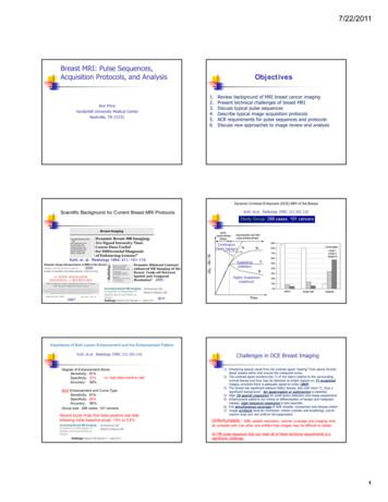
1. Review background of MRI breast cancer imaging 2. Present technical challenges of breast MRI 3. Discuss typical pulse sequences 4. Describl lbe typical image acquisition protocols 5. ACR requirements for pulse sequences and protocols 6. Discuss new approaches to image review and analysis Scientific Background for Current Breast MRI Protocols
MRI Physics Anthony Wolbarst, Nathan Yanasak, R. Jason Stafford . Introduction to MRI ‘Quantum’ NMR and MRI in 0D Magnetization, m(x,t), in a Voxel Proton Density MRI in 1D T1 Spin-Relaxation in a Voxel MRI Case Study, and Caveat Sketch of the MRI Device ‘Classical’ NMR in a Voxel
magnetic resonance imaging (MRI)-MRI image fusion in assessing the ablative margin (AM) for hepatocellular carcinoma (HCC). METHODS: A newly developed ultrasound workstation for MRI-MRI image fusion was used to evaluate the AM of 62 tumors in 52 HCC patients after radiofrequency ablation (RFA). The lesions were divided into two
The use of magnetic resonance imaging (MRI) is increasing globally, and MRI safety issues regarding medical devices, which are constantly being developed or upgraded, represent an ongoing challenge for MRI personnel. To assist the MRI community, a panel of 10 radiologists with expertise in MRI safety from nine high-volume academic centers .
5 yrs of anti-hormone therapy reduces the risk of: – breast cancer coming back somewhere else in the body (metastases / secondaries) – breast cancer returning in the same breast – a new breast cancer in the opposite breast – death from breast cancer The benefits of anti-hormone therapy last well
The Baldrige framework is used extensively as a foundation for internal systems, but there has been a substantial decrease in the number of manufacturing organizations applying for the award. This research study validates some of the reasons associated with that development. The Value of Using the Baldrige Performance Excellence Framework in Manufacturing Organizations Prabir Kumar .


