Dental Color Matching Instruments And Systems. Review Of Clinical And .
journal of dentistry 38s (2010) e2–e16 available at www.sciencedirect.com journal homepage: www.intl.elsevierhealth.com/journals/jden Review Dental color matching instruments and systems. Review of clinical and research aspects Stephen J. Chu a, Richard D. Trushkowsky b,*, Rade D. Paravina c a Department of Periodontology and Implant Dentistry, New York University College of Dentistry, New York, NY, United States Department of Cariology and Comprehensive Care, New York University College of Dentistry, 345 East 24th Street, New York, NY 10010, United States c Department of Restorative Dentistry and Biomaterials, University of Texas Dental Branch at Houston, Houston, TX, United States b article info abstract Article history: Objectives: To review current status of hand held systems for tooth color matching in vivo Received 8 May 2010 and corresponding research. Received in revised form Sources: ‘‘Medline’’ database from 1981 to 2010 were searched electronically with key words 23 June 2010 tooth, teeth, color and dentistry. Accepted 2 July 2010 Conclusion: Spectrophotometers, colorimeters and imaging systems are useful and relevant tools for tooth color measurement and analysis, and for quality control of color reproduction. Different measurement devices either measure the complete tooth surface providing a Keywords: ‘‘color map’’ or an ‘‘average’’ color of the limited area [3–5 mm] on the tooth surface. These Tooth instruments are useful tools in color analysis for direct or indirect restorations, communi- Teeth cation for indirect restorations, reproduction and verification of shade. Whenever possible, Color both instrumental and visual color matching method should be used, as they complement Color measurement each other and can lead towards predictable esthetic outcome. # 2010 Elsevier Ltd. All rights reserved. Colorimeters Spectrophotometers Digital camera 1. Introduction The interest in color research in dentistry has increased significantly over the past several decades. When keywords color and dentistry were used for Medline search, only 107 papers were found by 1970. In subsequent decades, the number of references increased as follows: 409 (1980), 1134 (1990), 2259 (2000) and 4062 (April 2010). Advancements in technology, computers, the Internet, and communication systems have greatly affected and shaped modern society. Commensurate with these strides are the advancements in contemporary dentistry. During the past half decade, the dental profession has experienced the growth of a new generation of technologies devoted to the analysis, communication and verification of shade. Shade determination for direct and indirect restorations has always been a challenge for the esthetic dentist. Clark in 1931 described this in the Color Problems in Dentistry.1 As opposed to subjective visual shade selection with not always quite controlled conditions and methods, and shade guides that exhibited significant shortcomings, several authors tried to objectively quantify tooth color in the past. This was done through identifying color problems in dentistry2; the importance of the quantity and quality of light required to properly analyse shade3 * Corresponding author. Tel.: 1 718 948 5808; fax: 1 718 948 4453. E-mail address: rt587@nyu.edu (R.D. Trushkowsky). 0300-5712/ – see front matter # 2010 Elsevier Ltd. All rights reserved. doi:10.1016/j.jdent.2010.07.001
e3 journal of dentistry 38s (2010) e2–e16 through studying correlation between extracted teeth and shade guides4 or the development of the early shade measuring instruments and shade guides.5 The late 1990s marked the birth of a new industry in dentistry, commercially available instrument-based color measurement systems, with the development of the ShadeScan system (Cortex Machina, Montreal, Canada).6 This was the first effort toward a shade analysis system for complete tooth surface measurement. Prior literature published by several authors described limited area measurement instruments, with an optical diameter of 3–5 mm, in the analysis of shade.7–9 Another study evaluated clinical application of the ShadeScan prototype which employed digital camera technology, in a case report comparing visual vs. instrument-based shade information in the restoration of a single maxillary central incisor.10 Today’s shade-matching technologies have been developed in an effort to increase the success of color matching, communication, reproduction and verification in clinical dentistry, and, ultimately, to increase the efficiency of esthetic restorative work within any practice. The aim of this paper is to provide a comprehensive review of the current state of shade-matching technologies and instrumentation, and their clinical and research application. 2. Rationale Dental shade-matching instruments have been brought to market to reduce or overcome imperfections and inconsistencies of traditional shade matching. The most commonly used shade-matching method is the visual method, whilst Vitapan Classical (Vita Zahnfabrik, Bad Säckingen, Germany) and its derivations are probably the most commonly used shade guides. The colored tabs of distinctive shades organize the empiric-based Vita chart.11–14 In addition, unequivocal findings were reported on color consistency amongst shade guides from the same manufacturer.15,16 Introduction of evidence-based Vitapan 3D-Master shade guides, Toothguide, Bleachedguide and particularly Linearguide by the same manufacturer correspond to color of human teeth and therefore increase chances for successful shade matching.17,18 Historically, assessing shade visually has been characterized by several innate difficulties: metamerism, suboptimal color matching conditions, tools and method as well as the receiver’s age fatigue, mood and drugs/medications.19 Despite these difficulties, the human eye can discern very small differences in color. However, the ability to communicate the degree and nature of these differences is lacking. The final color of an all-ceramic restoration is a merging of the underlying tooth structure or core and the ceramic material. The color of the final restoration cannot match the shade selected from a shade guide unless this modification is taken into account. Therefore, a stump or base tooth preparation shade needs to be obtained and transmitted to the technician.20 3. Overview Instruments for clinical shade-matching encompass spectrophotometers, colorimeters and imaging systems. As with any device, benefits and limitations exist, and the clinician must consider how the technology relates to expectations and needs. Intra-oral color measuring devices have been designed to primarily fit the needs of clinical dentistry, such as information on the corresponding shade tabs, tooth translucency, or information associated with color communication, reproduction and verification. This, together with price limitation dictated by the dental market, resulted in having scientific aspects, such as providing reflectance values or color formulation, less emphasized. Another significant difference compared to other, non-dental applications, are optical properties of human teeth—they are small, curved, multilayered, translucent and exhibit color transitions in all directions (gingival to incisal, mesial to distal and labial to lingual). This is why the accurate repositioning (measurement of the same area) is frequently of critical importance for either clinical and research use of dental color matching devices. The list of instruments and software for in vivo color matching and their properties is given in Table 1.21 In addition to the instruments listed in Table 1, there are numerous dental color matching products that are presently withdrawn from the market, of limited availability, or undergoing major redesigning. The list includes Chromascan (Sterngold, Stamford, CT, USA), Dental Color Analyzer (Wolf Industries, Vancouver, Canada), Identacolor II (Identa, Holbaek, Denmark), Digital Shade Guide DSG4 (A. Reith, Schorndorf, Germany), Ikam (Metalor Technologies, Attleboro, MA, USA), ShadeEye NCC Chroma Meter (Shofu Dental, Menlo Park, CA, USA), Beyond Insight Shade Taking Device (Beyond Dental & Health, Beijing, China), Shadescan (Cynovad, Montreal, Canada) and Vita Easyshade (Vita Zahnfabrik, Bad Säckingen, Germany). Table 1 – Instruments and software for color matching in dentistry: types, measurement area and relative cost ( 1000– 7500).21. Product ClearMatch CrystalEye Easyshade Compact Shade-X ShadeVision SpectroShade Micro Manufacturer Device type Measurement area Clarity Dental, Salt Lake City, UT Olympus America, Center Valley, PA Vident, Brea, CA X-Rite, Grandville, MI X-Rite, Grandville, MI MHT, Niederhasli, Switzerland Software, digital image analysis Imaging Spectrophotometer Spectrophotometer Spectrophotometer Imaging colorimeter Imaging Spectrophotometer Complete tooth image Complete tooth image 5-mm probe diameter 3-mm probe diameter Complete tooth image Complete tooth image Relative cost Low High Low Low Moderate Moderate
e4 journal of dentistry 38s (2010) e2–e16 4. Characteristics and clinical application 4.2. 4.1. Spectrophotometers Colorimeters measure tristimulus values and filter light in red, green and blue areas of the visible spectrum. Colorimeters are not registering spectral reflectance and can be less accurate than spectrophotometers (aging of the filters can additionally affect accuracy).31 ShadeVision (X-Rite, Grandville, MI) is an imaging colorimeter. Complete tooth image is provided through the use of three separate databases: for gingival, middle and incisal third. Virtual try-in feature enables virtual testing of color reproduction during fabrication.32 Spectrophotometers are amongst the most accurate, useful and flexible instruments for overall color matching and color matching in dentistry.22 They measure the amount of light energy reflected from an object at 1–25 nm intervals along the visible spectrum.23,24 A spectrophotometer contains a source of optical radiation, a means of dispersing light, an optical system for measuring, a detector and a means of converting light obtained to a signal that can be analysed. The data obtained from spectrophotometers must be manipulated and translated into a form useful for dental professionals. The measurements obtained by the instruments are frequently keyed to dental shade guides and converted to shade tab equivivalent.25 Compared with observations by the human eye, or conventional techniques, it was found that spectrophotometers offered a 33% increase in accuracy and a more objective match in 93.3% of cases.26 Crystaleye (Olympus, Tokyo, Japan) combines the benefits of a traditional spectrophotometer with digital photography. Through the development of optical and image processing technology, this product allows the practitioner to match tooth shade and color more accurately and simply compared with the traditional spectrophotometer.27 The significant benefit of this system is that ‘virtual shade tabs’ in the computers database can be cross-referenced and superimposed visually onto the natural tooth image to be matched giving the technician the ability to visualize the correct shade tabs. The digital image produced by the Crystaleye uses a 7-band LED light source, which results in a more precise depiction of color than the conventional systems used with digital cameras. Moreover, the image produced by the Crystaleye is taken from inside the oral cavity and consequently is devoid of the external light that can cause discrepancies. Vita Easyshade Compact (Vita Zahnfabrik, Bad Säckingen, Germany) is cordless, small, portable, cost efficient, battery operated, contact-type spectrophotometer that provides enough shade information to help aid in the color analysis process. Different measurement modes are possible with Easyshade Compact: tooth single mode, tooth area mode (cervical, middle and incisal shades), restoration color verification (includes lightness, chroma and hue comparison) and shade tab mode (practice/training mode).28 Shade-X (X-Rite, Grandville, MI) is also compact and cordless ‘‘spot’’ measurement’’ spectrophotometer with 3mm probe diameter, and keyed to the majority of popular shade guides. Shade-X have two databases to match the color of the dentin (more opaque) and the incisal tooth regions (more translucent).29 SpectroShade Micro (MHT Optic Research, Niederhasli, Switzerland) is an imaging spectrophotometer. It uses a digital camera/LED spectrophotometer combination. It has an internal computer with the analytical software. The tooth positioning guidance system, shown on the LCD touch screen, is used during color measurement. Images and spectral data can be saved on the internal memory and transferred to a computer.30 4.3. Colorimeters Digital cameras and imaging systems Digital Cameras. Most consumer video or digital still cameras acquire red, green and blue image information that is utilized to create a color image. The RGB color model is an additive model in which red, green and blue light are added together in various ways to reproduce a broad array of colors. Digital cameras represent the most basic approach to electronic shade taking, still requiring a certain degree of subjective shade selection with the human eye.33 Various approaches have been used to translate this data into useful dental color information. ClearMatch (Smart Technology, Hood River, OR) is a software system that uses high-resolution digital images and compares shades over the entire tooth with known reference shades.30 Similar to the software associated with color measuring devices, ClearMatch contains the color database of industry-standard shade guides.34 4.4. Interpretation and application of shade analysis data Complete tooth surface measurement [CTSM] devices give a color map of the gingival, body and incisal shades for the fabrication of direct or indirect restorations. These systems give a virtual shade overlay of the proposed tab onto the digital image on the computer screen of the tooth measured for visual reference and assessment by the clinician and/or technician. CTSM spectrophotometers, such as Crystaleye and SpectroShade, provide shade tab designation and the respective DE* values compared to shade tab values in the memory. However, these mappings are two dimensional and they do not necessarily take into account the shape, texture, thickness of the restoration, type of abutment and different core material (metal or ceramic).35 In addition, they ‘average out’ color data over the complete tooth surface or larger defined areas which can lead to inaccurate shade information. Limited area measurement devices provide 3–5 mm diameter areas of the tooth being measured. Therefore several areas of the tooth should be considered to obtain a representative evaluation of tooth shade. At a minimum, a limited area measurement of the gingival, body and incisal areas of the tooth [total of 3 measurements] should be assessed and recorded for the technician if an indirect restoration is prescribed. It was found that decreasing the window size when examining extracted teeth with a spectrophotometer and spectroradiometer resulted in lower CIE L*a*b* values.36 Small-window tooth color measurement may cause edge loss of the light due to a tooth’s translucency.37
journal of dentistry 38s (2010) e2–e16 Once the technology-based information is analysed, the information must be communicated to the technician for indirect restorations.38 The aforementioned instruments and technologies are predominantly intended for shade analysis, however, this information alone may be insufficient for the technician to adequately interpret the shade information provided. Subsequent reference photography is highly recommended for communication on tooth color. Digital photographs are an important adjunct to the laboratory technician and together with the shade or ‘‘color map’’ should be sufficient communication material to construct an acceptable restoration. Clinicians and technicians are frequently located in different areas. A digital camera permits the transfer of images from the clinician to the technician. The best way to reference shade information is by using shade tabs to communicate shade.39 A systematic protocol to referencing shade information as well as changes in shade between tabs is required. Shades identified in the digital map are arranged by gingival (G), middle (M) and incisal third (I). The shade tabs should be selected and photographs should be taken with each tab in its proper orientation in reference to the tooth. Camera and light settings and image format must be kept constant at all times for consistent shade color communication. In addition to the three basic shade tabs representing the respective G, M and I shades, two additional reference shade tab photographs should be included to graduate and calibrate shifts in hue, chroma and value between physical tabs visually in the laboratory, in the photographs and between the photos. One selected tab should be lighter in shade and one darker in shade in respect to the selected basic shades. The combination of accurate digital shade analysis information coupled with standardized reference shade communication photography can ensure a predictable esthetic restorative outcome of the final direct or indirect restoration. Indirect restorations can and should be verified visually and with the instrument system in the laboratory prior to being returned to the clinician. This verification process can streamline wasteful chair side time attributed to remakes due to incorrect shade.40,41 Clinical excellence backed with sound color science and its appropriate interpretation can probably make the critical difference between good to excellent.42 Examples of color matching, interpretation and application of shade analysis data are given in Figs. 1–6. 5. Research Besides the clinical applications, dental color measuring instruments and systems are increasingly used in research. The most frequent research topics associated with these instruments and systems are associated with evaluation of measurement uncertainties, comparison between visual and instrumental findings, visual color thresholds and evaluation on color compatibility, stability and interactions of human hard and soft oral tissues, head and neck tissues and dental materials. Although non-dental, professional bench-top color measurement instruments were used in many research projects in dentistry, this paper will focus mainly on these conducted using hand held devices designed for dental application. 5.1. e5 Measurement uncertainties In color science, evaluation of measurement uncertainties of shade-matching instruments is performed through precision and accuracy testing. The uncertainties associated with precision are most frequently associated with random errors, whilst the uncertainties associated with accuracy usually originate from systematic errors. Precision is tested by evaluation of repeatability (same method, operator or instrument) and reproducibility (different method, operator and/or instrument). Based on time interval, we are talking about short term (measurements in succession), medium term (hours) and long term measurements (weeks or longer). Based on the specimen manipulation, measurement can be performed with- and without replacement.43 Evaluation of measurement uncertainties encompasses the use of referent instrument, preferably professional, non-dental, color measuring instrument.37 In a study that compared five dental color measuring devices—ShadeScan, Easyshade, Ikam, IdentaColor II and ShadeEye, five group A Classical tabs were in vitro measured five times by two operators, whilst 25 upper right central incisors were measured in vivo by one operator. The best precision in vivo was recorded for Easyshade and Ikam, whilst performance of the other instruments was better in vitro than in vivo.12 In an another study, SpectroShade, ShadeVision, VITA Easyshade and ShadeScan were evaluated.31 High reliability (reproducibility?) and variability in accuracy was reported. One in vitro study found that repeatability and accuracy of a dental color measuring instrument (ShadeScan) was influenced by shade guide systems used for testing.44 When the precision of measurement of different tooth areas was evaluated, the middle third of each labial tooth surface exhibited the most consistent resuts.45 Dental color matching instruments also provide the information on best matching tabs from different shade guides.25,46,47 This application-specific color information is also of importance and therefore requires proper attention of dental researches. When VITA EasyShade and ShadeEye NCC were tested on extracted human teeth, there were discrepancies in shade guide designations provided by the instruments. Higher inter-device agreement was recorded for Vitapan Classical than for Vitapan 3D-Master.25 Digital imaging systems are becoming increasingly popular in determining the color of teeth. Precision and accuracy of these systems are influenced by the quality of camera and image processing method. Several studies reported that digital cameras, may be reliable instruments for determining the color of teeth and gingiva when combined with the appropriate calibration protocols.48–54 Clinical imaging and conventional image processing methods such as Adobe Photoshop and Corel Photo-Paint are suitable for many purposes in dentistry: lab communication, documentation, patient education and others. However, scientific imaging,55 appropriate methods of data processing and color science terminology is preferred in dental research. This, together with lack of the referent instrument (the instrument that has already been validated, not necessary by the same authors), diminished the validity of some studies on precision and accuracy of dental color matching instruments.
e6 journal of dentistry 38s (2010) e2–e16 Fig. 1 – Clinical application of CrystalEye spectrophotometer (Courtesy Dr. Shigemi Nagai). (a) Crystaleye captures the spectral image of the tooth, abutment, arch and full face image. (b) Laboratory report can be sent to the lab as a quick color note. (c) Color analysis of the ceramic crown (after 1st bake try-in) placed on the abutment. Excellent color match was confirmed in the cervical and body areas. Minor modification may be required in the incisal area to increase the value and yellowness. (d) Color difference DEs obtained in all 3 areas are considered to be excellent color match. L* value map indicates indistinguishable value distribution on #8 and #9.
journal of dentistry 38s (2010) e2–e16 e7 Fig. 1. (Continued ). 5.2. Comparison between visual and instrumental findings When the first dental color measuring instruments appeared, they exhibited slightly better accuracy compared to visual findings that were described as inconsistent.56 Since that time, the modernization of both visual and instrumental means for color matching in dentistry occurred.17,57 More recently, better results were reported with dental spectrophotometer than using the visual method in approximately 47% of the cases,58 which is in accordance with independent studies that documented the supremacy of spectrophotometric color matching compared to visual shade assessment.27,59 Another paper reported that the performance of Easyshade was comparable or better than that of dentists, whilst the agreement between visual and instrumental findings was qualified as good to very good.60 In another paper, it was found that the agreement amongst the observer groups was significantly better than that of each device and that color matching instruments did not reflect human perception.61 It was found that consensus amongst observers led to better shade-matching results with some shade guides, compared to results of individual observers,62 and that intraexaminer shade-matching agreement was mainly acceptable.63 Significantly higher visual-instrumental agreement was recorded for experienced dentists (compared to dental students and non-dental observers), regardless of shade guides and lighting conditions.64 The comparison between visual and instrumental findings is a very attractive topic as it reveals pros and cons of both methods. Visual color matching is subjective and influenced by variety of factors. However, this method is not inferior and should not be underrated. Actually, the all ‘‘objective’’ color measuring instruments have been developed based on the visual response of the ‘‘standard observer’’ and they are good only if they match that response. In addition, the numerically smallest DE* value does not necessarily correspond to the best match because of the uneven eye sensitivity to hue, value and chroma differences. Therefore, the answer whether to use visual or instrumental method for color matching in dentistry is: whenever if possible, use both, as they complement each other and can lead towards predictable esthetic outcome.39–41 5.3. Evaluation of visual color thresholds Perceptibility and acceptability visual thresholds can be quantified only by combining visual and instrumental color measurement methods. Although majority of studies used professional (non-dental) color measuring devices in threshold evaluation, this topic is of exceptional importance in interpretation of color differences in clinical dentistry and dental research. When the color difference between compared objects can be seen by 50% of observers (the other 50% will notice no difference), we are talking about the 50:50% perceptibility threshold.65 When the color difference is considered acceptable 50% of observers (the other 50% would consider it unacceptable), this corresponds to 50:50% acceptability threshold. A color match in dentistry is a color difference at or below the former threshold, an acceptable color match is a color difference at or below the later one.65 In dental literature, it is frequently interpreted that a DE* of 1 is the 50:50 perceptibility threshold under controlled conditions (50% of observers will notice the color difference and 50% will see no difference between compared objects),66 whereas a DE* of 2.767 and 3.368 were found to be 50:50 acceptability thresholds (50% of observers will accept the restoration and 50% will replace it because of color mismatch).
e8 journal of dentistry 38s (2010) e2–e16 Fig. 2 – Clinical application of EasyShade Compact spectrophotometer. (a) Instrument calibration. (b) Color measurement. (c) Color difference metric values as compared to the corresponding Vitapan Classical shade. (d) Color coordinate values and the corresponding Vitapan 3D-Master shade. Several other studies, more or less controlled, reported the variability of findings on visual color thresholds.57,69–71 This, in addition to the introduction of new color difference formulae (CIEDE2000), suggests systematic approach and standardization of methods. 5.4. shade guides exhibit moderate to pronounced discrepancy with results recorded for natural teeth. This discrepancy has been quantified by calculation of coverage error, the mean value of the minimal color differences amongst Color compatibility Color compatibility studies encompass comparisons amongst teeth, shade guides and dental materials. Evaluation of color of natural teeth is important for clinical dentistry since knowledge on color ranges and color distribution of natural teeth can provide guidelines for designing of dental materials that will enable better and easier match to natural teeth.13,72 Databases encompassing spectral properties of teeth, regardless whether created using dental or non-dental color measuring devices, are of particular validity.73 Two independent studies, both performed using Vita Easyshade, reported almost the same color coordinate ranges of natural teeth: L* 55.5–89.674 and L* 58.7–88.775; a* 4.2–7.374 and a* 3.6–7.075, and b* 3.6–38.974 and b* 3.7–37.3.75 Current Fig. 3 – Clinical application of Shade-X spectrophotometer.
journal of dentistry 38s (2010) e2–e16 e9 Fig. 4 – Clinical application of ShadeVision colorimeter. (a) The fractured veneer restoration on tooth #8 is to be replaced with a new veneer restoration. (b) The ShadeVision color mapping for tooth #9 to be matched with gingival, body and incisal shade reference tabs. (c) Veneer preparation tooth #8. It is important that an even thickness of tooth reduced is performed to insure color control of ceramic layering for predictable shade matching of the restoration. (d) Insertion of veneer restoration tooth #8 [Venus porcelain, Heraeus, Hanau, Germany] which matches the contralateral tooth satisfactorily and predictably without a remake. each tab in the shade guide and the database of human teeth.17,73–76 Although teeth exhibit color transitions in all directions, the correlation between colors of different regions on the labial tooth surface has been recorded. This study, performed using digital camera, suggested that color of a missing part of a tooth can be determined using the color of existing areas.77 The same team reported color correlation between maxillary incisors and canines, which may influence color matching of missing teeth.78 As far as the compatibility amongst materials is concerned, different comparisons have been performed. An Easyshade comparison between Vitapan Classical shade guide and four different veneering porcelain systems for metal ceramic restorations, revealed that differences were shade-dependent: A2 porcelain disks exhibited better match to corresponding VITA shade, followed by A3 and A3.5 disks.79 The same instrument was used to test the ability of a ceramic system to correctly reproduce the shade selected using two shade guides. Toothguide 3D-Master outperformed VITA Classical.80 When the quality of color match of different shade g
c Department of Restorative Dentistry and Biomaterials, University of Texas Dental Branch at Houston, Houston, TX, United States 1. Introduction The interest in color research in dentistry has increased significantly over the past several decades. When keywords color and dentistry were used for Medline search, only 107 papers were found by 1970.
DENTAL SCIENCES 1 Chapter 1 I Dental Assisting— The Profession 3 The Career of Dental Assisting 4 Employment for the Dental Assistant 4 The Dental Team 6 Dental Jurisprudence and Ethics 12 Dental Practice Act 12 State Board of Dentistry 12 The Dentist, the Dental Assistant, and the Law 13 Standard of Care 13 Dental Records 14 Ethics 14
Cigna Dental Care DMO Patient Charge Schedules 887394 09/15 CDT 2016 Covered under Procedure Code1 Dental Description and Nomenclature Cigna Dental 01 and 02 PCS Cigna Dental 03 PCS Cigna Dental 04 PCS Cigna Dental 05 PCS Cigna Dental 06 PCS Cigna Dental 07 PCS Cigna Dental 08 PCS Chair Time Per Y/N Minutes Code # (if different) Y/N Code # (if .
Mid-level dental providers, variously referred to as dental therapists, dental health aide therapists and registered or licensed dental practitioners, work as part of the dental team to provide preventive and routine dental services, such as cleanings and fillings. Similar to how nurse practitioners work alongside physicians, mid-level dental .
is a detailed list of dental services provided by a dental office and given to Delta Dental for payment. Delta Dental means Delta Dental Plan of Michigan, Inc., a service provider for dental benefits under the Michigan Dental Program. Delta Dental ID Card is a permanent (not monthly) card. We send
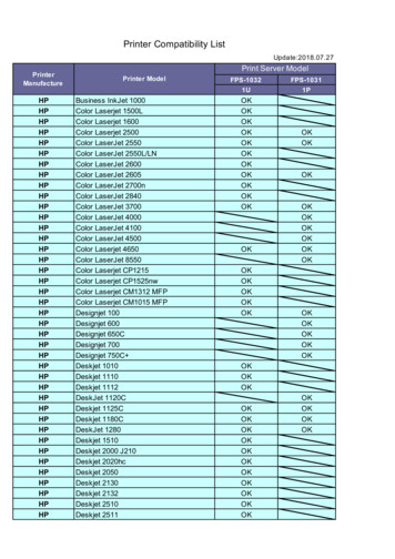
FPS-1032 FPS-1031 1U 1P HP Business InkJet 1000 OK HP Color Laserjet 1500L OK HP Color Laserjet 1600 OK HP Color Laserjet 2500 OK OK HP Color LaserJet 2550 OK OK HP Color LaserJet 2550L/LN OK HP Color LaserJet 2600 OK HP Color LaserJet 2605 OK OK HP Color LaserJet 2700n OK HP Color LaserJet 2840 OK HP Color LaserJet 3700 OK OK HP Color LaserJet 4000 OK HP Color LaserJet 4100 OK
o next to each other on the color wheel o opposite of each other on the color wheel o one color apart on the color wheel o two colors apart on the color wheel Question 25 This is: o Complimentary color scheme o Monochromatic color scheme o Analogous color scheme o Triadic color scheme Question 26 This is: o Triadic color scheme (split 1)
Dental Blue for Individuals. SM - a consumer-driven dental plan for individuals and their eligible dependents . Dental Blue for Seniors. SM - a consumer dental product for individuals and their spouse age 65 and older . Dental Blue For Federal Employee Program - offers federal employees a dental supplemental plan to
Jun 14, 2016 · active duty Soldiers treated at any of five dental clinics on Fort Bragg. These clinics included Davis Dental Clinic, Joel Dental Clinic, LaFlamme Dental Clinic, Pope Dental Clinic, and Smoke Bomb Hill Dental Clinic. For each appointment the appointment type, date, and dental wellness class


