The Cell Cycle
CAMPBELL BIOLOGY IN FOCUSURRY CAIN WASSERMAN MINORSKY REECE9The Cell CycleLecture Presentations byKathleen Fitzpatrick andNicole Tunbridge,Simon Fraser University 2016 Pearson Education, Inc.SECOND EDITION
Overview: The Key Roles of Cell Division The ability of organisms to produce more of theirown kind best distinguishes living things fromnonliving matter The continuity of life is based on the reproduction ofcells, or cell division 2016 Pearson Education, Inc.
In unicellular organisms, division of one cellreproduces the entire organism Cell division enables multicellular eukaryotes todevelop from a single cell and, once fully grown, torenew, repair, or replace cells as needed Cell division is an integral part of the cell cycle, thelife of a cell from its formation to its own division 2016 Pearson Education, Inc.
Figure 9.2100 m(a) Reproduction50 m(b) Growth anddevelopment20 m 2016 Pearson Education, Inc.(c) Tissue renewal
Concept 9.1: Most cell division results in geneticallyidentical daughter cells Most cell division results in the distribution ofidentical genetic material—DNA—to two daughtercells DNA is passed from one generation of cells to thenext with remarkable fidelity 2016 Pearson Education, Inc.
Cellular Organization of the Genetic Material All the DNA in a cell constitutes the cell’s genome A genome can consist of a single DNA molecule(common in prokaryotic cells) or a number of DNAmolecules (common in eukaryotic cells) DNA molecules in a cell are packaged intochromosomes 2016 Pearson Education, Inc.
Figure 9.320 m 2016 Pearson Education, Inc.
Eukaryotic chromosomes consist of chromatin, acomplex of DNA and protein Every eukaryotic species has a characteristicnumber of chromosomes in each cell nucleus Somatic cells (nonreproductive cells) have twosets of chromosomes Gametes (reproductive cells: sperm and eggs)have one set of chromosomes 2016 Pearson Education, Inc.
Distribution of Chromosomes During Eukaryotic CellDivision In preparation for cell division, DNA is replicatedand the chromosomes condense Each duplicated chromosome has two sisterchromatids, joined identical copies of the originalchromosome The centromere is where the two chromatids aremost closely attached 2016 Pearson Education, Inc.
Figure 9.4SisterchromatidsCentromere 2016 Pearson Education, Inc.0.5 m
During cell division, the two sister chromatids ofeach duplicated chromosome separate and moveinto two nuclei Once separate, the chromatids are calledchromosomes 2016 Pearson Education, Inc.
Figure 9.5-s1ChromosomesChromosomalDNA moleculesCentromereChromosomearm 2016 Pearson Education, Inc.
Figure 9.5-s2ChromosomesChromosomalDNA moleculesCentromereChromosomearmChromosome duplicationSisterchromatids 2016 Pearson Education, Inc.
Figure 9.5-s3ChromosomesChromosomalDNA moleculesCentromereChromosomearmChromosome duplicationSisterchromatidsSeparation of sisterchromatids 2016 Pearson Education, Inc.
Eukaryotic cell division consists of Mitosis, the division of the genetic material in thenucleus Cytokinesis, the division of the cytoplasm Gametes are produced by a variation of cell divisioncalled meiosis Meiosis yields nonidentical daughter cells that haveonly one set of chromosomes, half as many as theparent cell 2016 Pearson Education, Inc.
Concept 9.2: The mitotic phase alternates withinterphase in the cell cycle In 1882, the German anatomist Walther Flemmingdeveloped dyes to observe chromosomes duringmitosis and cytokinesis 2016 Pearson Education, Inc.
Phases of the Cell Cycle The cell cycle consists of Mitotic (M) phase, including mitosis and cytokinesis Interphase, including cell growth and copying ofchromosomes in preparation for cell division 2016 Pearson Education, Inc.
Interphase (about 90% of the cell cycle) can bedivided into subphases G1 phase (“first gap”) S phase (“synthesis”) G2 phase (“second gap”) The cell grows during all three phases, butchromosomes are duplicated only during theS phase 2016 Pearson Education, Inc.
Figure 9.6ERG1S(DNA synthesis)G2 2016 Pearson Education, Inc.
Mitosis is conventionally divided into five phases ProphasePrometaphaseMetaphaseAnaphaseTelophase Cytokinesis overlaps the latter stages of mitosis 2016 Pearson Education, Inc.
10 mFigure 9.7-1G2 of InterphaseCentrosomes(with Nucleolus Nuclearenvelope 2016 Pearson Education, Inc.PlasmamembraneProphaseEarly mitoticCentromerespindleAsterTwo sister chromatidsof one chromosomePrometaphaseFragmentsof esKinetochoremicrotubules
10 mFigure ome atone spindle pole 2016 Pearson Education, Inc.Telophase learenvelopeformingNucleolusforming
Figure 9.7-3G2 of InterphaseCentrosomes(with Nucleolus Nuclearenvelope 2016 Pearson Education, Inc.PlasmamembraneProphaseEarly mitoticCentromerespindleAsterTwo sister chromatidsof one chromosome
Figure 9.7-4PrometaphaseFragmentsof nuclearenvelopeKinetochore 2016 Pearson Education, microtubulesMetaphaseplateSpindleCentrosome atone spindle pole
Figure 9.7-5AnaphaseTelophase andCytokinesisCleavagefurrowDaughterchromosomes 2016 Pearson Education, Inc.NuclearenvelopeformingNucleolusforming
10 mFigure 9.7-6G2 of Interphase 2016 Pearson Education, Inc.
10 mFigure 9.7-7Prophase 2016 Pearson Education, Inc.
10 mFigure 9.7-8Prometaphase 2016 Pearson Education, Inc.
10 mFigure 9.7-9Metaphase 2016 Pearson Education, Inc.
10 mFigure 9.7-10Anaphase 2016 Pearson Education, Inc.
10 mFigure 9.7-11Telophase and Cytokinesis 2016 Pearson Education, Inc.
The Mitotic Spindle: A Closer Look The mitotic spindle is a structure made ofmicrotubules and associated proteins It controls chromosome movement during mitosis In animal cells, assembly of spindle microtubulesbegins in the centrosome, a type of microtubuleorganizing center 2016 Pearson Education, Inc.
The centrosome replicates during interphase,forming two centrosomes that migrate to oppositeends of the cell during prophase and prometaphase An aster (radial array of short microtubules)extends from each centrosome The spindle includes the centrosomes, the spindlemicrotubules, and the asters 2016 Pearson Education, Inc.
During prometaphase, some spindle microtubulesattach to the kinetochores of chromosomes andbegin to move the chromosomes Kinetochores are protein complexes that assembleon sections of DNA at centromeres At metaphase, the centromeres of all thechromosomes are at the metaphase plate, animaginary structure at the midway point betweenthe spindle’s two poles 2016 Pearson Education, Inc.
Video: Mitosis Spindle 2016 Pearson Education, Inc.
Figure 9.8AsterSisterchromatidsCentrosomeMetaphase icrotubules1 m0.5 m 2016 Pearson Education, Inc.Centrosome
Figure 9.8-1MicrotubulesChromosomes1 mCentrosome 2016 Pearson Education, Inc.
Figure 9.8-2KinetochoresKinetochoremicrotubules0.5 m 2016 Pearson Education, Inc.
In anaphase, sister chromatids separate and movealong the kinetochore microtubules toward oppositeends of the cell The microtubules shorten by depolymerizing at theirkinetochore ends Chromosomes are also “reeled in” by motorproteins at spindle poles, and microtubulesdepolymerize after they pass by the motor proteins 2016 Pearson Education, Inc.
Figure onMarkChromosomemovementMotorMicrotubule proteinChromosome 2016 Pearson Education, Inc.KinetochoreTubulinsubunits
Figure 9.9-1ExperimentKinetochoreSpindlepoleMark 2016 Pearson Education, Inc.
Figure eMotorproteinChromosome 2016 Pearson Education, Inc.KinetochoreTubulinsubunits
Nonkinetochore microtubules from opposite polesoverlap and push against each other, elongatingthe cell At the end of anaphase, duplicate groups ofchromosomes have arrived at opposite ends of theelongated parent cell Cytokinesis begins during anaphase or telophase,and the spindle eventually disassembles 2016 Pearson Education, Inc.
Cytokinesis: A Closer Look In animal cells, cytokinesis occurs by a processknown as cleavage, forming a cleavage furrow In plant cells, a cell plate forms during cytokinesis 2016 Pearson Education, Inc.
Animation: Cytokinesis 2016 Pearson Education, Inc.
Video: Cytokinesis and Myosin 2016 Pearson Education, Inc.
Figure 9.10(a) Cleavage of an animal cell(SEM)Cleavage furrowContractile ring ofmicrofilaments 2016 Pearson Education, Inc.100 mDaughter cells(b) Cell plate formation in a plant cell(TEM)Vesicles Wall offorming parent cellCellcell plateplate1 mNewcell wallDaughter cells
Figure 9.10-1Cleavage furrow 2016 Pearson Education, Inc.100 m
Figure 9.10-2Vesiclesformingcell plate 2016 Pearson Education, Inc.Wall ofparent cell1 m
Figure 9.11NucleusChromosomesNucleolus condensingChromosomes10 mPrometaphaseCell plateProphaseMetaphase 2016 Pearson Education, Inc.AnaphaseTelophase
Figure 9.11-1NucleusNucleolusChromosomescondensing10 mProphase 2016 Pearson Education, Inc.
Figure 9.11-2Chromosomes10 mPrometaphase 2016 Pearson Education, Inc.
Figure 9.11-310 mMetaphase 2016 Pearson Education, Inc.
Figure 9.11-410 mAnaphase 2016 Pearson Education, Inc.
Figure 9.11-5Cell plate10 mTelophase 2016 Pearson Education, Inc.
Binary Fission in Bacteria Prokaryotes (bacteria and archaea) reproduce by atype of cell division called binary fission In E. coli, the single chromosome replicates,beginning at the origin of replication The two daughter chromosomes actively moveapart while the cell elongates The plasma membrane pinches inward, dividing thecell into two 2016 Pearson Education, Inc.
Figure 9.12-s1Origin ofreplicationChromosomereplication begins. 2016 Pearson Education, Inc.E. coli cellTwo copiesof originCell wallPlasmamembraneBacterialchromosome
Figure 9.12-s2Origin ofreplicationChromosomereplication begins.One copy of the originis now at each end ofthe cell. 2016 Pearson Education, Inc.E. coli cellTwo copiesof originOriginCell wallPlasmamembraneBacterialchromosomeOrigin
Figure 9.12-s3Origin ofreplicationChromosomereplication begins.One copy of the originis now at each end ofthe cell.Replication finishes. 2016 Pearson Education, Inc.E. coli cellTwo copiesof originOriginCell wallPlasmamembraneBacterialchromosomeOrigin
Figure 9.12-s4Origin ofreplicationChromosomereplication begins.One copy of the originis now at each end ofthe cell.Replication finishes.Two daughtercells result. 2016 Pearson Education, Inc.E. coli cellTwo copiesof originOriginCell wallPlasmamembraneBacterialchromosomeOrigin
The Evolution of Mitosis Since prokaryotes evolved before eukaryotes,mitosis probably evolved from binary fission Certain protists (dinoflagellates, diatoms, and someyeasts) exhibit types of cell division that seemintermediate between binary fission and mitosis 2016 Pearson Education, Inc.
Figure 9.13ChromosomesMicrotubulesIntact nuclearenvelope(a) DinoflagellatesKinetochoremicrotubuleIntact nuclearenvelope(b) Diatoms and some yeasts 2016 Pearson Education, Inc.
Concept 9.3: The eukaryotic cell cycle is regulated bya molecular control system The frequency of cell division varies with the type ofcell These differences result from regulation at themolecular level Cancer cells manage to escape the usual controlson the cell cycle 2016 Pearson Education, Inc.
Evidence for Cytoplasmic Signals The cell cycle is driven by specific signalingmolecules present in the cytoplasm Some evidence for this hypothesis comes fromexperiments with cultured mammalian cells Cells at different phases of the cell cycle were fusedto form a single cell with two nuclei at differentstages Cytoplasmic signals from one of the cells couldcause the nucleus from the second cell to enter the“wrong” stage of the cell cycle 2016 Pearson Education, Inc.
Figure 9.14ExperimentExperiment 1SG1SSExperiment 2MG1ResultsMMG1 nucleus beganG1 nucleusmitosis withoutimmediatelychromosomeentered S phaseduplication.and DNA wassynthesized.Conclusion Molecules present in the cytoplasmcontrol the progression to S and M phases. 2016 Pearson Education, Inc.
Checkpoints of the Cell Cycle Control System The sequential events of the cell cycle are directedby a distinct cell cycle control system, which issimilar to a timing device of a washing machine The cell cycle control system is regulated by bothinternal and external controls The clock has specific checkpoints where the cellcycle stops until a go-ahead signal is received 2016 Pearson Education, Inc.
Figure 9.15G1 checkpointControlsystemG1MG2M checkpointG2 checkpoint 2016 Pearson Education, Inc.S
For many cells, the G1 checkpoint seems to be themost important If a cell receives a go-ahead signal at the G1checkpoint, it will usually complete the S, G2, andM phases and divide If the cell does not receive the go-ahead signal, itwill exit the cycle, switching into a nondividing statecalled the G0 phase 2016 Pearson Education, Inc.
Figure 9.16G1 checkpointG0G1G1G1SWithout go-ahead signal, cellenters G0.(a) G1 checkpointM G2With go-ahead signal, cellcontinues cell hout full chromosomeattachment, stop signal isreceived.(b) M checkpoint 2016 Pearson Education, Inc.G2checkpointMetaphaseWith full chromosomeattachment, go-ahead signal isreceived.
Figure 9.16-1G1 checkpointG0G1Without go-ahead signal, cellenters G0.(a) G1 checkpoint 2016 Pearson Education, Inc.G1With go-ahead signal, cellcontinues cell cycle.
Figure hout full chromosomeattachment, stop signal isreceived.(b) M checkpoint 2016 Pearson Education, Inc.G2checkpointMetaphaseWith full chromosomeattachment, go-ahead signal isreceived.
The cell cycle is regulated by a set of regulatoryproteins and protein complexes including kinasesand proteins called cyclins 2016 Pearson Education, Inc.
An example of an internal signal occurs at the Mphase checkpoint In this case, anaphase does not begin if anykinetochores remain unattached to spindlemicrotubules Attachment of all of the kinetochores activates aregulatory complex, which then activates theenzyme separase Separase allows sister chromatids to separate,triggering the onset of anaphase 2016 Pearson Education, Inc.
Some external signals are growth factors, proteinsreleased by certain cells that stimulate other cells todivide For example, platelet-derived growth factor (PDGF)stimulates the division of human fibroblast cells inculture 2016 Pearson Education, Inc.
Figure 9.17-s1ScalpelsA sample ofhuman connectivetissue is cutup into smallpieces.Petridish 2016 Pearson Education, Inc.
Figure 9.17-s2ScalpelsA sample ofhuman connectivetissue is cutup into smallpieces.PetridishEnzymes digestthe extracellularmatrix, resultingin a suspension offree fibroblasts. 2016 Pearson Education, Inc.
Figure 9.17-s3ScalpelsA sample ofhuman connectivetissue is cutup into smallpieces.PetridishEnzymes digestthe extracellularmatrix, resultingin a suspension offree fibroblasts.Cells are transferredto culture vessels.Without PDGF 2016 Pearson Education, Inc.
Figure 9.17-s4ScalpelsA sample ofhuman connectivetissue is cutup into smallpieces.PetridishEnzymes digestthe extracellularmatrix, resultingin a suspension offree fibroblasts.Cells are transferredto culture vessels.Without PDGF 2016 Pearson Education, Inc.PDGF is added tohalf the vessels.With PDGFCultured fibroblasts(SEM)10 m
Figure 9.17-1Cultured fibroblasts(SEM) 2016 Pearson Education, Inc.10 m
Another example of external signals is densitydependent inhibition, in which crowded cells stopdividing Most animal cells also exhibit anchoragedependence, in which they must be attached to asubstratum in order to divide Cancer cells exhibit neither density-dependentinhibition nor anchorage dependence 2016 Pearson Education, Inc.
Figure 9.18Anchorage dependence: cellsrequire a surface for divisionDensity-dependent inhibition:cells form a single layerDensity-dependent inhibition:cells divide to fill a gap andthen stop20 m(a) Normal mammalian cells 2016 Pearson Education, Inc.20 m(b) Cancer cells
Figure 9.18-120 m(a) Normal mammalian cells 2016 Pearson Education, Inc.
Figure 9.18-220 m(b) Cancer cells 2016 Pearson Education, Inc.
Loss of Cell Cycle Controls in Cancer Cells Cancer cells do not respond to signals that normallyregulate the cell cycle Cancer cells do not need growth factors to grow anddivide They may make their own growth factor They may convey a growth factor’s signal without thepresence of the growth factor They may have an abnormal cell cycle controlsystem 2016 Pearson Education, Inc.
A normal cell is converted to a cancerous cell by aprocess called transformation Cancer cells that are not eliminated by the immunesystem form tumors, masses of abnormal cellswithin otherwise normal tissue If abnormal cells remain only at the original site, thelump is called a benign tumor Malignant tumors invade surrounding tissues andundergo metastasis, exporting cancer cells to otherparts of the body, where they may form additionaltumors 2016 Pearson Education, Inc.
5 mFigure 9.19Breast cancer cell(colorized ercellGlandulartissueA tumor growsfrom a singlecancer cell. 2016 Pearson Education, Inc.Cancer cellsinvadeneighboringtissue.Cancer cells spreadthrough lymph andblood vessels toother parts of thebody.A small percentageof cancer cells maymetastasize toanother part of thebody.
Figure artissueA tumor growsfrom a singlecancer cell. 2016 Pearson Education, Inc.Cancer cellsinvadeneighboringtissue.Cancer cells spread throughlymph and blood vessels toother parts of the body.
Figure ellCancer cells spreadthrough lymph andblood vessels toother parts of thebody. 2016 Pearson Education, Inc.A small percentageof cancer cells maymetastasize toanother part of thebody.
5 mFigure 9.19-3Breast cancer cell(colorized SEM) 2016 Pearson Education, Inc.
Recent advances in understanding the cell cycleand cell cycle signaling have led to advances incancer treatment Medical treatments for cancer are becoming more“personalized” to an individual patient’s tumor 2016 Pearson Education, Inc.
Figure 9.UN01-1Control200A B CTreatedA B CNumber of cells1601208040002000200400600400600Amount of fluorescence per cell (fluorescence units)Data from K. K. Velpula et al., Regulation of glioblastoma progression by cordblood stem cells is mediated by downregulation of cyclin D1, PLoS ONE 6(3):e18017 (2011). doi:10.1371/journal.pone.0018017 2016 Pearson Education, Inc.
Figure 9.UN01-2Human glioblastomacell 2016 Pearson Education, Inc.
Figure 9.UN02PG1CytokinesisMitosisSG2MITOTIC eAnaphaseMetaphase 2016 Pearson Education, Inc.
Figure 9.UN03 2016 Pearson Education, Inc.
Figure 9.UN04 2016 Pearson Education, Inc.
In unicellular organisms, division of one cell reproduces the entire organism Cell division enables multicellular eukaryotes to develop from a single cell and, once fully grown, to renew, repair, or replace cells as needed Cell division is an integral part of the cell cycle, the life of a cell from its formation to its own division
May 02, 2018 · D. Program Evaluation ͟The organization has provided a description of the framework for how each program will be evaluated. The framework should include all the elements below: ͟The evaluation methods are cost-effective for the organization ͟Quantitative and qualitative data is being collected (at Basics tier, data collection must have begun)
Silat is a combative art of self-defense and survival rooted from Matay archipelago. It was traced at thé early of Langkasuka Kingdom (2nd century CE) till thé reign of Melaka (Malaysia) Sultanate era (13th century). Silat has now evolved to become part of social culture and tradition with thé appearance of a fine physical and spiritual .
On an exceptional basis, Member States may request UNESCO to provide thé candidates with access to thé platform so they can complète thé form by themselves. Thèse requests must be addressed to esd rize unesco. or by 15 A ril 2021 UNESCO will provide thé nomineewith accessto thé platform via their émail address.
̶The leading indicator of employee engagement is based on the quality of the relationship between employee and supervisor Empower your managers! ̶Help them understand the impact on the organization ̶Share important changes, plan options, tasks, and deadlines ̶Provide key messages and talking points ̶Prepare them to answer employee questions
Dr. Sunita Bharatwal** Dr. Pawan Garga*** Abstract Customer satisfaction is derived from thè functionalities and values, a product or Service can provide. The current study aims to segregate thè dimensions of ordine Service quality and gather insights on its impact on web shopping. The trends of purchases have
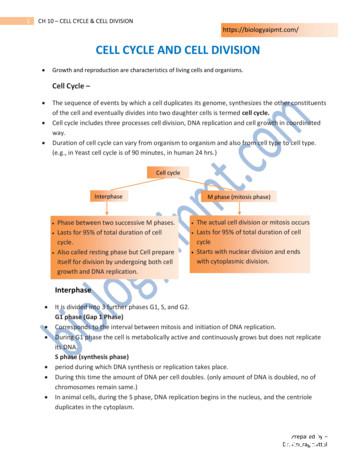
of the cell and eventually divides into two daughter cells is termed cell cycle. Cell cycle includes three processes cell division, DNA replication and cell growth in coordinated way. Duration of cell cycle can vary from organism to organism and also from cell type to cell type. (e.g., in Yeast cell cycle is of 90 minutes, in human 24 hrs.)
Chính Văn.- Còn đức Thế tôn thì tuệ giác cực kỳ trong sạch 8: hiện hành bất nhị 9, đạt đến vô tướng 10, đứng vào chỗ đứng của các đức Thế tôn 11, thể hiện tính bình đẳng của các Ngài, đến chỗ không còn chướng ngại 12, giáo pháp không thể khuynh đảo, tâm thức không bị cản trở, cái được
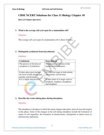
Class-XI-Biology Cell Cycle and Cell Division 1 Practice more on Cell Cycle and Cell Division www.embibe.com CBSE NCERT Solutions for Class 11 Biology Chapter 10 Back of Chapter Questions 1. What is the average cell cycle span for a mammalian cell? Solution: The average cell cycle span o


