Breast MR Protocols - Continuing Education
Breast MR ProtocolsBreast MR ImagingSuggested 1.5T and 3T Protocols forscanner vendors GE Healthcare Siemens HealthcareThis activity is jointly provided by:andACTIVITYAPPROVED BY This activity is supported by anunrestricted educational grantfrom Bracco Diagnostics Inc.Images courtesy of R. Edward Hendrick, PhD, FACR,and Roberta Strigel, MD, MS, FSBI.For additional CME/CE activities please visit www.ImagingEducation.com.
Breast MR ProtocolsBREAST MR PROTOCOLSFACULTYR. Edward Hendrick, PhD, FACRClinical ProfessorDepartment of RadiologyUniversity of Colorado-Denver, School of MedicineAurora, ColoradoRoberta Strigel, MD, MS, FSBIAssociate ProfessorDepartment of RadiologyUniversity of Wisconsin School of Medicine and Public HealthMadison, WisconsinCONTINUING EDUCATION INFORMATIONTARGET AUDIENCEThis activity has been designed for radiologists, radiologic technologists (RTs), medical physicists, radiologynurses, and other healthcare providers involved in breast magnetic resonance imaging (MRI).LEARNING GOAL/PURPOSETo provide radiologists, RTs, medical physicists, radiology nurses, and other healthcare providers involvedin breast MRI with protocols/sequences that can help to optimize imaging on General Electric (GE) andSiemens MR scanners while enhancing patient safety.EDUCATIONAL OBJECTIVESAfter completing this activity, participants should be better able to: Review requirements for breast MRI, including techniques used to optimize breast magnetic resonance(MR) protocols Implement 1.5T and 3T breast MR protocols that provide optimal visualization of breast cancers Discuss breast MR image interpretation including the use of computer-aided evaluation systemsSTATEMENT OF NEED/PROGRAM OVERVIEW Recent advances in MRI and MR technology have greatly improved the utility of this importantnoninvasive diagnostic modality for detecting and staging breast cancer, monitoring response totherapy, and guiding biopsies and surgical procedures using breast MRI. These improvements includehigher field strength magnets, dedicated breast coils, innovative pulse sequences, high-relaxivitycontrast agents, and optimized protocols Breast MRI is the clinical standard for screening patients at elevated risk for breast cancer and forstaging and extent of disease assessment due to the superior ability of this imaging modality to detectbreast cancers occult on other imaging modalities. The American College of Radiology (ACR) PracticeGuidelines for the Performance of Contrast-enhanced Magnetic Resonance Imaging of the Breast,Breast MRI Accreditation Program Requirements, and Practice Guideline for Performance of MagneticResonance Imaging-guided Breast Interventional Procedures provide important evidence-basedguidelines for radiologists and other healthcare personnel involved in breast MRI. To capitalize on theseadvances, radiologists, MRI technologists, medical physicists, and radiology nurses require educationthat enhances their understanding of improvements in MR technology and optimal breast MRI protocols1
Breast MR ProtocolsACCREDITATIONPhysician CreditThis activity has been planned and implemented in accordance with the accreditation requirementsand policies of the Accreditation Council for Continuing Medical Education (ACCME) through the jointprovidership of Medical Education Resources (MER) and ABC Medical Education LLC. MER is accreditedby the ACCME to provide continuing medical education for physicians.Credit DesignationMedical Education Resources designates this enduring activity for a maximum of 0.5 AMA PRA Category 1Credit . Physicians should claim only the credit commensurate with the extent of their participation in theactivity.Nursing CreditMedical Education Resources is accredited as a provider of continuing nursing education by the AmericanNurses Credentialing Center’s Commission on Accreditation.The continuing education activity provides 0.5 contact hour of continuing nursing education.MER is a provider of continuing nursing education by the California Board of Registered Nursing, Provider#CEP 12299, for 0.5 contact hour.Radiologic Technologist CreditThis activity is approved by the American Society of Radiologic Technologists (ASRT) for 0.5 Category ACE Credit. The ASRT is recognized by the American Registry of Radiologic Technologists as a RecognizedContinuing Education Evaluation Mechanism. The ASRT had no involvement in the development of thisactivity.INSTRUCTIONS FOR RECEIVING CONTINUING EDUCATION (CME/CE) CREDITThere is no fee for participating in and receiving CME/CE credit for this activity. The participants must1. Read the learning objectives and faculty disclosures2. Study the educational activity3. Complete the online posttest by recording the best answer to each questionPhysicians and NursesOriginal Release Date: December 2013Re-review Date: January 2022Expiration Date: January 2024Physicians CME Credit (0.5 AMA PRA Category 1 Credit )Nursing ANCC Credit (0.5 Contact Hour) Click on Physicians and Nurses Credit to complete the activity evaluation and posttest A statement of credit will be issued only upon receipt of a completed activity evaluation anda completed posttest with a score of 70% or better Statements of credit will be issued upon completion via e-mail Click on CE Credit to access the posttest A statement of credit will be issued only upon receipt of a completed posttest with a score of75% or better Statements of credit will be issued upon completion via e-mail2
Breast MR ProtocolsDISCLOSURE OF CONFLICTS OF INTERESTIt is the policy of MER to ensure balance, independence, objectivity, and scientific rigor in all of its educationalactivities. In accordance with this policy, MER identifies conflicts of interest with its instructors, contentmanagers, and other individuals who are in a position to control the content of an activity. Conflicts areresolved by MER to ensure that all scientific research referred to, reported, or used in a CME activityconforms to the generally accepted standards of experimental design, data collection, and analysis.The faculty reported the following financial relationships with commercial interests whose products orservices may be mentioned in this CME activity:Reported Financial RelationshipFacultyR. Edward Hendrick, PhD, FACRConsulting fees from GE Healthcare forwork on digital breast tomosynthesis.Roberta Strigel, MD, MS, FSBIGrants/research support from GE Healthcare.The content managers reported the following financial relationship with commercial interests whoseproducts or services may be mentioned in this CME activity:Content ManagerReported Financial RelationshipAndy Galler (ABC Medical Education)No financial relationships to disclose.Medical Education ResourcesNo financial relationships to disclose.DISCLAIMERThe content and views presented in this educational activity are those of the authors and do not necessarilyreflect those of Medical Education Resources (MER), ABC Medical Education (ABC) and/or BraccoDiagnostics Inc. (Bracco). The authors have disclosed if there is any discussion of published and/orinvestigational uses of agents that are not indicated by the FDA in their presentations. The suggestedprotocols presented here were developed for adult women. The opinions expressed in this educationalactivity are those of the faculty and do not necessarily represent the views of MER, ABC and/or Bracco.Before prescribing any medicine, primary references and full prescribing information should be consulted.Any procedures, medications, or other courses of diagnosis or treatment discussed or suggested in thisactivity should not be used by clinicians without evaluation of their patient’s conditions and possiblecontraindications on dangers in use, review of any applicable manufacturer’s product information, andcomparison with recommendations of other authorities. The information presented in this activity is notmeant to serve as a guideline for patient management.3
Breast MR ProtocolsBREAST MR PROTOCOLSBreast magnetic resonance imaging (MRI) requires maximizing sensitivity and specificity for breast cancerwhile minimizing scan time. Only with the use of modern MRI equipment, appropriate imaging sequences,proper patient positioning, appropriate contrast agent administration, and standardized image acquisitionand interpretation will sensitivity for breast cancer be maximized. Evaluation of lesion morphology andtemporal enhancement kinetics are required for accurate lesion characterization to improve specificity anddiagnostic accuracy. Longer overall scan times should be avoided, as they can lead to patient discomfortand motion, which in turn lead to artifacts and image degradation secondary to motion. See Table 1 for asummary of the prerequisites for maximizing sensitivity and specificity of breast MR imaging.1-3 In addition,the following text provides some additional details, as well as tips to consider, when performing breast MRI.Table 1: Summary of Prerequisites for Maximizing the Sensitivity and Specificity of Contrast-enhancedBreast MR Imaging1-3 High magnetic field strength (1.5T or greater) with a highly homogeneous magnetic field ( 1 ppm over 30 cm) Bilateral image acquisition with a prone-positioning bilateral breast coil (7 channels or greater) Unenhanced imaging with a T2-weighted pulse sequence to identify cysts and contribute to lesion characterization Multiphase contrast-enhanced imaging with a 3D T1-weighted spoiled gradient-echo pulse sequence Phase-encoding direction selected to minimize artifacts across breast tissue: phase-encoding right-to-left for axialimaging, head-to-foot for sagittal imaging (frequency-encoding anterior-posterior in either case) Intravenous administration of a gadolinium chelate at a dose of 0.1 mmol/kg and a rate of 2 mL/sec followed by a20 mL saline flush Homogeneous fat suppression across both breasts Thin-section acquisitions (section thickness of 3 mm or less, ideally closer to 1 mm) for the multiphase T1-weightedcontrast-enhanced series Pixel size of less than 1 mm in each in-plane direction for the multiphase T1-weighted contrast-enhanced series Temporal resolution (i.e., per-series imaging time) of less than 3 minutes for imaging both breasts in the multiphaseT1-weighted contrast-enhanced seriesProtocol SequencesThe American College of Radiology (ACR) Breast MRI Accreditation Program requires submission of abiopsy-proven breast carcinoma case acquired bilaterally with the following pulse sequences:2 Localizer or scout images, preferably obtained in all 3 perpendicular planes: axial, sagittal, and coronal A T2-weighted (T2W)/bright fluid series of both breasts, preferably with fat-suppression, to distinguishcysts from solid lesions A multiphase T1-weighted (T1W) series set acquired once before and multiple times after contrastagent administration, preferably acquired as a 3D (volume) gradient-echo (GRE) pulse sequence with fatsuppression, to identify the vascular bed and detect enhancing lesions in the breastIn addition, we recommend performing a 3D T1W non-fat-suppressed series prior to the multiphase series,to provide an overview of breast anatomy and to distinguish fat from water-based tissues (fibroglandulartissues, chest wall, and breast lesions).3The multiphase dynamic contrast-enhanced (DCE) sequences (i.e., pre- and postcontrast T1W, preferablywith fat suppression) are the most important images for identifying and characterizing lesions. The numberand length of the individual DCE sequences is variable, but each acquisition is required to have high spatialresolution (pixel size of 1 mm or less and slice thickness 3 mm or less) per ACR guidelines. It is essential thatthe pre- and postcontrast technical parameters are identical so that precontrast images can be subtractedfrom postcontrast images. This will provide a valid subtracted series from which other post-processed images(e.g., orthogonal plane reformatted images and maximum-intensity projections [MIPs]) can be reconstructed.The protocol sequences and sequence timing should be as consistent as possible from patient to patient,regardless of breast size or body habitus to maximize spatial resolution while including all breast tissue.4
Breast MR ProtocolsIt is important to be as efficient as possible, collecting the necessary information in the shortest exam time.Exams that take up to 45 minutes or longer are challenging for patients and increase the chance that thepatient will experience discomfort and move, causing motion artifacts and image misregistration. This isparticularly problematic for the DCE T1W series set, which is typically acquired last. Motion during the DCEseries set can significantly compromise kinetic evaluation, subtracted images, MIPs, and other reformattedimages. When appropriately constructed and performed on modern equipment, standard diagnostic breastMRI protocols should be completed within 30 minutes.CoilsPer the ACR, a dedicated, bilateral breast coil is required.2 Modern bilateral breast coils commonly have 7to 16 channels to improve signal uniformity and intensity.ContrastCurrently, seven extracellular fluid (ECF) gadolinium-based contrast agents (GBCAs) are being usedfor breast MRI (Table 2). Of these contrast agents, only gadobutrol (Gadavist) has been FDA-approvedspecifically for use in breast MRI. The other ECF agents shown in Table 2 are FDA-approved for use inimaging the central nervous system and other body areas/applications, but they are often used “off-label”for breast MRI.All of the available GBCAs exhibit the same mechanism of action; they increase signal intensity of lesionsagainst background tissue, a phenomenon known as T1-shortening. The degree to which a GBCA cancause T1-shortening is known as relaxivity (r1). At a given molar concentration of GBCA, a higher r1relaxivity translates into greater T1-shortening, and subsequently a greater effect of a GBCA on lesionconspicuity and breast cancer detection.3,4For breast MRI, both standard and high-relaxivity contrast agents should be administered at labeled dosesof 0.1 millimoles per kilogram (mmol/kg) of body mass and injected at a rate of 2 milliliters per second(mL/s), followed by a 20 mL saline flush injected at the same rate. Use of a power injector to inject both thecontrast agent and the saline flush is recommended.Most of the available ECF contrast agents are formulated at a concentration of 0.5 moles/liter (or0.5 millimoles/milliliter [mmol/mL]); only gadobutrol (Gadavist) is formulated at a higher concentration of1.0 mmol/mL. However, the recommended GBCA doses in breast MRI are the same despite the differencesin concentration: 2 mL per 10 kg of body weight for a 0.5 mmol/mL GBCA, and 1 mL per 10 kg of bodyweight for gadobutrol (Gadavist). Using a 150 lb ( 68 kg) woman as an example, approximately 14 mL ofa 0.5 mmol/mL concentration agent would be recommended, and 7 mL of the 1.0 mmol/mL concentrationagent gadobutrol (Gadavist) (i.e., half the volume).5
Breast MR ProtocolsTable 2: FDA-Approved Extracellular Gadolinium-Based Contrast Agents and Their Properties5-14Brand Name(Manufacturer)FDA Approval Date Generic Namer1 RelaxivityMolarity (mL mmol-1s-1)(M, moles at 1.5T/3.0T inper liter) Plasma at 37oCApproved IndicationsApproved DoseGadopentetatedimeglumineCNS, adults & pediatrics( 2 years of age);Head & neck, adults & pediatrics( 2 years of age); Body (excludingthe heart), adults & pediatrics( 2 years of age)0.1 mmol/kg0.54.25 / 3.76GadoteridolCNS, adults & pediatrics( 2 years of age);Head and neck, adultsAdults: 0.1 mmol/kg 2nd dose of0.2 mmol/kg up to30 min after 1st dose;pediatrics: 0.1 mmol/kg0.54.39 / 3.46Omniscan(GE Healthcare)1993GadodiamideCNS, adults & pediatrics(2-16 years of age);Body (excluding the heart),adults & pediatrics(2-16 years of age)0.1 mmol/kg0.54.47 / 3.89OptiMARK(Covidien)1999GadoversetamideCNS, adults;Liver, adults0.1 mmol/kg0.54.43 / 4.24GadobenatedimeglumineCNS, adults & pediatrics(including term neonates);MRA in adults, to evaluate knownor suspected renalor aorto-ilio-femoral occlusivevascular diseaseAdults & pediatrics 2 yearsof age: 0.1 mmol/kg;pediatrics 2 years of age:0.05-0.1 mmol/kg(ie, 0.1-0.2 mL/kg) (CNS)0.56.2 / 5.37Gadavist(Bayer Healthcare)2011GadobutrolCNS, adults & pediatrics(including term neonates);Assess presence and extentof malignant breast disease;MRA to evaluate known orsuspected supra-aortic orrenal artery disease in adult &pediatrics (including term neonates)0.1 mmol/kg1.04.61 / 4.46Dotarem(Guerbet)2013Clariscan(GE Healthcare)2013GadoteratemeglumineCNS, adults & pediatrics(including term neonates)*0.1 mmol/kg0.53.91 / 3.43Magnevist(Bayer co)2004CNS central nervous system; MRA magnetic resonance angiography.*Approval for use in term neonates is limited to Dotarem. Clariscan approved for pediatric patients aged 2 to 17 years.6
Breast MR ProtocolsImage InterpretationImage interpretation steps include:1. Identification of enhancing lesions separate from background parenchymal enhancement.2. Characterization of morphologic characteristics of identified lesions according to the latest version ofthe ACR Breast Imaging Reporting and Data System (BI-RADS) Atlas.153. Evaluation of temporal kinetics of identified lesions according to the ACR BI-RADS Atlas.4. Use of both morphology and kinetics to make a recommendation regarding level of suspicion accordingto the ACR BI-RADS Atlas.Computer-aided Evaluation (CAE) SystemsSeveral computer-aided evaluation (CAE) systems are available for breast MRI. CAE for breast MRI, unlikecomputer-aided detection (CAD) for mammography, does not direct the interpreting radiologist to potentiallysuspicious lesions; rather, it aids in evaluation of lesion kinetics and overall assessment of the degree ofsuspicion. CAE provides a convenient and efficient way to visually and quantitatively assess temporalkinetic information by providing color overlay maps and time-signal intensity curves of enhancing lesions.With modern CAE systems, images are sent from the MR system to the picture archiving and communicationsystem (PACS) and the CAE system, where color overlay maps, kinetic analyses, image subtractions, andmultiplanar and MIP reconstructions are performed. When the radiologist identifies a suspicious area ofenhancement, the CAE software provides information on lesion size, degree of early-phase enhancement,and the signal-intensity time course including the delayed-phase enhancement pattern (persistent, plateau,wash-out), which correlates with degree of suspicion of breast cancer.16Note that CAE systems impose a delay between receipt of images from the MR or PACS system andavailability of processed results to the radiologist. Importantly, CAE-subtracted images and temporal kineticevaluation are susceptible to patient motion between pre- and postcontrast images; if unrecognized asmotion artifacts, it is possible to misinterpret displaced tissue color maps or signal-intensity time curves asenhancing lesions. All or most CAE systems have motion correction algorithms, but they are not perfect,and evaluation by the interpreting radiologist requires assessment of the pre- and postcontrast images formotion to avoid these mistakes.Use of CAE temporal kinetic color overlay maps alone can lead to overestimation or underestimation ofthe degree of suspicion of a lesion due to the overlap in the kinetics of benign and malignant lesions, aswell as misinterpretation of artifacts created by patient motion. For example, unrecognized motion canresult in “wash-out” kinetics and overestimation of lesion suspicion, while the lack of the more concerningplateau or wash-out delayed-phase enhancement patterns on CAE can cause underestimation of the levelof suspicion for small, low-grade, or non-mass-like lesions such as ductal carcinoma in situ (DCIS). If alesion has suspicious morphologic features, the level of suspicion generated by the concerning morphologicfeatures should not be diminished by the presence of a less suspicious temporal kinetic signal-intensity timecourse, such as persistent delayed-phase enhancement.CAE color map thresholds for standard agents (such as gadopentetate dimeglumine [Magnevist]) shouldbe set at values of 50% and 100% signal enhancement above precontrast levels, whereas a high-relaxivityagent (such as gadobenate dimeglumine [MultiHance]), may require color map thresholds to be set at 100%and 200% signal enhancement levels. Systems that allow for real-time adjustment of CAE thresholds mayallow more flexibility (e.g., using several thresholds, such as 50%, 100%, 150%, and 200%). It is especiallyimportant to make the color map threshold adjustment if CAE images from an exam performed with a highrelaxivity agent are being compared to CAE images from an exam performed with a conventional-relaxivityagent in the same patient. There is usually no need to make additional adjustments to CAE thresholds whenchanging between breast MRI exams performed at 1.5T and 3T.7
Breast MR ProtocolsTips for Maximizing Sensitivity and SpecificityTo maximize sensitivity, it is important to acquire multiphase T1W images with high spatial resolution (i.e.,sub-millimeter in-plane resolution in the frequency- and phase-encoding directions) to obtain morphologicdetail, including lesion shape, margin, and internal enhancement pattern. It is also important to acquire thinslices; if the slices are not sufficiently thin, there is the risk of volume-averaging small, subtle lesions withbackground tissue, decreasing their lesion conspicuity. In addition, the use of thin slices (nearly isotropicvoxels, where the slice thickness is nearly as small as the in-plane resolution) allows image reconstructionin any plane while maintaining approximately the same spatial resolution as acquired in-plane images.On the other hand, pixel size and slice thickness should not be so small that signal-to-noise ratios suffer.Finally, radiologists should identify enhancing lesions on the early-phase postcontrast or subtracted images,where the signal difference between enhancing lesions and background parenchyma is greatest.To maximize specificity, kinetics should be obtained with a temporal resolution of 3 minutes or less (thatis, each series in the multiphase set should be acquired in 3 minutes or less). While there are studiesshowing that proper lesion time-intensity curve shape can be captured with 2 minute and 3 minute temporalresolution,17,18 there are no data to suggest that curve shapes are captured correctly with longer acquisitiontimes. Conversely, there is a trade-off between temporal resolution and signal-to-noise, just as there isbetween temporal resolution and spatial resolution.17 Using very rapid acquisitions, just like using excessivelysmall voxels, can reduce signal-to-noise ratios to the extent that enhancing lesions, especially non-masslike lesions, are not detected. More recent advanced MRI sequences allow for faster acquisitions withouttraditional compromises in spatial resolution using undersampling and advanced reconstruction techniquessuch as view-sharing and compressed sensing. However, all of these techniques have trade-offs, and mostare not routinely used in clinical practice.It is beneficial to evaluate the breasts in at least one additional imaging plane beyond the primary acquisitionplane (e.g., if the primary image acquisition plane is axial, be sure to also evaluate sagittal reformattedimages). Again, it is most beneficial to do this in the early-phase of contrast enhancement, where the contrastdifference between enhancing lesions and background parenchyma is greatest. Therefore, reconstructionof images in other planes from early-phase images acquired with isotropic or nearly isotropic voxels is mostbeneficial for this analysis and shortens the overall scan time (instead of acquiring additional postcontrastseries in additional scan planes after the multiphase T1W series has been acquired).Breast MR: Practical ConsiderationsThere are no additional MR safety considerations specific to breast MRI; the biggest safety hazard whileperforming breast MR, like other MR exams, is the inadvertent introduction of ferromagnetic materials (iron-,nickel-, or cobalt-containing metals) into the scanner room. Such metals may be implanted in the patient,worn by the patient, or inadvertently brought in by the patient, someone accompanying the patient, or by aphysician, technologist, or maintenance personnel. Ferromagnetic materials near the bore of the scannerare rapidly accelerated into the scanner bore and can severely injure the patient or others in the scanroom. Additional considerations include burns. The MR safe practice guidelines developed by the ACR toestablish industry standards for safe and responsible practices in clinical and research MR environmentsalso apply to breast MRI.19In addition, as for other contrast-enhanced MR exams, women undergoing breast MR exams may need tobe screened for adequate renal function (glomerular filtration rate) prior to performing the breast MR exam,per institutional and ACR guidelines.20Additional practical considerations include patient size and patient breast size. Larger patients may not fitinto the scanner itself, particularly when positioned on the breast coil. Very large or very small breasts maynot fit well in the breast coil, and good breast positioning, including as much breast tissue in the breastcoil as possible, is important for optimal image acquisition and interpretation.21 Technologists performingbreast MRI should take care to ensure that as much breast tissue as possible is positioned inside thebreast coils, that breast positioning is as symmetric as possible between the left and right breasts, andthat the nipple is positioned centrally and symmetrically in each breast. It is particularly important to makesure that the nipple is not folded inside other breast tissue. It also is important to stabilize the breast tominimize motion, while at the same time avoiding compression and deformation of the breast. Radiologictechnologists performing breast MR should be trained on proper breast positioning,21 and experiencedtechnologists and coil manufacturer applications specialists can be helpful in providing such training. Inaddition, the ACR Breast MRI Accreditation Program requirements for technologist experience and trainingshould be reviewed and followed.28
Breast MR ProtocolsAbbreviated Breast MRI (AB-MR)AB-MR is a new protocol for screening women at elevated risk of breast cancer.22,23 It is a short scanningprotocol consisting of scout images, a precontrast T1W series covering both breasts, contrast injection and abrief (20-45 second) delay to permit perfusion of contrast agent, followed by an identical postcontrast series.The entire acquisition protocol should require no more than 5 minutes of scanning.24 If a precontrast T2Wseries is acquired to assist the radiologist with differential diagnosis, it should be optimized to minimize and notexcessively increase scan time. Image post-processing should include subtraction of pre- from postcontrastimages and reconstruction of MIP images. Image acquisition techniques for pre- and postcontrast T1W imagesshould follow the same optimization procedures as those used for dynamic breast MRI, with the exceptionthat only a single postcontrast series is acquired. Initial clinical results for AB-MR show high sensitivity andspecificity, comparable to those of the full dynamic protocol used for diagnostic breast MRI.22,23,25-32 Furtherstudies are underway to study the utility of AB-MR in women with dense breasts.32Additional Tips for Technologists Performing Breast MRI:Every effort should be made to ensure the patient is comfortable in the prone positioning used for breast MRI.This includes proper head support, adequate padding between the sternal notch and the breast coil, where alarge portion of the patient’s weight is supported, and positioning arms and legs comfortably with pillows, asneeded. During patient positioning, make sure that IV contrast lines remain unkinked and positioned formaximum comfort and flow at the injection site Make sure that the appropriate patient positioning (head-first prone or feet-first prone) is enteredduring patient data entry. Incorrectly entering supine instead of prone, or reversing head-first andfeet-first, will reverse left and right on the images, which can lead to identification of lesions in the wrongbreast during interpretation. Breast MRI patients imaged on GE (and Aurora) scanners are typicallypositioned feet-first prone (FFP), while patients imaged on Siemens and most other manufacturers’scanners are typically positione head-first prone (HFP) Always inform the patient about the importance of not moving during scanning and not moving betweenpre- and postcontrast multiphase T1W series. This is best done prior to the precontrast series, ratherthan just prior to injection, to avoid causing the patient to startle and move between pre- and postcontrastseries, which affects the registration of subtracted images. Advice about the injection is best given priorto the precontrast series If fat suppression is being used for the multiphase T1W images, always check precontrast images foradequate fat suppression prior to injection of contrast agent. Some sites insert a very fast “fat-sat check”precontrast series to assess fat suppression prior to the multiphase series that includes the full precontrast,injection, and postcontrast series Always check postcontrast images as soon as possible to confirm that contrast agent injection wassuccessful. This is best done by comparing precontrast and postcontrast images viewed with similarwindow width and level settings and looking for the presence of contrast agent in the heart and internalmammary vessels. If contrast agent is not visible in postcontrast images, the patient
The continuing education activity provides 0.5 contact hour of continuing nursing education. MER is a provider of continuing nursing education by the California Board of Registered Nursing, Provider #CEP 12299, for 0.5 contact hour. Radiologic Technologist CreditFile Size: 1MB
4 Breast cancer Breast cancer: A summary of key information Introduction to breast cancer Breast cancer arises from cells in the breast that have grown abnormally and multiplied to form a lump or tumour. The earliest stage of breast cancer is non-invasive disease (Stage 0), which is contained within the ducts or lobules of the breast and has not spread into the healthy breast tissue .
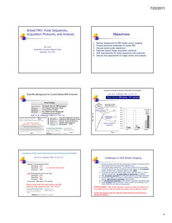
1. Review background of MRI breast cancer imaging 2. Present technical challenges of breast MRI 3. Discuss typical pulse sequences 4. Describl lbe typical image acquisition protocols 5. ACR requirements for pulse sequences and protocols 6. Discuss new approaches to image review and analysis Scientific Background for Current Breast MRI Protocols
5 yrs of anti-hormone therapy reduces the risk of: – breast cancer coming back somewhere else in the body (metastases / secondaries) – breast cancer returning in the same breast – a new breast cancer in the opposite breast – death from breast cancer The benefits of anti-hormone therapy last well
Breast cancer development In the United States, breast cancer is the most common cancer diagnosed in women (excluding skin cancer). Men may also develop breast cancer, but less than 1% of all people with breast cancer are men. Breast cancer begins when healthy cells in the breast change and grow uncontrollably, forming a mass called a tumor.
Breast Implants or Patient Educational Brochure - Breast Reconstruction with MENTOR MemoryShape Breast Implants, and a copy of Quick Facts about Breast Augmentation & Reconstruction with MENTOR MemoryShape Breast Implants. For MENTOR Saline-fi lled Implants, patients should receive a copy of Saline-Filled Breast Implants: Making an Informed Decision.
breast cancer, metastases, advanced breast cancer, secondary tumours, secondaries or stage 4 breast cancer. For most people with secondary breast cancer in the brain, breast cancer has already spread to another part of the body such as the bones, liver or lungs. However, for some people, the brain may be the only area of secondary breast cancer.
for-the-management-of-breast-cancer-v1.doc 8 . Organisation of breast cancer surgical services . The multidisciplinary team (MDT) Breast cancer care should be provided by breast specialists in each disciplineand multidisciplinary teams form the basis of best practice. All new breast cancer patients should be reviewed by a multi-disciplinary .
Standard can be used by an organization to assure interested parties that an appropriate environmental management system is in place. Guidance on supporting environmental management techniques is contained in other International Standards, particularly those on environmental management in the documents established by ISO/TC 207. Any reference to other International Standards is for information .


