Cancer Cell Perspective - National Cancer Institute
Cancer CellPerspectiveDragging Ras Back in the RingAndrew G. Stephen,1 Dominic Esposito,1 Rachel K. Bagni,1 and Frank McCormick1,2,*1LeidosBiomedical Research, Inc., Frederick National Laboratory for Cancer Research, P.O. Box B, Frederick, MD 21702, USA2UCSF Helen Diller Family Comprehensive Cancer Center, Room 371, 1450 3rd Street, P.O. Box 589001, San Francisco, CA 94158-9001, USA*Correspondence: r.2014.02.017Ras proteins play a major role in human cancers but have not yielded to therapeutic attack. Ras-driven cancers are among the most difficult to treat and often excluded from therapies. The Ras proteins have beentermed ‘‘undruggable,’’ based on failures from an era in which understanding of signaling transduction, feedback loops, redundancy, tumor heterogeneity, and Ras’ oncogenic role was poor. Structures of Ras oncoproteins bound to their effectors or regulators are unsolved, and it is unknown precisely how Ras proteinsactivate their downstream targets. These knowledge gaps have impaired development of therapeutic strategies. A better understanding of Ras biology and biochemistry, coupled with new ways of targeting undruggable proteins, is likely to lead to new ways of defeating Ras-driven cancers.Fifty years have passed since the transforming power of Rasgenes was first recognized. Harvey sarcoma virus, Kirsten sarcoma virus, and Rasheed sarcoma virus contain Ras genes (sonamed for their role in forming rat sarcomas; reviewed in Barbacid, 1987; Karnoub and Weinberg, 2008). These retrovirusesinitiated tumors efficiently and, using temperature-sensitive mutants, were shown to be necessary for tumor maintenance (Shihet al., 1979). They formed part of a fascinating collection ofretroviruses that was assembled in the 1970s, each able to transform cells in culture and in avian and rodent models. These experiments were, essentially, unbiased screens for genes thatcause cancer; the nature of the proteins that the genes encodedwas completely unknown. Remarkably, the majority of theseviruses encoded proteins that were later identified as components of the tyrosine kinase-Ras signaling pathway (Vogt,2012), even though the biochemical nature of these proteinswas unknown, and tyrosine kinase activity had not been discovered (Eckhart et al., 1979). Of the hundreds of mutant proteinsnow known to contribute to cancer that could have been identified in these assays, including those involved in DNA repair,cellular metabolism, RNA splicing, and the other hallmarks ofcancer (Hanahan and Weinberg, 2011), those in the tyrosinekinase-Ras pathway stand out as the major drivers and havebeen the richest source of targets of successful cancer therapies(Abl, epidermal growth factor receptor [EGFR], Her2/neu, B-Raf,Kit, ALK, etc.). These successes can therefore be attributed tothe central, dominant role of this pathway in cancer, as well asthe fortuitous abundance of druggable targets.However, specific therapies have not been developed formutant Ras proteins themselves or for the cancers that theydrive. Worse yet, tumors driven by Ras genes are excludedfrom treatment with other targeted therapies. Early efforts toblock Ras cancers by preventing Ras farnesylation, oncethought to be an essential posttranslational modification forRas activity, were thwarted by the unexpected presence of abackup system (geranylgeranyltransferase) that restored activityof K-Ras and N-Ras after farnesyltransferase treatment. Likewise, efforts to kill Ras cancers by blocking one of Ras’ majordownstream effectors, Raf kinase (Figure 1), ran into the unexpected discovery that, in Ras-transformed cells, Raf inhibitors272 Cancer Cell 25, March 17, 2014 ª2014 Elsevier Inc.activate the pathway rather than inhibit it (see below and discussion in Holderfield et al., 2013 and Lito et al., 2013). MAP kinasekinase (MEK) inhibitors and phosphatidylinositol 3-kinase (PI3K)inhibitors have not yet shown significant clinical activity in Rascancers, for reasons relating to feedback loops and poor therapeutic windows, among other issues discussed below.A convergence of urgent unmet clinical needs and advances indrug discovery has energized new efforts to target Ras cancerswithin academic centers and in the biopharmaceutical industry.To catalyze these renewed efforts, the National Cancer Instituterecently launched a national Ras program at Frederick NationalLaboratory for Cancer (see http://RasCentral.org), whose goalis to fill critical knowledge gaps that are essential to target Rascancers effectively and to engage the research communitytoward solving the Ras problem. Here, we will discuss some ofthese knowledge gaps, as well as recent advances and the challenges that lie ahead.Ras Mutations in CancerRas genes were the first oncogenes identified in human cancercells. In a series of classic experiments, the groups of Weinberg,Cooper, Barbacid, and Wigler independently identified the transforming genes from T24/EJ bladder carcinoma cells as H-Ras(Der et al., 1982; Parada et al., 1982; Santos et al., 1982; Taparowsky et al., 1982). More than 30 years later, Ras genes are wellestablished as the most frequently mutated oncogenes in humancancer (Table 1), though H-Ras itself is rarely one of them.Although these numbers are, by now, painfully familiar, theyunderscore major gaps in our knowledge of Ras biology. Mostobviously, we do not understand why K-Ras mutation is muchmore frequent in human cancer than N-Ras or H-Ras, eventhough each of these is a powerful transforming gene in modelsystems, and all forms are expressed widely in adult tissuesand in tumors.A simple explanation for the high frequency of K-Ras mutations, relative to H-Ras and N-Ras, is that the K-Ras proteinhas unique properties that favor oncogenesis. At first sight, thisseems unlikely because the Ras proteins are highly conserved,especially in their effector-binding regions where they are actually identical. However, K-Ras, but not N-Ras or H-Ras, confers
Cancer CellPerspectiveFigure 1. Simplified View of the RasPathwayRas proteins are converted from their GDP state totheir GTP state by GEFs, in response to upstreamsignals (Bos et al., 2007). GAPs convert Ras-GTPback to Ras-GDP. p120 GAP does this when recruited to activated RTKs. The signal that directsNF1 (neurofibromin)/SPRED to inactivate Ras isnot known. Several other GAPs are capable ofdownregulating Ras (Bos et al., 2007). Ras-GTPbinds and activates multiple downstream effectors. The group of proteins shown on the left includes potential effectors whose significance isless well understood relative to RalGDS, Rafkinases, and PI3Ks (Gysin et al., 2011). Proteinfamilies are represented as single proteins tosimplify the schematic; in addition, feedback loopsare not included.stem-like properties on certain cell types (Quinlan and Settleman, 2009). K-Ras-4B, the most highly expressed splice variantof K-Ras, binds calmodulin; H-Ras and N- Ras do not (Villalongaet al., 2001). We believe that this unique property of K-Ras-4Bconfers stem-like properties to cells expressing oncogenicK-Ras-4B proteins (M. Wang and F.M., unpublished data).Analysis of human syndromes caused by germline mutationsin H-Ras or K-Ras supports the idea that K-Ras is a strongeroncogene. Unexpectedly, humans can tolerate germline-activating mutations in H-Ras—the same activating mutations thatdrive somatic mutations. Costello syndrome, which is characterized by germline H-Ras mutations, is associated with a broadspectrum of developmental abnormalities and a high risk forrhabdomyosarcomas and neuroblastomas (reviewed in Rauen,2013). It is puzzling that these individuals do not succumbto malignancies associated with sporadic H-Ras mutations(Table 1). Although fully activating alleles of H-Ras can be tolerated, fully activated alleles of K-Ras may not. Variant alleles ofK-Ras that account for a small fraction of Noonan’s syndromeand cardiofaciocutaneous syndrome are weakly activated relative to their sporadic oncogenic counterparts (Schubbert et al.,2007).Further support for the idea that K-Ras has functions distinctfrom H-Ras and N-Ras comes from analyses of the roles ofRas genes in development. Mice that lack K-Ras die duringembryogenesis, whereas mice lacking H-Ras and/or N-Ras areviable (Johnson et al., 1997). However, replacing K-Ras withH-Ras at the K-Ras genomic locus allows mice to develop, suggesting that differential regulation of K-Ras and H-Ras geneexpression determines their relative importance in developmentrather than the properties of the proteins themselves (Potenzaet al., 2005). Furthermore, Balmain and colleagues discoveredthat these H-Ras knock-in mice develop tumors in response tocarcinogens at normal frequencies, except that they are nowdriven by H-Ras instead of K-Ras (To et al., 2008). These dataargue strongly that the locus is critical and that the specificRas paralog encoded at that locus does not affect the frequencyat which tumors arise. Equally important, they find that K-Ras4A, not K-Ras-4B, is necessary for lung tumor initiation, althoughK-Ras-4B is much more highly expressedduring progression. This supports theidea that K-Ras-4B is the more importanttarget in established tumors but raises the concern that K-Ras4A may have an important role in minor stem-like populationsof established tumors. These findings point toward an urgentneed to validate K-Ras-4A and K-Ras-4B as drug targets, amajor issue that has not yet been addressed.Different frequencies of K-Ras, N-Ras, and H-Ras mutationsin human tumors may also reflect differences in gene expressionresulting from differential codon usage; rare codons limit K-Rasexpression and thus allow more efficient oncogenesis by preventing oncogene-induced senescence (Lampson et al., 2013).In addition, different rates of DNA repair have been reportedfor the K-Ras gene relative to N-Ras and H-Ras (Feng et al.,2002).The underlying reasons for different frequencies of specificactivating mutations are not well understood either. Some ofthese differences reflect different mutagenic insults to thegenome; the G12C mutation, for example, is a hallmark of exposure to tobacco smoke and, accordingly, is the most commonmutation in K-Ras in lung cancer (reviewed in Prior et al., 2012;Table 2). Other differences in frequency may reflect different biological properties of mutant proteins. For example, G12C andG12V K-Ras mutations in lung adenocarcinoma preferentiallyactivate the RalGDS pathway, whereas G12D prefers the Raf/mitogen-activated protein kinase (MAPK) and PI3K pathways(Ihle et al., 2012). In addition, mutations at codon 61 have amore profound effect on intrinsic GTPase when these Rasproteins are bound to Raf kinase. This may drive a strongersignal through this effector pathway and account for higher frequency of N-Ras position 61 mutations in melanoma, a diseasefrequently driven by hyperactivation of Raf kinase through B-Rafmutations (Buhrman et al., 2010).From a clinical viewpoint, lung adenocarcinomas driven byK-Ras mutations at G12C and G12V have a worse outcomethan G12D, possibly because these mutations engage differentdownstream effectors as described above (Figure 1; Ihle et al.,2012). As MEK and PI3K inhibitors are tested in the clinic, it willbe important to ask whether Ras alleles respond differently tothese treatments. Patients suffering from cancers driven byany of these Ras mutations are excluded from treatment withCancer Cell 25, March 17, 2014 ª2014 Elsevier Inc. 273
Cancer CellPerspectiveTable 1. Frequency of Ras Isoform Mutations in Selected HumanCancersTable 2. Incidence of KRAS Mutations in Three Human CancersPrimary TissueTotal (%)Colorectal60,000KRAS (%)HRAS (%)NRAS (%)All KRAS G12CG12DG12VG13D5,700 25,000 15,700 13,600Pancreas710 171Lung45,600Colon351642Pancreas32,200Small intestine350 135Total new cases/year 137,800Biliary tract260228Endometrium17 1522Lung20Shown are the numbers of new cancer cases per year in the United Statesthat contain the most frequent KRAS mutant alleles. Data are based onestimated new case incidence values from the National Cancer Instituteand primary tumor mutation frequency data from COSMIC v.67.19 11Skin (melanoma)111820Cervix89219Urinary tract510116Data were compiled from the Catalogue of Somatic Mutations in Cancer(COSMIC) version 67. All human cancers that had total Ras mutation frequencies above 15% are listed.cetuximab (colorectal cancer) or erlotinib (lung adenocarcinoma)because these treatments are ineffective for cancers with theseRas mutations and may even increase rates of progression.Likewise, malignant melanomas with mutant N-Ras are excluded from treatment with vemurafenib. However, surprisingly,K-Ras-G13D-bearing colorectal cancers may show clinicalbenefit when treated with cetuximab. This result challenges ourunderstanding of how these Ras mutations actually function inclinical situations (De Roock et al., 2010).Even the prototypic oncogenes of Harvey and Kirsten sarcoma viruses are not fully understood; each has a codon 12 mutation, but each also carries a mutation of alanine 59 to threonine,which becomes phosphorylated by guanosine triphosphate(GTP). This must have helped Scolnick and colleagues (Shihet al., 1979) identify Ras’ crucial guanosine diphosphate (GDP)/GTP properties; without covalent phosphorylation, associationwith these nucleotides would have been very hard to detect.However, how phosphorylation at threonine 59 contributes toRas’ potent oncogenicity is unclear. This A59T mutation inhibitsRas-Raf interaction (Shirouzu et al., 1994) and is extremely rarein human cancer. These anecdotes simply remind us that after50 years, we still have a lot to learn about the biological andbiochemical functions of Ras proteins.Although K-Ras has emerged as by far the major Ras genemutated in human cancer, it is surprising that other activatingmutations in other members of the Ras superfamily, such asR-Ras or Rap proteins, occur very rarely. This is surprisingbecause these proteins share identical or near-identicaleffector-binding regions. However, only H-Ras, N-Ras, andK-Ras are capable of binding and activating Raf kinases, andthis unique property may well account for their predominanceas human oncogenes. In contrast, the closely related R-Ras proteins bind and activate PI3Ks but are rarely mutated in humancancer (Rodriguez-Viciana et al., 2004).Activating mutations in Ras genes, coupled with a long historyof Ras biology, implicate these mutant Ras proteins as majordrivers in many cancers. Loss of the Ras GTPase-activatingprotein (GAP) neurofibromin inculpates hyperactive wild-typeRas proteins as drivers in many more cancers. Somatic loss ofneurofibromin expression by mutation, deletion, or by othermeans occurs in about 14% glioblastoma, 13%–14% mela274 Cancer Cell 25, March 17, 2014 ª2014 Elsevier Inc.23,0009,200 11,9001,5001,000 19,500 11,50020029,700 53,700 39,100 15,300noma, 8%–10% lung adenocarcinoma, and at single-digit frequency in most other cancers (E.A. Collisson, personal communication). Neurofibromin must now be considered as a majortumor suppressor, along with p53 and phosphatase and tensinhomolog, in human cancers.Loss of neurofibromin is usually mutually exclusive with Rasmutation and receptor tyrosine kinase (RTK) activation, suggesting that these genetic events represent different ways of activating similar pathways. However, the precise consequencesof losing neurofibromin are not entirely clear. Levels of RasGTP are high in cells lacking neurofibromin, but which forms ofhyperactive wild-type Ras proteins are most important to themalignant phenotype is a more difficult question. Perhapselevated H-Ras, N-Ras, K-Ras-4A, and K-Ras-4B all contributeto some extent. However, neurofibromin is also a GAP forR-Ras proteins, and hyperactivation of these proteins can alsocontribute to the malignant phenotype because R-Ras proteinsactivate p110a, p110g, and p110d isoforms of PI3Ks (Marteet al., 1997; Huang et al., 2004).Recently, Legius and colleagues (Brems et al., 2007) discovered mutations in the Sprouty-related protein, SPRED1, in aform of neurofibromatosis type I (NF1) in which the neurofibromingene is wild-type. This disease is now called Legius syndrome(Brems et al., 2007). SPRED1 has a well-established pedigreeas a negative regulator of the Raf/MAPK pathway, though themechanism has been unclear. However, the fact that loss of neurofibromin is, to a significant extent, phenocopied by loss ofSPRED1, supports the idea that NF1 is a disease of hyperactiveRas and that the major function of neurofibromin is to turn Rasoff. The neurofibromin protein itself is over 2,800 amino acidsin length, and the GAP domain only accounts for about 300amino acids, raising the possibility that neurofibromin has otherfunctions that are not directly related to negative regulation ofRas. Most attempts to identify additional functions have failed,however, and it seems most likely that neurofibromin sensesan unidentified cellular metabolite and downregulates Rasaccordingly, just as p120 Ras-GAP senses phosphotyrosine residues, and downregulates Ras when it binds to these residues onactivated receptors in the plasma membrane (reviewed in Boset al., 2007). Whatever neurofibromin senses (if this model iscorrect) is likely to be conserved between S. cerevisiae and humans because the S. cerevisiae IRA1 and IRA2 proteins look verymuch like neurofibromin. Unfortunately, the complete lack of anyrecognizable domains or motifs outside the GAP domain and aSEC14 domain has not helped in identifying what these proteinsrecognize.
Cancer CellPerspectivein human cell lines resulted in a spectrum of responses, revealinga range of K-Ras dependencies (Singh et al., 2009). Assessmentof Ras dependency in 3D culture systems suggests that thisassay system is a more stringent measure of Ras dependency.These studies raise the question of what is the most relevantsystem to measure this essential parameter and, in general, responses to candidate therapeutics targeting K-Ras. Furthermore, the degree to which Ras genes are knocked down maybe critical. Genetic ablation is obviously different than smallinterfering RNA- or small hairpin RNA (shRNA)-mediated knockdown. It is also clear that knocking down activated Ras canlead to hyperactivation of upstream pathways, such as EGFRsignaling (Young et al., 2013). Presumably, these pathways aresuppressed in cells with activated Ras and rebound when thesuppressor is removed. Although this rebound effect may notbe sufficient to sustain a malignant phenotype, it may offset proapoptotic effects associated with oncogene inactivation.Figure 2. Structure Showing Small Molecule-Directed ElectrophilicAttack of K-Ras-G12CK-Ras-G12C (Protein Data Bank 4LUC A) is displayed in surface representation. The cocrystallized ligands, GDP and N-(1-[(2,4-dichlorophenoxy)acetyl]piperidin- 4-yl)-4-sulfanylbutanamide, are shown in stick mode. The location ofcalcium ion is shown as a green ball. Switch 1 (28–38) and switch 2 (57–63) arehighlighted by orange and red colors, respectively. Key ligand-interactingresidue (C12, V9, V7, F78, I100, M72, Q99, and R68) positions are coloredgreen. Position of C12 residue is shown in ball and stick (green). Note thatresidues 58 and 60 are part of both switch 2 and the key ligand-interactinggroup (shown in blue).By comparing proteins that bind to wild-type SPRED1 versusmutants from Legius syndrome, we found that neurofibrominbinds directly to SPRED proteins, via their EVH1 domains, andthat SPRED proteins bring neurofibromin to the plasma membrane (Stowe et al., 2012). SPRED proteins also bind to c-Kit,and perhaps to other RTKs, suggesting that neurofibromin regulates Ras locally in response to specific receptor signaling, ratherthan simply suppressing Ras throughout the plasma membrane.In this case, loss of neurofibromin may lead to local activation ofRas that is coupled to specific receptors, suggesting that inhibitors of these receptors might reverse the effects of neurofibromin loss. The recent Cancer Genome Atlas analysis of mutationsin lung cancer revealed an intriguing overlap between neurofibromin loss and Met amplification, suggesting a functionalconnection that merits further investigation (E.A. Collisson, personal communication).Validation of Ras as a TargetRas oncogenes can certainly initiate cancer in model organismsand probably do so in humans. However, their role in maintainingtumors is less clear. There is significant evidence that supportsK-Ras as a continued candidate for direct therapeutic targeting,dating back to the classic studies of temperature-sensitive mutants of Ras, by Scolnick, Lowy, and colleagues and includingmicroinjection studies with antibodies that block Ras activity(Kung et al., 1986) or block specific mutant alleles of Ras (Feramisco et al., 1985). Ablation of K-Ras in mouse models of lungadenocarcinoma (Fisher et al., 2001) or pancreas cancer (Yinget al., 2012) led to dramatic tumor regression, just as ablationof H-Ras leads to tumor regression in mouse models of melanoma (Chin et al., 1999). On the other hand, K-Ras knockdownDo K-Ras Therapies Have to Be Allele Specific?The most specific way to block oncogenic Ras would be to targetthe activating substitution itself. The first example was recentlypublished by Shokat and colleagues, who identified electrophiliccompounds that react covalently with cysteine-12 in G12Cmutant K-Ras (Ostrem et al., 2013). These compounds interactselectively with the GDP form of K-Ras-G12C protein (Figure 2)and bind at a pocket near switch 2 that had not been apparentfrom analysis of crystal structures. A similar approach led tothe identification of a GDP analog that covalently and specificallybinds G12C and renders this oncogenic protein inactive (Limet al., 2014). Perhaps other compounds could be identified thatinteract specifically with the G12D and G13D mutant forms usingsimilar strategies. These brilliant experiments remind us thatthese proteins are in dynamic and flexible states that mightpresent more opportunities for small molecule attack than waspreviously realized. Indeed, it is well established that Ras-GTPexists in two states, only one of which is active and each withdistinct binding properties for effectors, GAPs, and nucleotide(Geyer et al., 1996; Liao et al., 2008).The idea of targeting the GDP-bound form of an oncogenicmutant seems counterintuitive because we often think of oncogenic mutants as being locked in their GTP-bound states,signaling persistently downstream. However, codon 12 mutantsretain measurable intrinsic GTPase activity, even though they areall refractory to GAP-mediated GTPase stimulation. AlthoughGTP hydrolysis rates are slow, the GDP off rates are also slow,and indeed, oncogenic mutants often exist with similar levelsof GTP and GDP: if intrinsic GTPase and GDP off rates wereidentical, Ras proteins would be 50% GTP bound and 50%GDP bound. This presents an opportunity for targeting theGDP-bound state and trapping it in the off state and so preventing recharging with GTP.As an alternative to targeting specific Ras mutants, such asG12C, compounds could be developed that target individualRas isoforms but do not discriminate between wild-type andmutant Ras proteins. This could be achieved by targeting specific hypervariable regions at the C terminus where the Ras proteins differ most widely (Figure 3). The C-terminal hypervariableregion of K-Ras-4B is very different from the hypervariableregions of other Ras proteins and is involved in the specificCancer Cell 25, March 17, 2014 ª2014 Elsevier Inc. 275
Cancer CellPerspectiveFigure 3. Schematic Representation of the Ras IsoformsThe structures of the G domain of H-Ras, N-Ras, and K-Ras have been solvedand are virtually identical, but the structure of processed hypervariableregions has not been solved and is therefore depicted as a linear sequence.Lipid modifications with farnesyl (purple) and palmitoyl (orange) chains areshown.interaction of K-Ras-4B with calmodulin (Lopez-Alcalá et al.,2008). Because K-Ras-4B seems to be the major form ofK-Ras in established tumors, these specific biochemical properties may afford unique opportunities for therapeutic attack.Mouse models suggest that such compounds would be welltolerated because animals lacking any single isoform of Rasare viable (A. Balmain, personal communication).Targeting GDP/GTP Binding and ExchangeRas proteins bind GDP and GTP with picomolar affinity. It isgenerally accepted that oncogenic Ras proteins cannot beattacked with nucleotide analogs because high GTP concentrations make competition impossible. The high affinity for GTP isalso considered a barrier, though it is easy to imagine that analogs could be developed with equally high affinity. This approachto targeting Ras has therefore been abandoned. However, Rasproteins in their GTP state exist in complexes with effectors(Raf kinases, RalGDS, PI3K, other Ras-binding proteins), aswell as regulators (GAPs and guanine nucleotide exchange factors [GEFs]). The effects of most of these proteins on nucleotidebinding have not been measured. GEFs, of course, greatlyreduce the affinity for nucleotides, allowing GDP to be releasedrapidly and replaced by GTP. Although oncogenic mutants donot need GEFs to put them in the active state, they are still sensitive to GEF-mediated exchange and cycle through a complexstate in which nucleotide-free Ras protein is bound to the GEF;this may provide a potential opportunity for a mutant-specificnucleotide analog to bind. In support of this, we noted manyyears ago that antibodies directed against specific codon 12 mutants were effective at reversing transformation in cells, as citedabove, yet these antibodies do not bind to nucleotide-loadedRas (Clark et al., 1985). We therefore speculate that oncogenicRas exists in a nucleotide-free state frequently enough to makeit vulnerable to attack.276 Cancer Cell 25, March 17, 2014 ª2014 Elsevier Inc.Whether oncogenic Ras proteins are regulated at all by Sosand other GEFs has been surprisingly difficult to determine definitively, partly because there are many types of GEFs in mammalian cells. Furthermore, GEFs such as Sos have allosteric sites forRas binding as well as sites for GDP/GTP exchange, and it ishard to measure GTP loading on individual Ras isoforms in cells.However, it is clear that mutant Ras proteins are not 100% GTPbound, and GEFs could increase the fraction of Ras-GTP tosome extent. However, targeting Sos or other GEFs for treatingmutant Ras cancers does not appear an attractive proposition.Oncogenic mutants may or may not depend on GEFs, to somedegree, but wild-type Ras proteins most certainly do. Forthese reasons, recent efforts to target mutant Ras that ledto compounds that bind at the Sos-binding site may seemdisappointing (Maurer et al., 2012; Sun et al., 2012). However,the compounds that these groups discovered could be excellent starting points toward the discovery of compounds thathave selectivity for mutant forms of K-Ras or block effectorinteractions.Restoring GTP HydrolysisMutations at codons 12, 13, and 61 inhibit GAP-mediated GTPhydrolysis. As a result, mutant Ras proteins accumulate withelevated GTP-bound proportion. Trahey and McCormickdiscovered GAP while seeking to explain how relatively smallchanges in intrinsic GTPase between wild-type and mutantRas proteins accounted for profound differences in transformingactivity (Trahey and McCormick, 1987). Intrinsic rates of GTP hydrolysis are five orders of magnitude slower than rates catalyzedby GAPs and therefore do not contribute significantly to steadystate levels of Ras-GTP. However, once Ras proteins bind effectors, GAPs can no longer interact, and intrinsic GTPase maybecome important in determining how long Ras and its effectorsremain engaged. Indeed, effector binding may well affectintrinsic GTPase activity of Ras as it does for heterotrimeric Gproteins. If indeed intrinsic GTPase limits signal output, perhapsassays for compounds that stimulate intrinsic GTPase of Raseffector complexes may merit consideration. Mattos and colleagues recently showed that the Ras-binding domain of Raf(the RBD) has a profound effect on suppressing intrinsic hydrolysis rates of Ras Q61 mutants, but not wild-type Ras or G12Vmutants (Buhrman et al., 2010). They propose that suppressionof intrinsic GTPase stabilizes Ras-Raf complexes and increasessignal output to the MAPK pathway selectively; this accounts forthe preference of Q61 mutants over G12 mutants in melanoma, adisease that is clearly Raf-MAPK driven (Buhrman et al., 2010).In the 1980s, several groups, including those at Cetus andHoffmann La Roche, screened for compounds that restoreGTP hydrolysis to mutant Ras, in the presence or absence ofGAP. These screens failed to find compounds that increasedGTPase rates. Furthermore, as structures of Ras proteinsemerged, mostly from Wittinghofer’s group, it became clearthat codon 12 substitutions presented a steric block to GAPmediated GTP hydrolysis that could not be overcome by a smallmolecule. These studies were mostly based on G12V mutationsbecause these were the most widely used at that time. Whetherthe same conclusion can be applied to other mutations such asG12D or G13D remains to be seen because structures of theseproteins bound to GAP have not been solved.
Cancer CellPerspectiveThe approach of restoring GTP hydrolysis to mutant proteinsreceived a brief infusion of hope when Scheffzek and colleaguesshowed that G12V H-Ras could indeed hydrolyze a GTP analogdiaminobenzophenone-phosphoroamidate-GTP in which thearomatic amino group mimics the catalytic effects of GAP’sarginine finger (Ahmadian et al., 1999). A small molecule thatprovided this local charge might therefore trick mutant Rasinto GTP hydrolysis. At first sight, the GTD-/GTP-binding siteof Ras does not offer any room for such a molecule to bind.However, these is
cancer (Table 1), though H-Ras itself is rarely one of them. Although these numbers are, by now, painfully familiar, they underscore major gaps in our knowledge of Ras biology. Most obviously, we do not understand why K-Ras mutation is much more frequent in human cancer than N-Ras or H-Ras, even
Ovarian cancer is the seventh most common cancer among women. There are three types of ovarian cancer: epithelial ovarian cancer, germ cell cancer, and stromal cell cancer. Equally rare, stromal cell cancer starts in the cells that produce female hormones and hold the ovarian tissues together. Familial breast-ovarian cancer
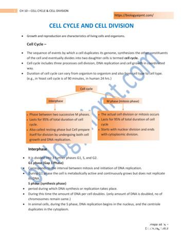
of the cell and eventually divides into two daughter cells is termed cell cycle. Cell cycle includes three processes cell division, DNA replication and cell growth in coordinated way. Duration of cell cycle can vary from organism to organism and also from cell type to cell type. (e.g., in Yeast cell cycle is of 90 minutes, in human 24 hrs.)
Part 6: Modeling the cell cycle in a normal cell Part 7: Modeling the cell cycle in a cancer cell Living Environment Major Understandings: Gene mutations in a cell can result in uncontrolled cell division, called cancer. Exposure of cells to certain chemicals and radiation increases mutations and thus increases the chance of cancer.
1. Teaching with a Multiple-Perspective Approach 8 . 2. Description of Perspectives and Classroom Applications 9 . 2.1 Scientific Perspective 9 . 2.2 Historical Perspective 10 . 2.3 Geographic Perspective 11 . 2.4 Human Rights Perspective 12 . 2.5 Gender Equality Perspective 13 . 2.6 Values Perspective 15 . 2.7 Cultural Diversity Perspective 16
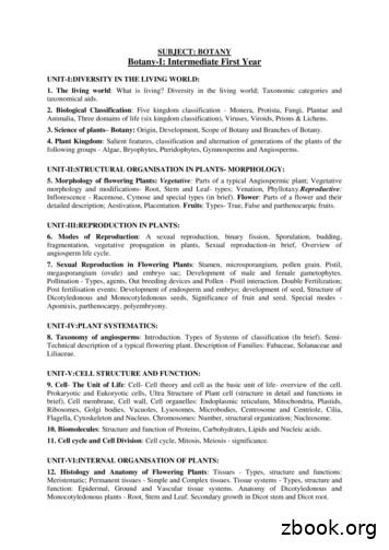
UNIT-V:CELL STRUCTURE AND FUNCTION: 9. Cell- The Unit of Life: Cell- Cell theory and cell as the basic unit of life- overview of the cell. Prokaryotic and Eukoryotic cells, Ultra Structure of Plant cell (structure in detail and functions in brief), Cell membrane, Cell wall, Cell organelles: Endoplasmic reticulum, Mitochondria, Plastids,
Cancer Cell Basics Difference between cancer cell and normal cell: Normal cell: Knows and stays in its place of origin Knows when to replicate and when to die 2009 Nobel Prize – telomeres help determine longevity Cancer cell: Does not know when to stop growing and proliferating Can travel (metastasize) from organ of origin to any place within the body
Non-small cell lung cancer is the most common type of lung cancer and accounts for 84% of cases. There are different types of non-small cell lung cancer, including: Adenocarcinoma - a cancer that forms in the outer parts of the lung. Squamous cell carcinoma - a cancer that forms from a cell lining the airway.
What does skin cancer look like? There are many different types of skin cancer (such as melanoma and basal cell skin cancer). Each type looks different. Also, skin cancer in people with dark skin often looks different from skin cancer in people with fair skin. A change on the skin is the most common sign of skin cancer. This may


