3D Cell Culture Protocol SeedEZ - Cell Seeding - LENA BIOSCIENCES
SEEDEZ PROTOCOLS USER GUIDELINES AND PROTOCOLS FOR THE SEEDING OF THREE-DIMENSIONAL CELL CULTURES INTO THE SEEDEZ support@lenabio.com www.lenabio.com
SEEDEZ IS A TRADEMARK OF LENA BIOSCIENCES, INC. FOR IN VITRO RESEARCH USE ONLY. NOT FOR USE IN DIAGNOSTIC OR THERAPEUTIC PROCEDURES. NOT FOR USE IN ANIMALS OR HUMANS. SEEDEZ IS PROTECTED BY ONE OR MORE ISSUED PATENTS AND PATENT PENDING APPLICATIONS. PURCHASE DOES NOT INCLUDE OR IMPLY ANY RIGHT TO RESELL THIS PRODUCT EITHER AS A STAND-ALONE PRODUCT OR AS A COMPONENT OF ANY OTHER PRODUCT, EITHER IN PART OR AS A WHOLE. A LICENSE IS REQUIRED TO USE THIS PRODUCT IN COMMERCIAL FOR FEE SERVICES. INFORMATION AND DATA CONTAINED IN THIS DOCUMENT ARE FOR GUIDANCE ONLY AND SHOULD NOT BE CONSTRUED AS A WARRANTY. ALL IMPLIED WARRANTIES ARE EXPRESSLY DISCLAIMED. ALL BUYERS OF THE PRODUCT ARE RESPONSIBLE FOR ENSURING THAT IT IS SUITABLE FOR THEIR END USE. THIS PUBLICATION IS A PUBLICATION OF LENA BIOSCIENCES, INC. THE INFORMATION AND DATA CONTAINED IN THIS DOCUMENT ARE SUBJECT TO CHANGE WITHOUT PRIOR NOTICE. WHILE EVERY PRECAUTION HAS BEEN TAKEN IN PREPARING THIS DOCUMENT, LENA BIOSCIENCES ASSUMES NO RESPONSIBILITY FOR ERRORS, OMISSIONS OR DAMAGES RESULTING FROM THE USE OF DATA AND INFORMATION CONTAINED IN THIS DOCUMENT. NO PART OF THIS PUBLICATION MAY BE REPRODUCED, TRANSMITTED, OR USED IN ANY OTHER MATERIAL IN ANY FORM OR BY ANY MEANS WITHOUT PRIOR WRITTEN PERMISSION FROM LENA BIOSCIENCES. 2018 LENA BIOSCIENCES, INC. ALL RIGHTS RESERVED.
Plating of 3D Cell Cultures Into the SEEDEZ December 2018 TABLE OF CONTENTS INTRODUCTION 2 USER GUIDELINES AND RECOMMENDATIONS 2 Mixed cell cultures 2 Adherent cells 2 Cells in an extracellular matrix or gel 2 Cell seeding in a sol-state gel suspension 2 Seeding of invasive cells on top of an ECM barrier for invasion assays or other uses 3 Cell seeding in a “sandwich” between two layers of extracellular matrix 3 Three-dimensional feeder layer 3 Stack-And-Culture 4 SEEDEZ -SANDWICH cultures 4 Multilayered tissue models, SEEDEZ -STACK 4 Side-By-Side Culture 4 Migration, invasion, chemo-invasion and angiogenesis assays 5 SEEDING METHODS AND OPTIMAL DISPENSING VOLUME 5 Spot-A-Culture 5 EXAMPLE 1: EXAMPLE 2: EXAMPLE 3: Matrigel spots dispensed using a positive displacement micropipette 6 Matrigel spots dispensed using a standard laboratory micropipette 7 Spread of high protein Matrigel from two different lots 8 Optimal dispensing volume for spot cultures 8 How to determine optimal dispensing volume for your application? 9 What to expect? 9 EXAMPLE 4: The spread of 25 µl MatrigelTM spots in SC-C048 SEEDEZ substrate 10 Wick-A-Culture/ Dip-And-Culture 10 PROTOCOL: The seeding of 3D mixed cultures into Poly-D-Lysine coated SEEDEZ 12 Poly-D-Lysine (PDL) coating 12 Maintenance and dissociation of P0-harvested mixed glial cells prior to seeding 13 Seeding 14 1-Week culturing results 15
PROTOCOL: The seeding of 3D mixed cultures in an extracellular matrix into uncoated SEEDEZ 17 Matrigel aliquoting and storage of single use vials 17 Maintenance and dissociation of P0-harvested mixed glial cells prior to seeding 18 Seeding 19 2-Week culturing results 21 PROTOCOL: The seeding of adherent cells in a diluted ECM into uncoated and coated SEEDEZ 23 Matrigel aliquoting and storage of single use vials 23 Poly-D-Lysine coating 23 Maintenance and dissociation of P0-harvested glial cells prior to seeding 24 Seeding 25 1-Day culturing results 26 10-Day culturing results in a serum-free medium followed by starving 28 SeedEZTM Cell Seeding Protocols 1
INTRODUCTION Cells seeded into the SEEDEZ may be of various origins and sources. Cells may be primary cells, secondary cells and cell lines. Please continue to use protocols normally used for cell maintenance prior to seeding. If cell lines or secondary cells are used, maintain cells in flasks until they are ready to be seeded into the SEEDEZ . USER GUIDELINES AND RECOMMENDATIONS To prepare cells for seeding, continue with protocols used for plating cells. This may include cell dissociation from a flask, followed by centrifuging and the re-suspension of cell pellet in a medium in which cells will be seeded into the SEEDEZ . Count live and dead cells using automatic counters, or hemocytometer and Trypan Blue exclusion. Depending on the equipment available, seed cells into the SEEDEZ using micropipettes, automated pipettes, dip-in or wick-in method as shown in Lena Biosciences video tutorials. Please visit www.lenabio.com for more information. MIXED CELL CULTURES To seed mixed cell cultures into the SEEDEZ , resuspend each cell type separately and then join them together prior to seeding. Always have in mind what are desired total live cell density and the ratio of heterogeneous cell types at seeding. ADHERENT CELLS To seed adherent cell types, pre-coat SEEDEZ with cell adhesive molecules and follow protocols used for the coating of cell ware disposables. You may also seed cells in a diluted or undiluted sol-state extracellular matrix (ECM) or hydrogel into uncoated SEEDEZ , or you may pre-coat SEEDEZ and then seed cells in undiluted or diluted ECM or hydrogel. CELLS IN AN EXTRACELLULAR MATRIX OR GEL If you previously worked with 3D gel-based cell cultures or extracellular matrix barriers, continue with protocols used for cell seeding in a gel you commonly use. The following are the most common seeding methods: A. Cell seeding in a sol-state gel suspension. B. The seeding of invasive cells on top of an extracellular matrix barrier for invasion assays. C. Cell seeding in a “sandwich” between, for example, a layer of Collagen I and a layer of Matrigel. SEEDEZ allows for cell seeding using all three approaches. CELL SEEDING IN A SOL-STATE GEL SUSPENSION Cells may be seeded in a sol-state gel suspension; or SEEDEZ may be coated with cell adhesive ligands or extracellular matrix (ECM) constituents, followed by addition of a cell suspension in a sol-state gel or ECM. SeedEZTM Cell Seeding Protocols 2
SEEDING OF INVASIVE CELLS ON TOP OF AN ECM BARRIER FOR INVASION ASSAYS OR OTHER USES If the objective is to seed invasive cells on top of the extracellular matrix (ECM) barrier, dispense sol-state ECM into the SEEDEZ first, let the ECM gel and then add cells. In this approach cells sit on top of the ECM at the start of experiment. You may also seed cells in a sol-state ECM suspension into the SEEDEZ . In contrast with previous approach, this provides for truly 3D cell migration/invasion assay, representative of in vivo conditions, in which cells are embedded in the extracellular matrix (ECM) from the assay start to its end. CELL SEEDING IN A “SANDWICH” BETWEEN TWO LAYERS OF EXTRACELLULAR MATRIX If you previously used “sandwich” cultures, you may have had only one cell layer between two layers of extracellular matrix at plating. If SEEDEZ is used, 3D cell culture will be truly 3D at seeding, comprising multiple layers of cells embedded in a 3D matrix. To achieve this, you may first coat the SEEDEZ with chosen molecules (Collagen I, for example) and then dispense cells in a sol-state ECM suspension such as Matrigel. Alternatively, you may use two SEEDEZ substrates comprising gelled extracellular matrix to seed cells in a sandwich between the substrates, or you may seed cells in the third SEEDEZ substrate sandwiched by two SEEDEZ substrates. THREE-DIMENSIONAL FEEDER LAYER SEEDEZ is a versatile tool which allows to plate different cell types at a user convenience and in a desired sequence. For example, you may seed one cell type first and then add a different cell type. Different cell types may also be seeded at the same time and one cell type may be a feeder cell. This is important for culturing of difficult to culture cell types, while still providing for metabolic support by way of feeder cells which are typically cultured in 2D. Feeder cells may be, for example, astrocytes supporting neurons or inactivated fibroblasts metabolically supporting stem cells. The SEEDEZ is unique; it enables to add three-dimensionality even to a feeder cell layer while culturing or co-culturing other cells in 3D. To provide for 3D-distributed feeder cells in a co-culture, or to provide for conditioned medium for another 3D culture of dissociated cells or cell aggregates, one of the following approaches may be used: A. Non-contact co-culture method Coat a SEEDEZ substrate, if needed, and seed feeder cells. Coat another SEEDEZ substrate, if needed, and seed fastidious cells or cell aggregates. Place the first SEEDEZ substrate in a culture well. Place the second SEEDEZ substrate in a well insert in the same well at a desired point in time. B. Contact co-culture method Coat the SEEDEZ , if needed, and seed feeder cells. Next, seed fastidious cells or cell aggregates at a time you choose. Alternatively, coat SEEDEZ , and seed fastidious cells / cell aggregates together with feeder cells. For contact co-culturing in a single SEEDEZ substrate with the feeder cells seeded first, followed by seeding of fastidious cells, you may spot cultures side-by-side or spot-a-secondculture on top of the first culture spot. THE SEEDEZ SUBSTRATE SHOULD NOT BE SATURATED WHEN YOU ARE ADDING THE SECOND CULTURE SPOT. Alternatively, feeder cells may be seeded in one SEEDEZ substrate and fastidious cells in another SEEDEZ substrate and then overlapped. SeedEZTM Cell Seeding Protocols 3
C. Conditioned medium Coat a SEEDEZ substrate, if needed, and seed feeder cells. Coat another SEEDEZ substrate, if needed, and seed fastidious cells or cell aggregates. Keep the substrates in separate wells. Add conditioned medium to the well seating the second substrate using an amount of medium from the well seating the first substrate. If you wish to keep feeder layers planar, you may still do so. First, plate the feeder layer. Next, coat the SEEDEZ substrate if needed, and seed difficult to culture cell types. At a desired time, place the SEEDEZ substrate over the planar feeder layer (contact co-culture) or place it in an insert in the same well (noncontact co-culture). STACK-AND-CULTURE 3D cell cultures embedded in different SEEDEZ substrates may be overlapped. To do this, simply place one or more SEEDEZ substrates on top of each other. You may overlap cultures to the extent you wish, and you may overlap them at any time. IF CULTURES ARE NOT BROUGHT TOGETHER AT THE TIME OF CELL SEEDING AND THE OBJECTIVE IS TO STUDY CELL MIGRATION, INVASION, CHEMO-INVASION OR ANGIOGENESIS, YOU MAY NEED TO ADD A LAYER OF EXTRACELLULAR MATRIX BETWEEN THE SEEDEZ SUBSTRATES TO PROVIDE FOR GOOD SUBSTRATE-TO-SUBSTRATE ADHESION. SEEDEZ -SANDWICH CULTURES You may seed cells in a sol-state gel between two SEEDEZ substrates; the two SEEDEZ substrates may contain gel and/or cells. Alternatively, seed cells in a SEEDEZ substrate such that the SEEDEZ is sandwiched between two layers of gel, a layer of gel and another SEEDEZ substrate, or between two SEEDEZ substrates. MULTILAYERED TISSUE MODELS, SEEDEZ -STACK You may use a stack of SEEDEZ substrates to model tissues normally comprising multiple layers; a neocortex, or cerebral cortex for example. Each tissue layer typically comprises layer-specific cell types and multiple layers of cells all of which may be mimicked by a culture in the SEEDEZ substrate. Use one SEEDEZ substrate to model a tissue layer. Remember this is a tissue layer, not a layer of cells, which you may mimic by a 3D cell culture in the SEEDEZ . Next, place the SEEDEZ substrates on top of each other in the order in which the tissue layers are organized in the tissue. You may also add “tissue layers” using cells or cells in an extracellular matrix seeded between the SEEDEZ substrate(s), or above and/or below the SEEDEZ substrate(s), as necessary. SIDE-BY-SIDE CULTURE Side-by-side culturing refers to a method of co-culturing in which a user may spot a culture, followed by spotting of another cell culture in the same SEEDEZ substrate. This allows to add one or more cell subpopulations to a cell population, or to seed different cell populations for the purpose of modeling tissue heterogeneity in health or disease; for example, normal, neoplastic and tumor cell mass. Cell populations and sub-populations may include cells from various origins and sources and may be from donors of various ages and/or stages of disease progression. Cells may be normal cells, diseased cells, cancer cells, stem cells etc. TO ACHIEVE THE MOST CONSISTENT AND REPRODUCIBLE RESULTS, ALL CELL POPULATIONS AND SUB-POPULATIONS SHOULD BE SEEDED FIIRST AND THEN MEDIUM ADDED. SeedEZTM Cell Seeding Protocols 4
MIGRATION, INVASION, CHEMO-INVASION AND ANGIOGENESIS ASSAYS SEEDEZ allows you to tailor cell migration, invasion, chemo-invasion and angiogenesis assays to your experimental goals and research objectives. It also provides for greater experimental flexibility; flexibility which cannot be achieved using a fixed insert, with a fixed membrane, and with a fixed extracellular matrix barrier. While this is not the most extensive list of all SEEDEZ uses in this context, SEEDEZ may be used to enable or provide one or more of the following: A. Control of extracellular matrix (ECM) composition or gel concentration in one or more SEEDEZ substrates. B. Addition and embedding of a test compound into the SEEDEZ . C. Addition of a test compound to a sol-state ECM followed by embedding into the SEEDEZ . Test compounds may be matrix metalloproteinase inhibitors, angiogenic inhibitors, actin polymerization inhibitors, chemokines, chemoattractants, or other modulators of cell motility, or another cell function. D. Seeding of different cell types into the same or different SEEDEZ substrates with the objective of joining them; for example, the seeding of invasive cells in one substrate and the seeding of normal cells in another substrate with or without extracellular matrix. E. Seeding of cells ECM test compound into the SEEDEZ . F. Addition of extracellular matrix on top or below the SEEDEZ substrate(s). G. “Spotting” of different cultures or spotting of different cell types in the same SEEDEZ substrate. For example, spot invasive cells and spot non-invasive cells in an extracellular matrix into a SEEDEZ substrate. H. Spot-a-CultureTM and Spot-a-DrugTM in the same or different SEEDEZ substrate. I. Development of custom assays by overlapping SEEDEZ substrates. IF THE OBJECTIVE IS TO ALLOW CELLS FROM ONE SEEDEZ SUBSTRATE TO INVADE OR MIGRATE INTO THE ABOVE OR BELOW SEEDEZ SUBSTRATE, THEN THE ADHESIVE LAYER BETWEEN THE SEEDEZ SUBSTRATES SHOULD BE A LAYER OF EXTRACELLULAR MATRIX USED IN ONE OR BOTH SUBSTRATES. IF THE OBJECTIVE IS TO ALLOW A TEST COMPOUND FROM ONE SEEDEZ SUBSTRATE TO DIFFUSE INTO ANOTHER SEEDEZ SUBSTRATE CONTAINING CELLS IN A HYDROGEL, THEN REDUCE OR OMIT ADDITION OF CULTURE MEDIUM TO THE WELL DURING SHORT-TERM DRUG DIFFUSION STUDIES IN A HUMIDIFIED INCUBATOR. SEEDING METHODS AND OPTIMAL DISPENSING VOLUME You may use three seeding approaches to embed any cells in any sol state suspension into the SEEDEZ : A. Spot-a-CultureTM B. Wick-a-CultureTM C. Dip-and-CultureTM SPOT-A-CULTURE SeedEZTM Cell Seeding Protocols 5
You may spot cultures using any micropipette available. However, when dispensing cells in a sol-state gel, and viscous or otherwise difficult to dispense reagents, you may want to consider using a positive displacement micropipette to reduce losses in pipetting and to obtain the most consistent results. The following examples show how proteinaceous Matrigel extracellular matrix spots look like when dispensed using both a positive displacement micropipette (see Example 1) and a standard laboratory micropipette (see Example 2). Lena Biosciences video tutorials also teach how easy it is to plate consistent cultures even when these cultures are dispensed in a Matrigel extracellular matrix sol-state suspension. EXAMPLE 1: MICROPIPETTE MATRIGEL SPOTS DISPENSED USING A POSITIVE DISPLACEMENT A B 16 mg/ml Matrigel 16 mg/ml Matrigel 8 mg/ml Matrigel 8 mg/ml Matrigel Fig. 1 Growth factor reduced (GFR) Matrigel at a final concentration of 16 mg/ml and 8 mg/ml dispensed using a hand-held positive displacement micropipette (Gilson Microman). A Front view of the samples. B Back view of the samples. Properly chilled Matrigel cell suspension is difficult to plate consistently in 3D and in high-throughput onto standard cell ware disposables. Typically, cultures spread during pipetting and transfer to incubator before Matrigel gels. Certain coatings such as Poly-D-Lysine make Matrigel cell cultures spread even more. Example 1 and Example 2 demonstrate that consistent Matrigel cultures can be formed with ease using SEEDEZ even without automated dispensing equipment. Formed cell cultures are three-dimensional, consistent, reproducible, and suitable for vigorous handling, as they are wicked by and self-contained in respective SEEDEZ substrates. Matrigel is an extracellular matrix comprising structural proteins such as Laminin, Collagen IV, and Entactin which present cultured cells with adhesive peptide sequences they often encounter in their natural environment. When using Matrigel , a common laboratory procedure is to dispense small volumes of chilled (4 C) Matrigel onto plastic tissue culture labware, or SEEDEZ , using frozen pipette tips. This is because Matrigel is a thermo-reversible hydrogel which is in sol state at 4 C, and gels at higher temperatures. If Matrigel is not chilled enough, then it may start gelling before it fully permeates the SEEDEZ substrate (see Fig. 1A, the first substrate in the first row); however, even in such state, the viscous solution permeated the SEEDEZ . In contrast, without the SEEDEZ , the shape and characteristic dimensions of 3D cell culture is difficult to reproduce culture-to-culture if plated onto standard labware under this condition. SeedEZTM Cell Seeding Protocols 6
EXAMPLE 2: MICROPIPETTE A Fig. 2 MATRIGEL SPOTS DISPENSED USING A STANDARD LABORATORY B Growth factor reduced (GFR) Matrigel at a final concentration of 8 mg/ml dispensed using a standard laboratory micropipette and standard pipette tips. A Front view of the samples. B Back view of the samples. For a fixed dispensing volume, the reproducibility and the spread of culture spots, gel spots, or drug spots in the SEEDEZ depend on chemical and physical properties of dispensed reagent, and its sensitivity to environmental parameters and their temporal and spatial variations, among other factors. For the most consistent results, please examine the specification sheet and the application requirements for the reagent you are dispensing into the SEEDEZ . For Matrigel, manufacturer recommends use of frozen pipette tips and cell ware disposables to prevent sol-state ECM gel from gelling prior to and during dispensing. As can be seen from the Example 1 and Example 2, properly chilled Matrigel (4 C) yields reproducible spots even when dispensed using standard laboratory micropipette and standard pipet tips, if delivered into the SEEDEZ , and even when only pipet tips and not the SEEDEZ were pre-chilled. Batch-to-batch variations in the composition of a reagent you wish to spot may also contribute to spot variability. While SEEDEZ makes these differences less apparent and produces geometrically more consistent cultures, the cultures will not be identical at seeding if the composition of a suspension in which cells are seeded is changed. For example, culture spread may be greater or smaller than what you found in the previous reagent batch for otherwise identical conditions at seeding. If Matrigel is used, slight variations in composition may be present batch to batch. To investigate this possibility, Growth Factor Reduced Matrigel from two different lots was tested, as shown in the Example 3. However, significant differences in Matrigel spread, if dispensed into the SEEDEZ were not observed. For a given volume of dispensed solution, factors which yield the greatest variations in culture dimensions (in x, y and z) and cell distribution across these dimensions at seeding are: A. Losses in pipetting. B. Relative change in position of the pipet tip from the center of the substrate. * *In the Examples 1-3, Matrigel was dispensed manually with the pipet tip touching approximately the center of the SEEDEZ substrates. SeedEZTM Cell Seeding Protocols 7
EXAMPLE 3: SPREAD OF HIGH PROTEIN MATRIGEL FROM TWO DIFFERENT LOTS Matrigel: Lot A A Frozen SeedEZ (kept at -20 oC) prior to Matrigel dispensing Room temperature SeedEZ (25 oC) prior to Matrigel dispensing B Matrigel: Lot B Frozen SeedEZ Kept at -20oC Room temperature SeedEZ at 25oC Matrigel was dispensed using a hand-held positive displacement micropipette, approximately near the center of the SeedEZ substrates. The dispensed volume was approximately 110% of the total substrate volume or approximately 120% of its void volume. Fig. 3 A. B. Reproducibility of Matrigel spread in the SeedEZ substrates. The spread of high protein concentration, 16 mg/ml GFR Matrigel into the SeedEZ as a function of Matrigel lot and the SeedEZ substrate temperature. Close-up view of the SeedEZ substrates showing Matrigel spread from Lot A as a function of the SeedEZ substrate temperature prior to Matrigel plating. 3D CELL CULTURES WILL BE MORE CONSISTENT, THAT IS, THE CULTURES WILL BE OF MORE CONSISTENT SHAPE, SPREAD, THICKNESS AND CELL DISTRIBUTION IN X, Y AND Z DIMENSIONS, CULTURE-TO-CULTURE AND BATCH-TO-BATCH IF YOU USE SEEDEZ FOR ALL YOUR 3D CELL CULTURE NEEDS. OPTIMAL DISPENSING VOLUME FOR SPOT CULTURES You may spot any volume of cells in any sol-state suspension. However, perfectly circular culture spots are obtained only when the dispensed volume is approximately twice the void volume of the SEEDEZ substrate. For spot cultures plated into the SEEDEZ SC-C048 substrate this volume is approximately 50 µl. An optimal dispensing volume is the volume that meets both of the following conditions: 1. A volume necessary to entirely wet and saturate the SEEDEZ substrate. 2. A volume which is after dispensing self-contained within the SEEDEZ substrate. SeedEZTM Cell Seeding Protocols 8
The first condition provides for a 3D cell culture in which cells are distributed throughout the SEEDEZ substrate. The second condition saves cells and reagents by minimizing the amount of cell suspension that leaves the SEEDEZ substrates if the substrate was over-saturated by it. LENA BIOSCIENCES MAKES NO WARRANTIES THAT 50 µL IS AN OPTIMAL DISPENSING VOLUME FOR SC-C048 SEEDEZ SUBSTRATE. THIS VALUE DEPENDS ON CHEMICAL AND PHYSICAL PROPERTIES OF THE REAGENT, MICROPIPETTE USED AND LOSSES IN PIPETTING, THE SEEDEZ COATING (IF ANY), THE SURFACE TREATMENT AND COATING (IF ANY) OF THE CELLWARE IN WHICH THE SEEDEZ IS PLACED, AMONG OTHER FACTORS. YOU MAY NEED LOWER OR HIGHER DISPENSING VOLUME THAN 50 µL TO CONSISTENTLY SATURATE SEEDEZ SC-C048 SUCH THAT DISPENSED VOLUME IS SELF-CONTAINED BY THE SUBSTRATE. HOWEVER, IT IS RECOMMEND THAT 50 µL BE USED AS A STARTING POINT FOR SC-C048. HOW TO DETERMINE OPTIMAL DISPENSING VOLUME FOR YOUR APPLICATION? To find optimal dispensing volume for your application, we recommend the following procedure: 1. Dispense 50 µl of reagent you will be using at cell seeding in each of the six SC-C048 SEEDEZ replicates. Do so in the same environment in which you will be plating cells, using the same micropipette, cell disposable in which SEEDEZ will be placed, and using the same coating applied to SEEDEZ prior to cell seeding, if any. 2. If 50 µl saturates SC-C048 SEEDEZ substrate but results in excess liquid surrounding the substrate, reduce dispensing volume to 40 µl and test it with six SEEDEZ replicates. 3. If 40 µl saturates the substrates and you cannot see excess liquid when you raise the SEEDEZ substrate using tweezers, then use 40 µl as your dispensing volume. 4. Repeat step 3 using cell suspension. You may need to adjust dispensing volume one more time. The procedure may need to be repeated more than twice to ensure consistency or to further reduce dispensing volume by repeating steps 1-4 in 5 µl increments. If the substrates are not fully saturated with 50 µl in step 1, increase dispensing volume to 60 µl in step 2. If the substrates are still not saturated, repeat steps 1-3 by increasing dispensing volume by 10 µl each time until the substrates are fully saturated, or until you see excess liquid. You may need to repeat this procedure more than twice to ensure consistency or to optimize dispensing volume in 5 µl increments. ALTERNATIVE SOLUTION FOR DIFFICULT TO DISPENSE REAGENTS: IF YOUR REAGENT IS DIFFICULT TO DISPENSE WITHOUT SIGNIFICANT LOSSES IN PIPETTING, USE WICK-IN OR DIP-IN METHOD. THE SEEDEZ WICKS MOST SOL-STATE GELS AND CELL CULTURE REAGENTS, AND PRODUCES CONSISTENT CULTURES, GEL SHAPES, OR DOSE FORMS IN HIGH-THROUGHPUT WITHOUT EXPENSIVE EQUIPMENT OR COMPLEX PROTOCOLS. WHAT TO EXPECT? For the majority of cell suspensions and other reagents normally used in cell culture applications, dispensing volume less than the optimal volume produces an elliptically shaped, rather than a circularly shaped spot. However, the shape of the spot is reasonably consistent across the SEEDEZ substrates so long as the SeedEZTM Cell Seeding Protocols 9
dispensed volume is constant and losses in pipetting negligible; this makes 3D cultures and side-by-side 3D cultures consistent even if they are not perfectly circular. As can be seen in the Example 4, the shape of 25 µl spot is elliptical, but consistent (dispensing was done manually without precise control over the position of the pipet tip relative to that of the center of the substrate). However, the closer the dispensed volume to the optimal dispensed volume, the more circular the spot becomes. EXAMPLE 4: SUBSTRATE THE SPREAD OF 25 µL MATRIGEL T M SPOTS IN SC-C048 SEEDEZ A B Measured spread of 25 µl of 16 mg/ml Matrigel Vertical axis: The average diameter of spread footprint in mm SeedEZ at 25oC SeedEZ at -20 oC prior to Matrigel plating prior to Matrigel plating C 25 µl of high-protein concentration GFR Matrigel was dispensed using a hand-held positive displacement micropipette, approximately near the center of SeedEZ substrates. Matrigel spread was measured using calipers with three measurements per spread. For both sample conditions, the Matrigel spread in SeedEZ SC-C048 was statistically indistinguishable at diameters of 8.11 0.71 mm and 8.09 0.72 mm, for the SeedEZ samples kept at 25 oC and -20 oC, respectively, prior to gel plating. Fig. 4 The spread of Matrigel spots for sub-optimal dispensing volume as a function of the SC-C048 temperature. A. 16 mg/ml Matrigel spots for sub-optimal (25 µl) dispensing volume. B. Measured Matrigel spread as a function of the SeedEZ substrate temperature. C. Close-up view of elliptically shaped air-dried Matrigel spots. WICK-A-CULTURE/ DIP-AND-CULTURE If you are making a large number of 3D cell cultures using dissociated cells, and you do not have automated equipment or a multichannel pipette, use the wick-in or dip-in seeding method. Wick-a-culture refers to a cell seeding method in which the SEEDEZ is placed in contact with the sol-state cell suspension, which the SEEDEZ wicks until it is saturated as shown in Fig. 5A. Dip-in seeding method is analogous to the wick-in method with the exception that the SEEDEZ substrate is fully immersed into the cell suspension. Regardless of whether the SEEDEZ was partly or completely immersed into the cell suspension, for rapid seeding of many substrates you may dip them all at once; in a reagent reservoir, for example. This seeding SeedEZTM Cell Seeding Protocols 10
approach makes reproducible and consistent 3D cell cultures (Figs. 5B-5C) easier and faster, and without expensive equipment or complex protocols. A B C Fig. 5 Wick-a-Culture and Dip-and-Culture method. A. Wick-a-Culture seeding method using
CELL SEEDING IN A "SANDWICH" BETWEEN TWO LAYERS OF EXTRACELLULAR MATRIX If you previously used "sandwich" cultures, you may have had only one cell layer between two layers of extracellular matrix at plating. If SEEDEZ is used, 3D cell culture will be truly 3D at seeding, comprising multiple layers of cells embedded in a 3D matrix.
2 Cell culture techniques A.R. BAYDOUN 2.1 Introduction 2.2 The cell culture laboratory and equipment 2.3 Safety considerations in cell culture 2.4 Aseptic techniques and good cell culture practice 2.5 Types of animal cell, characteristics and maintenance in culture 2.6 Stem cell culture 2. 7 Bacterial cell culture
cell biologist to maintain the integrity of the micro-scale . in vivo. cell environment within simple . in vitro. models. Summary: changing cell culture environment impacts on cell behaviour . Traditional 2D Cell Culture Alvetex 3D Cell Culture Normal . In Vitro. Environment General dimensions and physical differences
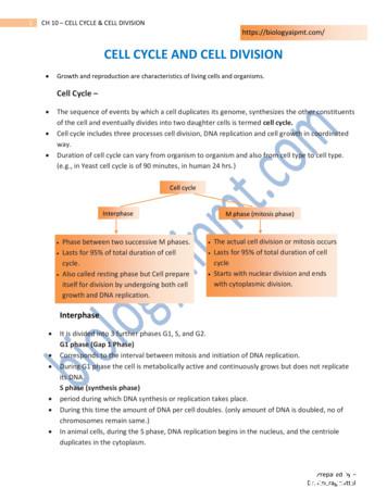
of the cell and eventually divides into two daughter cells is termed cell cycle. Cell cycle includes three processes cell division, DNA replication and cell growth in coordinated way. Duration of cell cycle can vary from organism to organism and also from cell type to cell type. (e.g., in Yeast cell cycle is of 90 minutes, in human 24 hrs.)
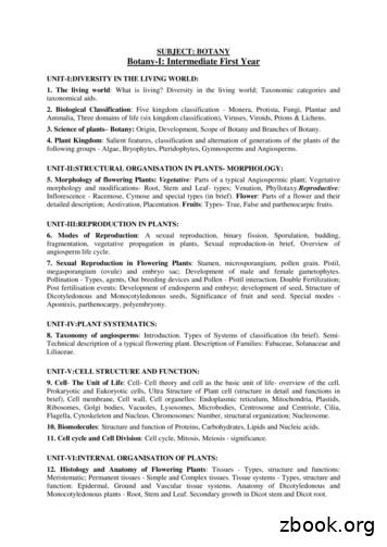
UNIT-V:CELL STRUCTURE AND FUNCTION: 9. Cell- The Unit of Life: Cell- Cell theory and cell as the basic unit of life- overview of the cell. Prokaryotic and Eukoryotic cells, Ultra Structure of Plant cell (structure in detail and functions in brief), Cell membrane, Cell wall, Cell organelles: Endoplasmic reticulum, Mitochondria, Plastids,
EGP Exterior Gateway Protocol OSPF Open Shortest Path First Protocol IE-IRGP Enhanced Interior Gateway Routing Protocol VRRP Virtual Router Redundancy Protocol PIM-DM Protocol Independent Multicast-Dense Mode PIM-SM Protocol Independent Multicast-Sparse Mode IGRP Interior Gateway Routing Protocol RIPng for IPv6 IPv6 Routing Information Protocol PGM
The RAFT 3D Cell Culture System. A) Cell/collagen mix within a microplate well. B) Specialized RAFT plate showing absorbers. C) Absorbers remove medium to concentrate the cell/collagen mix. D) Concentrated cell/ collagen mix. E) Cell/collagen layer showing absorbed medium. F) Final cell/collagen layer in the microplate well prior to
The Cell Cycle The cell cycle is the series of events in the growth and division of a cell. In the prokaryotic cell cycle, the cell grows, duplicates its DNA, and divides by pinching in the cell membrane. The eukaryotic cell cycle has four stages (the first three of which are referred to as interphase): In the G 1 phase, the cell grows.
2. AngularJS looks in the template for the ngApp directive which designates our application root. 3. Loads the module associated with the directive. 4. Creates the application injector 5. Compiles the DOM treating the ngApp directive as the root of the compilation AngularJS: beginner's Guide - part 1


