Why? Model 1 – Cell Mediated Response
ImmunityHow does our immune system protect us from disease?Why?One way in which organisms maintain homeostasis is by detecting foreign cells and particles like pathogens and cancer cells. Once the pathogen is detected and identified, other systems in the organism’s bodycan attack the invader, thus keeping the organism healthier. Cells of the human immune system are finelytuned to recognize and respond quickly to disease-causing organisms.Model 1 – Cell Mediated Response12ThTh3ImmunityTh41
1. In Model 1 a pathogen (virus, bacteria, foreign protein, parasite) has entered the bloodstream ofan individual. Draw the symbol that represents the pathogen.2. One response of the human immune system is endocytosis of a pathogen by a phagocyte (a typeof white blood cell). Refer to Model 1.a. Which diagram in the cell mediated response illustration shows this process?b. Draw the symbol that represents the phagocyte.3. Another type of white blood cell that is involved in the cell mediated response is a helper T-cell.a. Draw the symbol that represents the helper T-cell in Model 1.b. In your drawing above, circle the specialized surface proteins on the helper T-cell.4. According to Model 1, are all helper T-cells the same? Justify your answer with specific evidencefrom Model 1.5. The following statements are labels for the cell mediated process in Model 1. A piece of the pathogen is presented on the surface of the phagocyte.The helper T-cell disperses a chemical signal to activate other immune response systems.The helper T-cell binds to the piece of pathogen presented on the phagocyte.Pathogen is broken apart by chemicals in the phagocyte.a. Determine the order of the statements by referring to Model 1.b. With your group, decide where each statement belongs in the diagram and label Model 1appropriately. Note that more than one statement may match a diagram and some diagramsmay not match any statement.6. According to Model 1, do the helper T-cells interact with the free pathogens in the blood?2POGIL Activities for AP* Biology
Read This!The pieces of pathogen that are presented on the surface of a cell are called antigens. Cells that presentantigens on their surface, such as the phagocyte in Model 1, are called antigen-presenting cells (APC).They activate helper T-cells. After being activated by an antigen, the helper T-cell will begin a phase ofrapid cell division. The resulting daughter cells may be effector Th cells or memory Th cells. The memoryTh cells will stay in the body for several years, ready to respond to the pathogen if it should ever infiltratethe body again.7. Do all types of helper T-cells bind to all antigens? Justify your answer with specific evidence fromModel 1.8. Add labels to Model 1 for the “antigen” and “antigen-presenting cell.”Model 2 – Humoral Response1BImmunityBB23453
9. B-cells are a third type of white blood cell that is involved in immunity.a. Draw the symbol that represents a B-cell in Model 2.b. What is the name of the immune system response that involves B-cells?10. According to Model 2, are all B-cells the same? Justify your answer with specific evidence fromModel 2.11. In diagram 2 of Model 2, binding occurs between an antigen and a B-cell. How is this interaction different from the binding that occurs between antigens and helper T-cells?12. Consider diagrams 3–5 in Model 2. Write descriptions similar to those in Question 5 for each ofthese steps in the humoral response.13. Is a B-cell an antigen-presenting cell? Justify your reasoning.14. According to Model 2, do B-cells ever interact with pathogens that have infected a cell? Justifyyour reasoning with specific evidence from Model 2.15. Predict what would happen if a B-cell like the one shown in diagram 5 of Model 2 were to runinto a helper T-cell like the ones in Model 1.4POGIL Activities for AP* Biology
Model 3 – Adaptive Immunity16. Label the following items in Model 3.PathogenB-CellHelper T-cell17. Describe the interaction between the B-cell and the helper T-cell in Model 3.18. When a B-cell is activated by the interaction with a helper T-cell it begins to produce and disperse antibodies.a. Draw the symbol that represents an antibody in Model 3.b. According to Model 3, what does the antibody do to pathogens?19. How might the interaction between the antibody and pathogens affect the pathogen’s ability toinfect its host?Read This!After the B-cell is activated by the helper T-cell, the B-cell enters a phase of rapid cell division. Some ofthe daughter cells become plasma cells that make even more antibody molecules, some reaching a rate of2,000 molecules per second. The other daughter cells become memory B-cells that will stay in the bodyfor several years, ready to respond to the pathogen if it should ever enter the body again.Immunity5
Model 4 – Immune Response to a Pathogen90Amount of the Antibody Present8070605040302010First Encounterwith thePathogen!"0TimeSecond Encounterwith the Same Pathogen20. What does the y-axis of the graph in Model 4 represent?21. How many times did the organism in Model 4 encounter the same pathogen?22. Using Model 4, compare the amount of the antibody generated by the B-cells after the firstencounter with the antigen to the amount of the antibody generated in the second encounterwith the antigen.23. Refer to Model 4.a. Compare the time needed to reach the peak amount of antibody production for the first andsecond encounters.b. How does your answer to part a explain the fact that we get sick the first time we encounter avirus, but we do not get sick the second time we encounter the same virus?6POGIL Activities for AP* Biology
24. People who are allergic to bee stings are actually having a response to the antibodies produced bytheir immune system when they are stung. This is called anaphylaxis. Most people who end uphaving a bee sting allergy did not have anaphylaxis the first time they were stung. It is only uponthe second sting, or subsequent stings that they have an allergic response. Use what you havelearned in this activity to explain this phenomenon.25. Consider all the different types of cells mentioned in this activity that participate in the immuneresponse. Which cells are responsible for the response to the second pathogen exposure illustratedin Model 4? Justify your reasoning.26. Consider Models 1 and 2, and the interaction that helper T-cells and B-cells have with thepathogen.a. Why is the process in Model 1 called a “cell mediated” response?b. Why is the process in Model 2 called a “humoral” response? Note: Blood was once referred toas one of the humors of the body.Immunity7
Extension Questions27. Consider the life cycle of a cell. When the memory cells of the immune system are not activatedor responding to a pathogen exposure, what phase of the cell cycle are they likely in? Justify yourreasoning.Read This!Edward Jenner, an English country doctor, is credited with giving the first relatively safe vaccine. He noticed that girls who milked cows developed sores on their hands that were similar to the sores of smallpoxvictims. These sores were called cowpox, but the girls did not seem to be sick and they did not becomeill with the dreaded human disease, smallpox. Legend states that Jenner purposely infected a young boywith scrapings from cowpox sores and then exposed the boy to smallpox. The boy did not become ill, andthe practice of vaccinations moved rapidly into mainstream medicine. The word vaccination comes fromthe Latin word for cow, vacca. Today, thanks to intensive vaccination practices in the last half of the 20thcentury, smallpox is no longer a dreaded human disease. Since then, vaccines for other diseases like polioand measles have been developed, and we no longer have widespread deaths from these diseases.28. Use what you learned in this activity to explain why the girls observed by Jenner did not get sickfrom smallpox.29. Using the information from this activity and the Read This! box, write a definition for the termvaccine, using the terms antigen, antibody, and memory B-cells.30. The common cold is a viral disease. So is AIDS, which is caused by the Human Immunodeficiency Virus, or HIV. Effective vaccines against these viral diseases have not been developeddespite years of research and work by dedicated scientists. One reason for this is the rapid mutation rate of these viruses, leading to new surface proteins in a very short time. Return to Model 2to speculate about how this rapid change in the cold virus and HIV make vaccinations difficultto develop.31. Propose a reason why you must get vaccine “booster” shots every few years.8POGIL Activities for AP* Biology
2 Activities for AP* Biology POGIL 1. In Model 1 a pathogen (virus, bacteria, foreign protein, parasite) has entered the bloodstream of an individual. Draw the symbol that represents the pathogen. 2. One response of the human immune system is endocytosis of a pathogen by a phagocyte (a type of white blood cell).
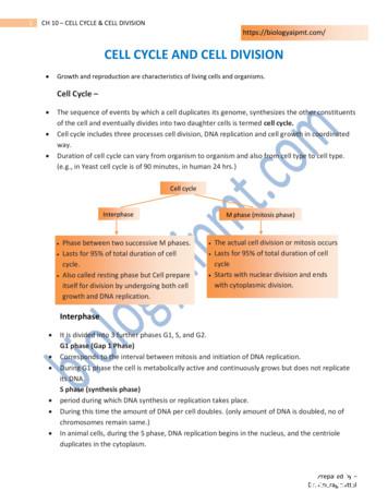
of the cell and eventually divides into two daughter cells is termed cell cycle. Cell cycle includes three processes cell division, DNA replication and cell growth in coordinated way. Duration of cell cycle can vary from organism to organism and also from cell type to cell type. (e.g., in Yeast cell cycle is of 90 minutes, in human 24 hrs.)
UNIT-V:CELL STRUCTURE AND FUNCTION: 9. Cell- The Unit of Life: Cell- Cell theory and cell as the basic unit of life- overview of the cell. Prokaryotic and Eukoryotic cells, Ultra Structure of Plant cell (structure in detail and functions in brief), Cell membrane, Cell wall, Cell organelles: Endoplasmic reticulum, Mitochondria, Plastids,
transfer system in oat cv. JO-1 using sonication-assisted Agrobacterium-mediated transformation (SAAT) and the vacuum-infiltration-assisted Agrobacterium-mediated transformation (VIAAT). The influence of different explants, sonication, and vacuum infiltration were evaluated in Agrobacterium-mediated genetic .
Metal-mediated synthetic organic chemistry has become very important in this context through the development of transition metal-mediated cross-coupling reactions. A schematic representation of a metal-mediated cross-coupling reaction is shown in Figure 2. Figure 2. Schematic representation of a metal-mediated cross-coupling reaction, a
3D Cell Model Project You will be creating a 3-dimensional model to represent either a plant cell or an animal cell. 1. Choose either a plant or animal cell. Below are the organelles required to be included in your model: Plant Cell Animal Cell Cell Membrane Cell Membrane Nucleus Nucleus Cytoplasm Cytoplasm Mitochondria Mitochondria
The Cell Cycle The cell cycle is the series of events in the growth and division of a cell. In the prokaryotic cell cycle, the cell grows, duplicates its DNA, and divides by pinching in the cell membrane. The eukaryotic cell cycle has four stages (the first three of which are referred to as interphase): In the G 1 phase, the cell grows.
Many scientists contributed to the cell theory. The cell theory grew out of the work of many scientists and improvements in the . CELL STRUCTURE AND FUNCTION CHART PLANT CELL ANIMAL CELL . 1. Cell Wall . Quiz of the cell Know all organelles found in a prokaryotic cell
Stent Type Stent Design Free Cell Area (mm2) Wallstent Closed cell 1.08 Xact Closed cell 2.74 Neuroguard Closed cell 3.5 Nexstent Closed cell 4.7 Precise Open cell 5.89 Protégé Open cell 20.71 Acculink Open cell 11.48 Stent Free Cell Area Neuroguard IEP Carotid Stent


