Midbrain Dopamine Neurons Signal Aversion In A Reward .
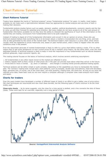
RESEARCH ARTICLEMidbrain dopamine neurons signalaversion in a reward-context-dependentmannerHideyuki Matsumoto1,2, Ju Tian1,2, Naoshige Uchida1,2,Mitsuko Watabe-Uchida1,2*1Center for Brain Science, Harvard University, Cambridge, United States;Department of Molecular and Cellular Biology, Harvard University, Cambridge,United States2Abstract Dopamine is thought to regulate learning from appetitive and aversive events. Herewe examined how optogenetically-identified dopamine neurons in the lateral ventral tegmentalarea of mice respond to aversive events in different conditions. In low reward contexts, mostdopamine neurons were exclusively inhibited by aversive events, and expectation reduceddopamine neurons’ responses to reward and punishment. When a single odor predicted bothreward and punishment, dopamine neurons’ responses to that odor reflected the integrated valueof both outcomes. Thus, in low reward contexts, dopamine neurons signal value prediction errors(VPEs) integrating information about both reward and aversion in a common currency. In contrast,in high reward contexts, dopamine neurons acquired a short-latency excitation to aversive eventsthat masked their VPE signaling. Our results demonstrate the importance of considering thecontexts to examine the representation in dopamine neurons and uncover different modes ofdopamine signaling, each of which may be adaptive for different environments.DOI: 10.7554/eLife.17328.001*For correspondence: mitsuko@mcb.harvard.eduCompeting interest: Seepage 21Funding: See page 21Received: 27 April 2016Accepted: 23 September 2016Published: 19 October 2016Reviewing editor: Rui M Costa,Fundação Champalimaud,PortugalCopyright Matsumoto et al.This article is distributed underthe terms of the CreativeCommons Attribution License,which permits unrestricted useand redistribution provided thatthe original author and source arecredited.IntroductionDopamine is thought to be a key regulator of learning from appetitive as well as aversive events(Schultz et al., 1997; Wenzel et al., 2015). It has been proposed that dopamine neurons act as ateaching signal in the brain by signaling the discrepancy between the values of actual and predictedrewards, that is, reward prediction error (RPE) (Bayer and Glimcher, 2005; Cohen et al., 2012;Hart et al., 2014; Roesch et al., 2007; Schultz, 2010; Schultz et al., 1997). Although accumulatingevidence supports this idea with respect to rewarding and reward-predicting events (Bayer andGlimcher, 2005; Cohen et al., 2012; Eshel et al., 2015; Hart et al., 2014; Roesch et al., 2007;Schultz, 2010; Schultz et al., 1997), how dopamine neurons integrate information about aversiveevents remains highly controversial.Pioneering work by Wolfram Schultz and colleagues introduced the idea that dopamine neuronssignal RPE. This work demonstrated that dopamine neurons in the midbrain of monkeys exhibit ahighly specific set of responses to reward (Mirenowicz and Schultz, 1994). When the animalreceives reward unexpectedly, dopamine neurons fire a burst of action potentials. If a sensory cuereliably predicts reward, however, dopamine neurons decrease their response to reward, andinstead burst to the cue. Finally, if an expected reward is omitted, dopamine neurons pause their firing at the time they usually receive reward (Hollerman and Schultz, 1998; Schultz et al., 1997).Subsequently, the idea of RPE coding by dopamine neurons has been substantiated by furtherexperiments in a variety of species including monkeys (Bayer and Glimcher, 2005; Hollerman andSchultz, 1998; Waelti et al., 2001), rats (Flagel et al., 2011; Pan et al., 2005; Roesch et al., 2007),Matsumoto et al. eLife 2016;5:e17328. DOI: 10.7554/eLife.173281 of 24
Research articleNeuroscienceeLife digest There are many types of learning; one type of learning means that rewards andpunishments can shape future behavior. Dopamine is a molecule that allows neurons in the brain tocommunicate with one another, and it is released in response to unexpected rewards. Mostneuroscientists believe that dopamine is important to learn from the reward; however, there aredifferent opinions about whether dopamine is important to learn from punishments or not.Previous studies that tried to examine how dopamine activities change in response to punishmenthave reported different results. One of the likely reasons for the controversy is that it was difficult tomeasure only the activity of dopamine-releasing neurons.To overcome this issue, Matsumoto et al. used genetically engineered mice in which shining ablue light into their brain would activate their dopamine neurons but not any other neurons. Tinyelectrodes were inserted into the brains of these mice, and a blue light was used to confirm thatthese electrodes were recording from the dopamine-producing neurons. Specifically if the electrodedetected an electrical impulse when blue light was beamed into the brain, then the recorded neuronwas confirmed to be a dopamine-producing neuron.Measuring the activities of these dopamine neurons revealed that they were indeed activated byreward but inhibited by punishment. In other words, dopamine neurons indeed can signalpunishments as negative and rewards as positive on a single axis. Further experiments showed that,if the mice predicted both a reward and a punishment, the dopamine neurons could integrateinformation from both to direct learning.Matsumoto et al. also saw that when mice received rewards too often, their dopamine neuronsdid not signal punishment correctly. These results suggest that how we feel about punishment maydepend on how often we experience rewards.In addition to learning, dopamine has also been linked to many psychiatric symptoms such asaddiction and depression. The next challenge will be to examine how the frequency of rewardschanges an animal’s state and responses to punishment in more detail, and how this relates tonormal and abnormal behaviors.DOI: 10.7554/eLife.17328.002mice (Cohen et al., 2012; Eshel et al., 2015) and humans (D’Ardenne et al., 2008). This signal isproposed to underlie associative learning (Rescorla and Wagner, 1972), and bears a striking resemblance to machine learning algorithms (Sutton and Barto, 1998).Many of the previous studies that characterized dopamine responses used rewarded outcomeswith varying degrees of predictability. Comparatively fewer studies have used aversive stimuli in thecontext of prediction errors. Among studies that have used aversive stimuli, these provide differingreports as to how dopamine neurons respond to aversive stimuli (Fiorillo, 2013; Schultz, 2015;Wenzel et al., 2015).It is thought that the majority of dopamine neurons are inhibited by aversive stimuli(Mileykovskiy and Morales, 2011; Mirenowicz and Schultz, 1996; Tan et al., 2012; Ungless et al.,2004). However, a number of electrophysiological recording studies have reported that dopamineneurons are activated by aversive stimuli both in anesthetized (Brischoux et al., 2009; Coizet et al.,2006; Schultz and Romo, 1987) and awake animals (Guarraci and Kapp, 1999; Joshua et al.,2008; Matsumoto and Hikosaka, 2009), although the proportions and locations of aversion-activated neurons differed among these studies. The differences in the results between these studiescould be due to the heterogeneity of dopamine neurons or to differences in experimental conditions(e.g. type of aversive stimuli; type of anesthesia). Furthermore, another study using fast-scan cyclicvoltammetry found that dopamine neurons are excited during successful avoidance of aversive stimuli (Oleson et al., 2012), which could be ’rewarding’. Therefore, some of the excitatory responses toaversive stimuli may not be due to aversiveness alone.Some of these discrepancies could correspond to differences in dopamine signaling dependingon the projection target. Roitman et al. (2008) monitored dopamine dynamics in the nucleusaccumbens using cyclic voltammetry while the animal received intra-oral administrations of a sucroseor quinine solution (Roitman et al., 2008). This study found that these stimuli caused oppositeMatsumoto et al. eLife 2016;5:e17328. DOI: 10.7554/eLife.173282 of 24
Research articleNeuroscienceresponses: dopamine release was increased by sucrose and decreased by quinine(McCutcheon et al., 2012), suggesting that at least the majority of dopamine neurons projecting tothe nucleus accumbens are inhibited by aversive stimuli. Matsumoto and Hikosaka (2009) examinedthe diversity of dopamine neurons in context of prediction error. They showed that dopamine neurons that are activated by the prediction of aversive stimuli are located in the lateral part of the substantia nigra pars compacta (SNc), supporting the notion that dopamine subpopulations are spatiallysegregated (Matsumoto and Hikosaka, 2009). Consistent with this finding, Lerner et al. (2015)showed, using calcium imaging with fiber photometry, that SNc neurons projecting to the dorsolateral striatum are activated by aversive stimuli (electric shock) whereas those projecting to the dorsomedial striatum are inhibited (Lerner et al., 2015). Lammel et al. (2011) provided further evidencefor spatial heterogeneity by showing that dopamine neurons projecting to the medial prefrontal cortex, located in the medial ventral tegmental area (VTA) exhibited a form of synaptic plasticity(AMPA/NMDA ratio) in response to aversive stimuli (formalin injection) whereas dopamine neuronsprojecting to the dorsolateral striatum did not (Lammel et al., 2011) although how these neuronschange their firing patterns in response to aversive stimuli remains unknown.In contrast to the above findings suggesting that dopamine neurons are heterogeneous withrespect to signaling aversive events, Schultz, Fiorillo and colleagues have argued that dopamine neurons largely ignore aversiveness (Fiorillo, 2013; Schultz, 2015; Stauffer et al., 2016). One argumentis that the excitation of dopamine neurons caused by aversive stimuli may be due to a ’generalization’ or ’spill-over’ effect of rewarding stimuli. Specifically, Mirenowicz and Schultz (1996) showedthat when rewarding and aversive stimuli are predicted by similar cues (e.g. in a same sensorymodality), aversion-predicting cues increase their tendency to activate dopamine neurons (’generalization’) (Mirenowicz and Schultz, 1996). Kobayashi and Schultz (2014) showed that in a highreward context, cues that predict a neutral outcome (e.g. a salient picture) increased their tendencyto activate dopamine neurons compared to the neutral cues in a low reward context(Kobayashi and Schultz, 2014). Based on these and other observations (Fiorillo et al., 2013;Nomoto et al., 2010), they proposed that the early response reflects attributes such as stimulusgeneralization and intensity, and the later response reflects the subjective reward value and utility(Schultz, 2016; Stauffer et al., 2016).One influential paper by Fiorillo (2013) concluded that dopamine neurons represent predictionerrors with respect to reward but not aversiveness (Fiorillo, 2013). That is, dopamine neurons ignoreaversive events. Recording from non-human primates, Fiorillo used three pieces of evidence to support this claim: First, dopamine neurons’ responses to aversive outcomes (air puff) were indistinguishable from their responses to neutral outcomes. Second, although most dopamine neuronsreduced their reward responses when the reward was predicted, their response to aversive eventswas unaffected by prediction. Third, dopamine neurons did not integrate the value of aversiveevents when combined with rewarding events. From these results, the author proposed that thebrain represents reward and aversiveness independently along two dimensions (Fiorillo, 2013). As aresult, the author proposed that different molecules regulate different types of reinforcement learning: dopamine for reward and a different molecule for aversiveness. If proven true, these ideas arefundamental in understanding how the brain learns from reward and aversion. However, it remainsto be clarified whether these observations can be generalized.The conclusions in many of the studies cited above relied upon indirect methods such as spikewaveforms and firing properties (Ungless and Grace, 2012) in order to identify dopamine neurons.These identification methods differed among studies and have recently been called into question(Lammel et al., 2008; Margolis et al., 2006; Ungless and Grace, 2012). The ambiguity of cell-typeidentification criteria across studies makes it difficult to consolidate data on dopamine signaling. Forexample, Ungless et al. showed that some neurons in the VTA that were excited by aversive eventsand identified as dopaminergic using standard electrophysiological criteria were revealed not to bedopaminergic when they were examined with juxtacellular labeling (Ungless et al., 2004). Furthermore, Schultz has argued that some previous recording studies may not have targeted areas rich indopamine neurons (Schultz, 2016).To circumvent this problem, we tagged dopamine neurons with a light-gated cation channel,channelrhodopsin-2 (ChR2) and unambiguously identified dopamine neurons based on theirresponses to light (Cohen et al., 2012). In the present study, we monitored the activity of identifieddopamine neurons using a series of behavioral tasks designed to determine how dopamine neuronsMatsumoto et al. eLife 2016;5:e17328. DOI: 10.7554/eLife.173283 of 24
Research articleNeuroscienceencode prediction of aversive events in addition to reward. Our results demonstrate that, in contrastto the proposal by Fiorillo (2013), dopamine neurons in VTA indeed are able to encode completeVPE, integrating information about both appetitive and aversive events in a common currency.Importantly, the ability of dopamine neurons to encode VPE depends on both reward contexts andthe animal’s trial-by-trial behavioral state.ResultsIdentification of dopamine neurons and task designsWe recorded the spiking activity of total 176 neurons in the VTA using tetrodes while mice performed classical conditioning tasks (Table 1). To identify neurons as dopaminergic, we optogenetically tagged dopamine neurons (Cohen et al., 2012). We then used a method developed previously(Stimulus-Associated spike Latency Test [SALT]) (Eshel et al., 2015; Kvitsiani et al., 2013; Tian andUchida, 2015) to determine whether light pulses significantly changed a neuron’s spike timing(p 0.001, Figure 1). To ensure that spike sorting was not contaminated by light artifacts, we compared the waveforms between spontaneous and light-evoked spikes, as described previously(Cohen et al., 2012). Dopamine neurons were mostly recorded from the central and posterior partof the lateral VTA including the parabrachial pigmented nucleus (PBP), parainterfascicular nucleus(PIF) and paranigral nucleus (PN) (Figure 1G,K,O). We obtained 72 optogenetically-identified dopamine neurons in total (5 4 neurons per mouse; mean S.D.; n 14 mice).We devised several different tasks to characterize dopamine activities in response to mild aversiveair puff (Table 1). ’Mixed prediction task’ (low reward context) was designed to examine interactionbetween the prediction of reward and the prediction of aversiveness. ’Low reward probability task’(low reward context) and ’high reward probability task’ (high reward context) were specificallydesigned to test the effects of reward probability on dopamine responses: two task conditions differed only with respect to reward probabilities. ’High reward probability task 2’ (high reward context) was originally conducted to replicate the diverse responses to aversive stimuli in dopamineneurons, which were reported in multiple previous studies. The effects of reward contexts were alsoexamined with the mixed prediction task and the high reward probability task 2.In the present study, we first focused on the characterization of dopamine activities in low rewardcontexts (Figures 2–4). Then, we compared dopamine activities between low and high reward contexts (Figure 5). Finally, we examined dopamine activities in relation to behaviors in different contexts (Figure 6).Dopamine neurons integrate values of both valences, appetitive andaversiveA previous study reported that dopamine neurons do not integrate information about aversivenessalong with reward-related information when rewarding liquid and an air puff are delivered to aTable 1. Summary of task conditions.CS (% outcome)TaskOdor AOdor BOdor COdor DOutcome (Reward CS) (Nothing CS) (Air puff CS) (Reward and air puff CS) Reward trials (%) Free reward (%)Mixed prediction taskWaterAir puff0Low reward probability taskWater20Air puff0090High reward probability taskWater9000Air puffHigh reward probability task 2 WaterAir puff250025075750000909000008013276306307DOI: 10.7554/eLife.17328.004Matsumoto et al. eLife 2016;5:e17328. DOI: 10.7554/eLife.173284 of 24
Research articleNeuroscienceFigure 1. Optogenetic identification of dopamine neurons in the ventral tegmental area (VTA). (A) Voltage trace from 10 pulses of 10 Hz lightstimulation (cyan bars, top) of a representative dopamine neuron. A spontaneous spike and a light-triggered spike were magnified at the bottom. (B)Responses from this neuron to 10 Hz (left) and 50 Hz (right) stimulation. (C) Isolation of an identified dopamine neuron from noise and other units. (D)Histogram of p values testing whether light-activation induced significant changes in spike timing (n 62 neurons) in the mixed prediction task. The pvalues were derived from SALT (Stimulus-Associated spike Latency Test; see Materials and methods). Neurons with p values 0.001 and waveformcorrelations 0.9 were considered identified (grey). P values and waveform correlations were calculated using light stimulation with all the frequencies(1–50 Hz). (E) Probability of light-evoked spike as a function of stimulation frequency for each dopamine neuron (grey) and the average acrossdopamine neurons (blue circles and bars, median and interquartile range). (F) Histograms of the mean (left) and S.D. (right) spike latency to lightstimulation with all the frequencies (1–50 Hz) for 26 identified dopamine neurons. (G) Reconstruction of the positions of individual dopamine neuronsrecorded in the mixed prediction task. Each circle represents a lesion site from individual animals used in the mixed prediction task. Each horizontal lineon the track (indicated by a vertical line over the lesion site) indicates estimated recording positions of individual dopamine neurons. LabeledFigure 1 continued on next pageMatsumoto et al. eLife 2016;5:e17328. DOI: 10.7554/eLife.173285 of 24
Research articleNeuroscienceFigure 1 continuedstructures: parabrachial pigmented nucleus of the VTA (PBP), parainterfascicular nucleus of the VTA (PIF), paranigral nucleus of the VTA (PN), rednucleus (RN), substantia nigra pars compacta (SNc), and substantia nigra pars reticulata (SNr). Scale bar, 1 mm. (H–J) Optogenetic identification ofdopamine neurons recorded in high and low reward probability tasks (29 dopamine neurons identified out of 73 neurons). Conventions are the same asin D–F. (K) Reconstruction of the positions of individual dopamine neurons recorded in high (red) and low (cyan) reward probability tasks. Conventionsare the same as in G. (L–N) Optogenetic identification of dopamine neurons recorded in high reward probability task 2 (17 dopamine neuronsidentified out of 41 neurons). (O) Reconstruction of the positions of individual dopamine neurons recorded in high reward probability task 2 (magenta).DOI: 10.7554/eLife.17328.003monkey at the same time (Fiorillo, 2013). However, this method may produce complex interactionsbetween the two different outcomes. To test how reward and aversion interact and affect dopamineresponses, we devised a ’mixed prediction’ paradigm (Figure 2) in which a single odor (Odor D inFigure 2A, conditioned stimulus, CS) predicted both a rewarding and a mildly aversive event in acomplementary and probabilistic manner: a reward (water) was delivered in 25% of the trials and anaversive event (air puff) was delivered in the remaining 75% of the trials. For comparison, weincluded the following trial types: Odor A predicted water in 25% of trials (nothing in 75%), Odor Cpredicted air puff in 75% of trials (nothing in 25%), and Odor B predicted no outcome. Each behavioral trial began with the odor CS (1 s), followed by a 1-s delay and an unconditioned stimulus (US).We chose higher probability for air puff than water in order to balance the strength of positive andnegative values in the task; we suspect that the magnitude of the negative value of mild air puff ismuch smaller than the magnitude of the positive value of water, which could cause us to overlook asmall effect of predicted air puff on the CS response.We first asked whether the recorded dopamine neurons were inhibited or excited by odor cues(CSs) that predicted different outcomes. We found that the vast majority of the neurons were inhibited by the air puff-predicting CS while excited by the reward-predicting CS (Figure 2B–D). On average, the firing rate during the CS period was significantly lower for the air puff-predicting CS thanfor the CS predicting nothing, while it was higher for the reward-predicting CS than for the CS thatpredicted nothing (Figure 2E). A similar tendency was observed using data from two animals insteadof three (i.e. leaving one animal out of three) (Figure 2—figure supplement 1). Among 26 identifieddopamine neurons, 85% (22 neurons) were significantly modulated by these three odors (p 0.05,one-way ANOVA), and 59% (13 of 22 significant neurons) showed the monotonic CS value coding(water nothing air puff). These results suggest that the firing of identified dopamine neuronswas negatively modulated by the stimulus predicting aversive events.We next examined whether prediction of aversion in addition to reward changed the response ofdopamine neurons. In contrast to the previous study (Fiorillo, 2013), we found that the majority ofneurons showed an intermediate response to the CS predicting both water and air puff (Odor D)compared to the CSs predicting water only (Odor A) or air puff only (Odor C) (Figure 2B,F,G). As apopulation, the net response to these CSs increased monotonically according to the values of bothwater and air puff, with the CS response to Odor D falling in between that of Odor A and Odor C(Figure 2H). 89% (23 of 26 neurons) of identified dopamine neurons were significantly modulated bythese three odors (p 0.05, one-way ANOVA), and 65% (15 of 23 significant neurons) showedthe monotonic CS value coding (water water and air puff air puff). These results indicate thatVTA dopamine neurons combine values for both reward and punishment along a one-dimensionalvalue axis.Dopamine neurons signal prediction errors for aversionIt has been shown that dopamine neurons’ responses to reward are greatly reduced when thereward is predicted, a signature of prediction error coding (Schultz et al., 1997). We replicatedthese findings here even in low reward probability conditions (20–25%, Figure 3—figure supplement 1; see Materials and methods). We next examined whether these dopamine neurons show prediction error coding for aversive events. To address this question, we occasionally delivered air puffduring inter-trial intervals without any predicting cues. These responses to unpredicted air puff werecompared to the responses to air puff in trials when air puff was predicted by an odor cue. We foundthat the inhibitory response to an air puff was significantly reduced when the air puff was predictedby an odor cue (Figure 3A–D). To further examine whether dopamine neurons showed predictionMatsumoto et al. eLife 2016;5:e17328. DOI: 10.7554/eLife.173286 of 24
Research articleNeuroscienceFigure 2. Dopamine neurons integrate values of both valences, reward and aversion. (A) Task design in the mixedprediction task. (B) Mean S.E.M. of firing rates of optogenetically-identified dopamine neurons during all fourtrial conditions; reward (blue), nothing (black), punishment (red), and both reward and punishment (magenta). (C)Scatter plot of the mean responses during the CS epoch (0–1 s, indicated by a solid black line in B) for rewardversus nothing. The baseline firing rate ( 1–0 s from odor onset) was subtracted for each neuron. Black filledcircles indicate neurons with significant difference between responses to the CS predicting reward and thatpredicting nothing (unpaired t test, p 0.05). (D) Scatter plot of the mean responses during the CS epoch fornothing versus punishment. Black filled circles indicate neurons with significant difference between responses tothe CS predicting nothing and that predicting punishment. (E) Comparison of the responses of individual neurons(n 26) during CS (0–1 s) predicting reward (blue), nothing (black) and punishment (red). For all box plots, thecentral mark is the median, the edges of the box are the 25th and 75th percentiles, and the whiskers extend to themost extreme data points not considered outliers (points 1.5 interquartile range away from the 25th or 75thpercentile), and outliers are plotted individually as plus symbols. **t(25) 3.7, p 0.001, paired t test. One outlier 5 Hz in response to Odor A is not represented. (F) Scatter plot of the mean responses during the CS epoch forreward versus reward and punishment. Black filled circles indicate neurons with significant difference betweenresponses to the CS predicting reward and that predicting reward and punishment. (G) Scatter plot of the meanresponses during the CS epoch for reward and punishment versus punishment. Black filled circles indicate neuronsFigure 2 continued on next pageMatsumoto et al. eLife 2016;5:e17328. DOI: 10.7554/eLife.173287 of 24
Research articleNeuroscienceFigure 2 continuedwith significant difference between responses to the CS predicting punishment and that predicting rewardand punishment. (H) Comparison of the responses during CS predicting reward (blue), both reward andpunishment (magenta), and punishment (red). 1t(25) 2.5, p 0.02; ***t(25) 4.4, p 2.0 10 4, paired t test. Oneoutlier 5 Hz in response to Odor A is not represented.DOI: 10.7554/eLife.17328.005The following figure supplement is available for figure 2:Figure supplement 1. Comparison of CS responses using dopamine neurons from two animals instead of three.DOI: 10.7554/eLife.17328.006error coding for aversive events, we compared the firing rate during the outcome period in air puffomission trials with that in trials that predict nothing. We found that the omission of a predicted airpuff slightly but significantly increased firing rates, compared to no change in nothing trials(Figure 3E–H) although we observed variability in air puff omission responses. Together, theseresults demonstrate that dopamine neurons signal prediction errors for aversive events in additionto rewarding events. These results indicate that dopamine neurons have the ability to signal VPEsfor both appetitive and aversive events, supporting previous work by Matsumoto and Hikosaka(Matsumoto and Hikosaka, 2009) and contrasting with previous work by Fiorillo (Fiorillo, 2013).Homogeneous response function of dopamine neuronsAlthough we found that most dopamine neurons were inhibited by air puff (mildly aversive event),there was a considerable variability in the extent to which individual dopamine neurons were inhibited. Does this diversity support a functional diversity across dopamine neurons in the lateral VTA?In a previous study, dopamine neurons in the lateral VTA exhibited neuron-to-neuron variability inthe magnitude of response to a given size of reward (Eshel et al., 2016). Despite this variability inresponsivity, the response functions of individual dopamine neurons were scaled versions of eachother, indicating a remarkable homogeneity. One consequence of this scaled relationship is thatneurons that responded strongly to a given size of reward were more greatly suppressed by rewardexpectation. In other words, reward expectation suppressed a neuron’s reward response in proportion to the size of its response to unexpected reward.Does the same relationship hold for inhibitory responses to air puff? To address this question, weexamined the correlation between aversion-related responses in dopamine neurons (Figure 4, Figure 4—figure supplement 1). We indeed found a similar relationship: dopamine neurons that werestrongly inhibited by air puff also exhibited a larger prediction-dependent reduction of theirresponses to air puff (Figure 4A, Pearson’s r 0.69, p 1.9 10 6). In other words, the ratiobetween individual dopamine neurons’ responses to unpredicted versus predicted air puff was preserved across neurons. In addition, similar to reward responses (Eshel et al., 2016), inhibitoryresponses to the air puff-predicting CS were correlated with prediction-dependent reduction ofresponses to air puff US (Figure 4B, Pearson’s r 0.39, p 0.016). These results indicate that theresponse function was preserved across dopamine neurons in the case of aversive stimuli.We next examined the relationship between responses of dopamine neurons to reward and toaversion. We compared responses of dopamine neurons to unpredicted water and unpredicted airpuff (Figure 4C). We observed no obvious unique clusters across neurons, supporting the notionthat there was no clear subpopulation of dopamine neurons specialized in signaling reward versusaversion in the lateral VTA. Rather, we found that most of dopamine neurons were inhibited byunpredicted aversive stimuli and excited by unpredicted rewarding stimuli. Interestingly, we did notfind any negative or positive correlation of neurons’ responses to water and air puff; the proportionof the response magnitudes in response to reward versus aversion was diverse across neurons. Thes
Midbrain dopamine neurons signal aversion in a reward-context-dependent manner Hideyuki Matsumoto1,2, Ju Tian1,2, Naoshige Uchida1,2, Mitsuko Watabe-Uchida1,2* 1Center for Brain Science, Harvard University, Cambridge, United States; 2Department of Molecular and Cellular Biology, Harvard University, Cambridge, United States
C. Dopamine receptors and regulation of gene expression 201 D. Development of transgenic animals in the study of dopamine receptor physiology 203 E. Clinical and pharmacological implications of multiple dopamine receptors 204 IX. Dopamine Receptors in the Pituitary 205 X. Peripheral Dopamine Receptors 205 A. Dopamine receptors in blood vessels 205
to Midbrain Dopamine Neurons Mitsuko Watabe-Uchida,1 Lisa Zhu,1 Sachie K. Ogawa,1 Archana Vamanrao,1 and Naoshige Uchida1,* . in contrast to activation of VTA dopamine neurons by reward values, and whether there are indeed direct projections from the striatum to dopamine
midbrain that release dopamine target the striatum, an area of the brain that is responsible for motor control. These neurons also release other neurotransmitters, but the identity of these other chemicals is not known, and the details of the interaction between the neurons and the striatum are poorly understood.
ways in the midbrain (23). The midbrain neurons stimulated with electric shock are the pain projection neurons which con- nect the primary fibers activated by peripheral (e.g., foot shock) stimulation with the highest levels of the brain's pain systems. That is, these midbrain reticular fibers are higher
hypothesized that increased error-related midbrain fMRI activation would positively correlate with higher dopamine D2 receptor availability. In a second sample of subjects, pharmacological manipulation during fMRI was accomplished with an indirect dopamine agonist (oral methylphenidate). As dopamine exerts inverted U-shaped effects on cognition,57
midbrain DA neurons in vivo results from tonic activation of NMDA receptors by endogenous excitatory amino acids (Cher-gui et al. 1993). As a first approximation, in this study we have focused only on the NMDA-induced burst firing mechanism and its modulation by electrical coupling. There is no consensus regarding the mechanisms of burst
of strategies in games. Because risk and ambiguity aversion have similar e ects in games (making ‘safe’ strategies appear relatively more attractive), and are positively correlated, studies that focus only on risk aversion or ambiguity aversion in
Pipe Size ASTM Designation (in) (mm) (D2310) (D2996) 2 - 6 50 - 150 RTRP 11FX RTRP 11FX-5430 8 - 16 200 - 400 RTRP 11FX RTRP 11FX-3210 Fittings 2 to 6-inch Compression-molded fiberglass reinforced epoxy elbows and tees Filament-wound and/or mitered crosses, wyes, laterals and reducers 8 to 16-inch Filament-wound fiberglass reinforced epoxy elbows Filament-wound and/or mitered crosses, wyes .


