Doctor 017
11NameDoctor017Doctor 017Doctor 017Mohammad Al-Salem0
This lecture will discuss the blood supply of the brain stem and the lesions related to theblood supply.We’ll begin by briefly discussing The circle of WillisFormed by 2 arteries in the cranial cavity which are the internal carotid artery and thebasilar artery formed by the twovertebral arteries after entering throughthe foramen magnum.In this picture to the right you can seethe major branches from the circle ofWillis.The part which is of interest to us forthe supply of the brain stem is lowerpart (the two vertebral arteries formingthe basilar)1 Page
Branches from the vertebral (like the anterior spinal and posterior inferior cerebellarartery) and branches from the basilar will be the main blood supply of the medullaoblongata and the pons, while the midbrain will be supplied by the posterior cerebralartery and superior cerebellar artery and the basilar.In this picture on the right you can see the course of the vertebralartery, which originates from the subclavian artery and movesthrough the transverse foramina of cervical vertebra and eventually(at c1 vertebra) it curves upward forward and medially and thenenters the foramen magnum and forms the basilar artery when the 2sides unite.The second picture below is a posterior view of the arteries andshows the 2 vertebral arteries and these arteries give the anteriorspinal artery, a single artery which movesalong the anterior median fissure of thespinal cord and medulla, it has two rootseach one from the vertebral artery on eachside.The vertebral artery also gives a branchcalled the posterior inferior cerebellarartery (PICA) and this gives a branch calledthe posterior spinal artery.This is a picture of the brainstem and thearteries, you can see the two vertebral arteries, the vertebral gives the anterior spinal aswell as PICA -posterior inferior cerebellar artery and (PICA gives the posterior spinal),these arteries will supply the medulla oblongata (vertebral, ASA,PICA,PSA).2 Page
You can the basilar artery which moves along the basilar groove on the pons and givespontine arteries which supply the pons. The basilar artery then divides into two posteriorcerebral arteries which receive the posterior communicating artery to complete the circleof Willis.The main branches of the basilar artery are:1- anterior inferior cerebellar artery(AICA) supplies inferior surface of thecerebellum.2- Pontine arteries.3- Superior cerebellar arteries that branchoff the basilar before its bifurcation intothe posterior cerebral arteries (suppliessuperior surface of cerebellum and pons).3 Page
Blood supply of the medulla oblongataThe above cross section is at the level of closed medulla (the cavity is the central canal)and the other is at the level of open medulla (the cavity is the 4th ventricle), and you cansee the arteries that supply the medulla oblongata.Starting anteriorly above, you can see the anterior median fissure and a cross section ofthe anterior spinal artery which supplies the midline structures (notice the sections on theleft, the ASA supplies the purple), the area slightly lateral to it (green) is supplied by thevertebral artery. The most lateral and posterior parts are supplied by PICA (a branch ofvertebral) and posterior spinal artery (a branch of PICA).Notice that the PSA supplies the posterior aspect on the lower level or closed medulla butwhen you ascend to open medulla the PSA doesn’t contribute to supply.To sum up, midline structures are supplied by the ASA, more lateral to it the vertebralartery and most lateral and posterior structures by PICA (open medulla), and PSAcontributes to posterior structures in closed medulla.4 Page
Medial medullary syndrome (Dejerine syndrome)It is caused by a lesion in anterior spinal arterywhich supplies the area close to the midline at.Notice in the picture the dark area is thelocation where there is loss of blood supply.Symptoms (related to the structures on themidline): Contralateral hemiparesis (weakness inmuscles, paralysis may happen based onseverity)Notice that the anterior aspect of the midline isoccupied by the pyramids (corticospinal tracts),it is contralateral because at this leveldecussation hasn’t happened yet. Contralateral loss of proprioception, finetouch and vibratory sense due to damage to themedial lemniscus (remember that decussationhappened so the medial lemniscus on the leftcarries information about the right side of the body). Deviation of the tongue to the ipsilateral side when it is protruded (hypoglossal root ornucleus injury).This syndrome is characterized by Alternating hemiplegiaNote: Alternating hemiplegia means;1- The upper and lower limbs are paralyzed in the contralateral side of lesion uppermotor neuron lesion (decussation).2- while the face is paralyzed in the ipsilateral side of lesion lower motor neuron lesion(no decussation).Symptoms related to cranial nerve (ipsilaterally)The white area in the second picture represents the lesion which is at the midline.5 Page
Lateral medullary syndrome (Wallenberg syndrome) or PICA syndromeIt is caused by a lesion in PICA whichsupplies the area close to lateral areas.The dark area is affected (supplied by PICA),in the radiograph it is the are with a red arrowon it.Symptomscontralateral loss of pain and temperaturesensation from the body (anterolateral system,decussation already happened at this level sothe ALS on the left carries information aboutthe right side of the body).ipsilateral loss of pain and temperaturesensation from the face (involvement ofspinal trigeminal tract and nucleus).vertigo and nystagmus (vestibular nuclei).Nystagmus is irregular movements of theeyeballs (the vestibular nucleus connected tothe cranial nerves supplying the eye muscles).loss of taste from the ipsilateral half of the tongue (solitary tract and nucleus).Nucleus tractus solitarius is a sensory nucleus for 2 types of sensations, visceral sensoryand taste. This nucleus receives taste sensations from the same side through 3 cranialnerves (7th and 9th and 10thhoarseness and dysphagia (nucleus ambiguus or roots of cranial nerves IX and X)Nucleus ambiguus is a motor nucleus for 3 cranial nerves (9th, 10th and 11th) and has thelower motor neurons supplying the muscles of the larynx and pharynx. (muscles areaffected ipsilaterally)Ipsilateral Horner syndrome (hypothalamospinal fibers)Rem: lateral (medullary) reticulospinal tract has descending autonomic regulating fibersprovide a pathway by which the hypothalamus can control the sympathetic and sacralparasympathetic outflow. If these fibers are cut then symptoms similar to Hornersyndrome will develop like ptosis, miosis (constriction of pupil) and anhidrosis, allrelated to sympathetic injury.6 Page
Vascular lesions of the posterior spinal arterySymptomsipsilateral loss of proprioception and vibratory sense (relatedto PCML system specifically nucleus gracilis and cuneatus).ipsilateral loss of pain and temperature sensation from theface (lateral to the nucleus cuneatus is the trigeminal nucleusand is affected).Blood supply of ponsRemember that the basilar artery moves throughthe basilar groove on the pons and gives theanterior inferior cerebellar artery and superiorcerebellar and pontine arteries and ends bydividing into two posterior cerebral arteries.The following is a cross section of the pons:7 Page
The blood supply of pons will be discussed at 2 levels, the inferior level (caudal part)closer to the pontomedullary junction and the superior level (midpontine) where we cansee the trigeminal nuclei.Generally, the pons will be supplied by paramedian branches (from basilar), from therename they’re close to the midline so the structures at the midline will be supplied bythese branches (purple structures in the figure), the lateral structures (green and blue) aresupplied by the circumferential branches (some sources divide them into short and longcircumferential branches). AICA also contributes to supply of lateral part (blue) with thecircumferential arteries.At the upper level (midpontine, level of trigeminal nucleus) branches from the superiorcerebellar arteries aid in supply of the posterior part with the circumferential branches.Foville syndromeDue to occlusion of the paramedial branchesSymptoms ipsilateral abducens nerve paralysis (the abducentnucleus is found posteriorly close to the floor of the4th ventricle and the abducent nerve moves anteriorlyand emerges from the pontomedullary junction closeto the midline).8 Page
contralateral hemiparesisThe anterior part of the purple color is the basilar part which contains corticospinal fibersand these fibers decussate in the lower part of the medulla so fibers on the right supplyleft side (symptoms related to long tracts are contralaterally and symptoms related tocranial nerves ipsilaterally). variable contralateral sensory loss reflecting various degrees of damage to the mediallemniscus.Millard-Gubler syndrome (or just Gubler syndrome)If the area of damage is shifted somewhat laterally to include the root of the facial nervealong with corticospinal fibers, the patient has a contralateral hemiparesis and anipsilateral paralysis of the facial muscles.Syndrome of the midpontine baseDue to occlusion of the paramedial branches andshort circumferential branches. Corticospinal fibers (which are passing throughthe basilar part) are affected causing contralateralhemiparesis. Sensory and motor trigeminal roots (trigeminalnuclei, the motor nuclei medially and slightlylaterally the sensory nucleus) are affected causingipsilateral loss of pain and thermal sense andparalysis of the masticatory muscles. Fibers of the middle cerebellar peduncle (ataxia).(Syndrome of the midpontine base hallmark ofbrainstem vascular lesions, ipsilateral cranial nerve sign coupled with a contralateral longtract sign).9 Page
Blood supply of the midbrain Basilar artery (gives direct branches tothe midbrain in addition to the followingbranches):-quadrigeminal artery (this artery couldarise from both the basilar artery at thebifurcation and the posterior cerebellarartery)-superior cerebellar arteries Internal carotid: anterior choroidal artery(not seen in this picture go back to the 2ndpic in page 7 to see it) Posterior cerebral artery (is divided intoparts, in the pic you can see P1 and P2):medial posterior choroidal artery.10 P a g e
The parts closer to the midline (purple) are supplied by paramedian branches from thebifurcation of the basilar artery. Anterolateral (blue) parts are supplied by circumferentialbranch of the quadrigeminal and posterior choroidal arteries.Posterolateral parts (green) are supplied by medial posterior choroidal arteries.The posterior part (yellow) which is the tectum, is supplied by the quadrigeminal arteryand superior cerebellar artery.The difference between the 2 levels shown in the picture (above at the level of superiorcolliculus and below at the level of inferior colliculus) is that in the superior section themost lateral (red) parts are supplied by the Thalamogeniculate artery, a branch of theposterior cerebral artery.Weber syndromeDue to occlusion of vessels servingthe medial portions of the midbraininvolving the oculomotor nerve andthe crus cerebri.Symptoms: Ipsilateral paralysis of allextraocular muscles except thelateral rectus (supplied by theabducent) and superior oblique (by the trochlear). Paralysis of the contralateral extremities.Corticospinal fibers in midbrain pass through crus cerebri and the crus cerebri on theright is related to the left side of the body ( )عدنا الفكرة الف مرة decussation happens inferiorto this level Ipsilateral dilatation of pupil (oculomotor nerve has parasympathetic fibers supplyingthe constrictor pupillae muscle, so dilation occurs when damaged). Contralateral weakness of the facial muscles of the lower half of the face.This goes against the principle we explained multiple times, and this is theexplanation:The crus cerebri contains the corticonuclear fibers going to the motor nucleus of thefacial nerve and these fibers are upper motor neurons.11 P a g e
Cranial nerve nuclei receive bilateral corticoneclear fibers (nucleus on the right receivesfibers from both right and left cortex), the part of motor nucleus related to the lower facereceives fibers only from the contralateral cortex, this is why the weakness in lower faceis contralateral but if the lesion was in a lower level like one of the previously mentionedit would be ipsilaterally because it is at the level of lower motor neuron, the same conceptapplies for the tongue. Contralateral deviation of the tongue when it is protrudedThe tongue is supplied by the hypoglossal nerve and its nucleus receives bilateral fibersfrom the both cortexes, the part of nucleus related to the genioglossus muscle receivesfibers only from the contralateral cortex.Weber syndrome hallmark of brainstem vascular lesions, ipsilateral cranial nerve signcoupled with a contralateral long tract sign.Claude syndromeDue to occlusion of vesselsserving the central area of themidbrain which includes theoculomotor nerve and the rednucleus.Symptoms: ipsilateral paralysis of mosteye movements; the eye isdirected down and out (laterally), because 2 muscles are spared, superior oblique andlateral rectus and these muscles cause the eye to be directed that way. Ipsilateral dilatation of pupil (oculomotor nerve has parasympathetic fibers supplyingthe constrictor pupillae muscle, so dilation occurs when damaged) contralateral ataxia, tremor, and incoordinationCaused by involvement of the red nucleus whichreceives input from the cerebellum (cerbellorubraltract) and even the levels slightly below the rednucleus which have the superior cerebellarpeduncle decussation.The last lesion is Benedikt syndrome (basicallythe previous 2 syndromes together).12 P a g e
TONSILLAR HERNIATIONThere is a part of the cerebellum calledtonsils, when it is pushed out of its normallocation the condition is called tonsillarherniation (notice in the picture the directionof herniation which is downward towards theforamen magnum), this will cause pressureon the medulla oblongata in that area.Causes:any mass in the posterior cranial fossa(tumor, hemorrhage)increase in intracranial pressureThe major concern in acute herniation isdamage to the ventrolateral reticular area(heart rate and respiration)Symptoms:(caused either directly by pressure from the herniation or indirectly through occlusion tothe arteries that supply the medulla)sudden change in heart rate and respirationhypertensionhyperventilationrapidly decreasing levels of consciousness (part of the reticular formation is connected tothe reticular nuclei which project to the cortex and are responsible for keeping you alert)If sever, deathIn addition to variable amounts of sensory and motor deficits according to the severity.13 P a g e
Arnold-Chiari PhenomenonCongenital anomaly in which there is a herniationof the tonsils of the cerebellum and the medullaoblongata through the foramen magnum into thevertebral canal.It is less severe, and some people may beasymptomatic but as people get older symptomsmight start appearing (symptoms are similar toabove).If a person is diagnosed with this there is surgicaltreatment and prognosis is great, however intonsillar herniation treatment is directed tohemorrhage or tumor causing the herniation and soit is more difficult.Central herniationNotice the direction of herniation.Cause: space occupying lesion in the hemisphere(supratentorial compartment, above the tentoriumcerebri) elevates intracranial pressure and forces thediencephalon downward through the tentorial notchand into the brainstem affecting the midbrainmainly.Symptoms: change in respiration, eye movementsare irregular.As the damage progresses downward into thebrainstem, there is significant change in respirationTachypnea and apneaprofound loss of motor and sensory functions.probable loss of consciousnessDecorticate posture may occur as the pressure affects the fibers heading to the brainstem(UMN), where the lower limbs are extended and upper limbs are flexed but as herniationdevelops decerebrate may occur and this is a bad sign because it means the lesion is closeto the vital centers.14 P a g e
Upward Cerebellar HerniationA mass in the posterior cranial fossa may forceportions of the cerebellum upward through thetentorial notch (upward cerebellar herniation)and compress the midbrain rather than causingtonsillar herniation.The result may be occlusion of branches of thesuperior cerebellar artery with resultantinfarction of cerebellar structures or obstructionof the cerebral aqueduct and hydrocephalus.accumulation of fluids will lead to an increasein intracranial pressure causing vomiting, headache, lethargy, decreased levels ofconsciousness.Uncal HerniationMovement of the uncus(anteromedial part of the temporallobe) downward over the edge of thetentorium cerebelli, causing pressureon the midbrain.Early signs:dilated pupil ipsilateral to theherniation (involvement ofoculomotor)abnormal eye movements ipsilateral to the herniation (oculomotor nerve)double vision ipsilateral to the herniation (loss of synchrony of movement of the eyes).Weakness of the extremities (corticospinal fiber involvement) opposite to the dilatedpupil.Later:15 P a g e
respiration is affected16 P a g e
Millard-Gubler syndrome (or just Gubler syndrome) If the area of damage is shifted somewhat laterally to include the root of the facial nerve along with corticospinal fibers, the patient has a contralateral hemiparesis and an ipsilateral paralysis of the facial muscles. Syndrome of the midpontine base Due to occlusion of the paramedial branches and
C1000-017 C1000-017 Dumps C1000-017 Braindumps C1000-017 Real Questions C1000-017 Practice Test C1000-017 dumps f
Doctor Eduardo Velázquez Girón Doctor Mario German González Tenorio Doctor Juan Carlos Caicedo Doctor Willy Paul Stangl Herrera Doctor Alex Estrada Juri - Doctor Orlando Ávila Neira - Doctor Jaime Castro Plaza Doctor Rodrigo Bayrón Ríos .
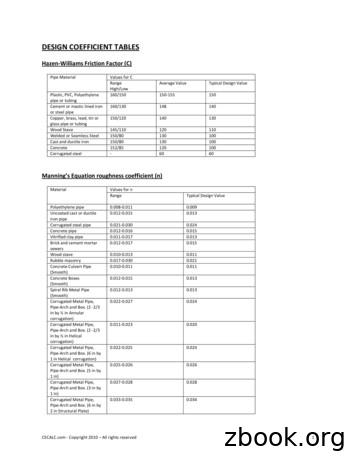
Uncoated cast or ductile iron pipe 0.012-0.015 0.013 Corrugated steel pipe 0.021-0.030 0.024 Concrete pipe 0.012-0.016 0.015 Vitrified clay pipe 0.011-0.017 0.013 Brick and cement mortar sewers 0.012-0.017 0.015 Wood stave 0.010-0.013 0.011 Rubble masonry 0.017-0.030 0.021 Concrete Culvert Pipe .
Directive 017: Measurement Requirements for Oil and Gas Operations (May 2020) i. Directive 017 . Release date: May . 12, 2020 Effective date: May . 12, 2020 Replaces previous edition issued December 13, 2018 . Measurement Requirements for Oil and Gas Operations . Contents
Emissions Unit Name and Description Date of Installation COMBINED HEAT & POWER PLANT [NES] FBE-PNP-E1 017-0040 -5-0019 53 MM Btu/hr (4.5-MW) Centaur 50 dual (NG/No.2 fuel oil) fired combustion turbine (CT) generator e/w Dry SoLoNo X Injectors March 2015 FBE-PNP-E2 017-
catalog number 30 zzz plgzhvwzkhho frp . e-6 (2-valve) pa18001-001 mp7 pa1mp7101-001 mp8 pa1mp8101-001 cummins n14 pa1n14221-017 isx (piston less) pa1isx141-017 isb pa1isb606-026 detroit diesel series 60 (11 liter) pa1s60102-033 series 60 (12.7 liter) pa1s60106-017 . all pai parts 1 year. caterpillar
This May, the University will award over 8,912 degrees. Of these, approximately 6,335 will be Bachelor's degrees, 1,877 Master's degrees, 2 Doctor of Juridical Science, 146 Juris Doctor degrees, 83 Master of Laws degrees, 75 Doctor of Pharmacy degrees, 52 Doctor of Dental Medicine degrees, 102 Doctor of
spaces. The Floor Doctor attachment can be used to clean hard surface floors in your home. Lightweight and easy-to-use, this hard floor tool effectively cleans and dries floors in seconds. To order the Floor Doctor or the Universal Hand Tool for your Rug Doctor machine, call 1-800-Rug Doctor (1-800-784-3628) or visit www.rugdoctor.com.


