Dimerization Of Cell-penetrating Buforin II Enhances Antimicrobial .
Lee and Yang Journal of Analytical Science and -8(2021) 12:9Journal of Analytical Scienceand TechnologyRESEARCH ARTICLEOpen AccessDimerization of cell-penetrating buforin IIenhances antimicrobial propertiesHyunhee Lee1 and Sungtae Yang1,2*AbstractAntimicrobial peptides (AMPs) that selectively permeabilize bacterial membranes are promising alternatives toconventional antibiotics. Dimerization of AMP is considered an attractive strategy to enhance antimicrobial andmembrane-lytic activity, but it also increases undesired hemolytic and cytotoxic activity. Here, we prepared Lyslinked homodimers of membrane-permeabilizing magainin II and cell-penetrating buforin II. Dimerization did notsignificantly alter conformational behavior, but it had a substantial impact on antimicrobial properties. We foundthat while the magainin II dimer showed increased antimicrobial and cytotoxic effects, the buforin II dimerconferred much greater antibacterial potency without exhibiting cytotoxic activity. Interestingly, the buforin II dimerwas highly effective against several antibiotic-resistant bacterial isolates. Membrane permeabilization experimentsindicated that the magainin II dimer rapidly disrupted both anionic and zwitterionic membranes, whereas thebuforin II dimer selectively disrupted anionic membranes. Like the monomeric form, the buforin II dimer wasefficiently translocated across lipid bilayers. Therefore, our results suggest that the dimerization of cell-penetratingbuforin II not only disrupts the bacterial membrane, but also translocates it across the membrane to targetintracellular components, resulting in effective antimicrobial activity. We propose that dimerization of intracellulartargeting AMPs may present a superior strategy for therapeutic control of pathogenic bacteria.Keywords: Antimicrobial peptide, Cell-penetrating peptide, Buforin II, Dimerization, Peptide-membrane interactionIntroductionAntimicrobial peptides (AMPs) are ubiquitous amongunicellular and multicellular organisms and are responsible for first-line host defenses against invading pathogens (Haney et al. 2019; Hilchie et al. 2013; Zhang andGallo 2016). Resistance to AMP remains at very lowlevels, making it a promising candidate to overcomeantibiotic-resistant bacterial infections (Biswaro et al.2018; de Breij et al. 2018; Kumar et al. 2018; Lohner2016; Mahlapuu et al. 2016). AMPs of multicellular organisms have a killing and/or inhibitory effect on a widerange of microorganisms, including gram-positive andgram-negative bacteria. It is widely accepted that mostAMPs kill bacterial cells by permeabilizing the negatively* Correspondence: styang@chosun.ac.kr1Department of Medical Science, Graduate School, Chosun University,Gwangju 61452, South Korea2Department of Microbiology, School of Medicine, Chosun University,Gwangju 61452, South Koreacharged cytoplasmic membrane (Guha et al. 2019; Matsuzaki 2019; Sani and Separovic 2016; Stone et al. 2019).In contrast to the membrane-lytic mechanism, it hasbeen proposed that some AMPs act intracellularly bybinding to DNA or altering enzyme activity (Le et al.2017; Lee et al. 2019). For example, magainin II is awell-known membrane-permeabilizing antimicrobialpeptide, whereas buforin II exerts potent antimicrobialactivity without causing membrane lysis (Imura et al.2008; Takeshima et al. 2003). Indeed, previous confocalfluorescence microscopic studies showed that magaininII is localized to the bacterial surface, whereas buforin IIaccumulates mainly in the cytoplasm (Park et al. 2000).The self-association of membrane-active peptides appears to be a crucial parameter for antimicrobial modeof action in either membrane permeabilization or peptide translocation across the membrane to target intracellular components (Petkov et al. 2019; Yang et al.2006a). Many studies have shown that the dimerization The Author(s). 2021 Open Access This article is licensed under a Creative Commons Attribution 4.0 International License,which permits use, sharing, adaptation, distribution and reproduction in any medium or format, as long as you giveappropriate credit to the original author(s) and the source, provide a link to the Creative Commons licence, and indicate ifchanges were made. The images or other third party material in this article are included in the article's Creative Commonslicence, unless indicated otherwise in a credit line to the material. If material is not included in the article's Creative Commonslicence and your intended use is not permitted by statutory regulation or exceeds the permitted use, you will need to obtainpermission directly from the copyright holder. To view a copy of this licence, visit http://creativecommons.org/licenses/by/4.0/.
Lee and Yang Journal of Analytical Science and Technology(2021) 12:9of membrane-permeabilizing peptides significantlychanges their biological and biophysical properties(Gunasekera et al. 2020; Lorenzon et al. 2019). For example, parallel and antiparallel magainin II dimers havegreater biological activity and greater ability to interactwith membranes than the monomeric form does (Mukaiet al. 2002). The pore formed by the magainin II dimeris characterized by a larger pore diameter and longerlifetime than that of the monomer (Hara et al. 2001).Although dimerization of AMPs may be a promisingstrategy to improve antimicrobial potency, dimerizationof membrane-permeabilizing AMPs increases cytotoxicity against mammalian cells (Gunasekera et al. 2020).These unwanted cytotoxic effects should be minimizedor eliminated. On the other hand, although dimerizationof cell-penetrating peptides leads to enhanced cellularuptake and drug delivery (Hoyer et al. 2012), little isknown about their antimicrobial properties.In this study, we synthesized membranepermeabilizing magainin II, cell-penetrating buforin II,and their counterpart homodimeric peptides with abranched Lys core; sequences are listed in Table 1. Wetested their antibacterial activities against gram-positiveand gram-negative bacteria as well as cytotoxicity againstmammalian cells, and we evaluated the roles ofdimerization in their antimicrobial properties. Interactions of the peptides with the lipid bilayer were investigated by circular dichroism and fluorescencespectroscopy in membrane mimetic environments. Weshowed that the Lys-linked buforin II dimer exertedgreater antibacterial potency against both gram-positiveand gram-negative bacteria, including several antibioticresistant bacterial isolates, without inducing cytotoxicityagainst mammalian cells. Our findings indicated thatboth membrane-permeabilizing and cell-penetratingabilities of the buforin II dimer seem to influence moreefficient and selective killing of pathogenic bacteria.Materials and methodsMaterials and microorganismsN-α-fluoren-9-yl-methoxycarbonyl (Fmoc) amino acidswith orthogonal side chain protecting groups and FmocLys(Fmoc)-OH (for the dimeric peptides) were purchased from Novabiochem (Läufelfingen, Switzerland).Table 1 Amino acid sequences and cytotoxic activities ofpeptides used in this studyPeptidesAmino acid sequencesHC50aLC50bMagainin IIGIGKWLHSAKKFGKAFVGEIMNS 200 200K-(magainin II)2(GIGKWLHSAKKFGKAFVGEIMNS)2K14.32.7Buforin IITRSSRAGLQWPVGRVHRLLRK 200 200K-(buforin II)2(TRSSRAGLQWPVGRVHRLLRK)2K 200 200aPeptide concentration (μM) causing 50% hemolysisPeptide concentration (μM) causing 50% growth inhibition of RAW 264.7 cellsbPage 2 of 7For peptide synthesis, reagents and solvents were purchased from Applied Biosystems (Foster City, CA, USA).After RP-HPLC purification (above 99% purity), thecorrect molecular weights were confirmed by MALDITOF-MS (Shimadzu, Japan). Phospholipids were obtained from Avanti Polar Lipids (Alabaster, AL, USA). Amembrane potential-sensitive fluorescent dye, 3,3′dipropylthiadicarbocyanine iodide [DiSC3(5)] was purchased from Molecular Probes, Inc. (Eugene, OR, USA).Microorganisms were purchased from the Korean Collection for Type Cultures (Daejon, Korea).Circular dichroism (CD) spectroscopyThe CD spectra of peptides were collected with a J-715spectrophotometer (Jasco, Japan) as previously described(Yang et al. 2019). The spectra are expressed as themean residue ellipticity [θ] versus wavelength. The ellipticity [θ] given in units of deg cm2 dmol-1 was calculatedusing the following formula: [θ] [θ]obs MRW/10lc,where MRW mean residue molecular weight of thepeptide, c is the concentration of the sample, and l is thelength of the cell.Antimicrobial, hemolytic, and cytotoxic activityThe antimicrobial activity of peptides against grampositive and gram-negative bacteria, including antibioticresistant pathogens, was measured by the broth microdilution method, as previously described (Lee et al. 2019).Briefly, single colonies of bacteria cultured on a LB agarplate were inoculated into LB medium and incubatedovernight at 37 C. A 50 μl of this culture was transferred into fresh tube of 10 ml LB medium and grown tomid-log phase. A set of serial dilutions of peptides (100μl) were added to 100 μl of 2 106 CFU/ml in 96-wellmicrotiter plates (Falcon). After incubation at 37 C for16 h, the minimal inhibitory concentration (MIC) wasdefined as the lowest peptide concentration that completely inhibited bacterial growth by measuring opticaldensity (OD) at 600 nm. The MICs were the average oftriplicate measurements in three independent assays.The hemolytic activities of peptides were determined bymonitoring the release of hemoglobin from human redblood cells, as described previously (Yang et al. 2019).Complete hemolysis was induced by 0.1% Triton X-100.Cytotoxicity of peptides against RAW 264.7 cells wasmeasured with a 3-(4, 5-dimethylthiazol-2-yl)-2, 5diphenyltetrazolium bromide (MTT) assay, as previouslydescribed (Yang et al. 2019). Cell viability was determined as the ratio of the absorbance at 570 nm forpeptide-treated cells compared to untreated cells.Membrane depolarization induced by peptidesThe membrane depolarization activities of peptides weredetermined using a membrane potential-sensitive
Lee and Yang Journal of Analytical Science and Technology(2021) 12:9fluorescent dye DiSC3(5), as previously described (Yanget al. 2006b). Briefly, Staphylococcus aureus cells culturedto mid-log phase were resuspended in 5 mM HEPES buffer. Upon addition of DiSC3(5) to the suspension of S.aureus, the fluorescence had quenched and stabilized.Addition of peptides (2 mM) increased the fluorescenceintensity due to membrane depolarization. After 5 min,gramicidin D was added to observe the maximum fluorescence intensity due to complete dissipation of the membrane potential. The DiSC3(5) fluorescence change wasmonitored on a Shimadzu RF-5301 spectrofluorometer atan excitation/emission wavelength of 620/670 nm.Membrane disruption induced by peptidesLarge unilamellar vesicles (LUVs) were generated by theextrusion method, and the membrane disruption induced by peptides was determined using the fluorescentdye calcein, as previously described (Lee et al. 2019).Briefly, peptides were added to calcein-containing LUVscomposed of anionic PC/PG (1:1) or zwitterionic PC.Membrane-lytic activity of peptides was defined as thepercent leakage from 100 μM LUVs after 10 min incubation with peptides (2 μM for PC/PG (1:1) and 20 μM forPC vesicles). Fluorescence changes due to calcein releasefrom LUVs were monitored on a Shimadzu RF-5301spectrofluorometer at excitation/emission wavelength 490/520 nm. Triton X-100 was added in order to showthe maximum fluorescence intensity due to completemembrane permeability.Ability of peptides to translocate into vesiclesLUVs were generated in HEPES buffer (150 mM NaCl,pH 7.4) with chymotrypsin (200 mM), as describedPage 3 of 7previously (Lee et al. 2019). External chymotrypsin wasinactivated by the addition of a trypsin-chymotrypsin inhibitor (200 mM). Fluorescence transfer from the Trpresidue of peptides to the dansyl group on membranesby peptide translocation into vesicles was monitored ona Shimadzu RF-5301 spectrofluorometer at an excitation/emission wavelength of 280/510 nm.ResultsCircular dichroism (CD) studiesCD spectroscopy was used to investigate structural differences between monomeric and dimeric peptides inaqueous buffer as well as in membrane-mimetic environments (Fig. 1). In aqueous buffer, all peptides had almostcompletely random coil structures. In contrast, bothmonomeric and dimeric magainin II assumed typical αhelices in membrane-memetic SDS micelles, as characterized by double minima around 208 and 222 nm.Compared to membrane-permeabilizing magainin II,cell-penetrating buforin II showed a negative peak at204 nm, indicating a somewhat different structure froma typical α-helix. However, the overall CD spectral patterns of the monomers were similar to those of dimers,suggesting that dimerization did not significantly alterthe structure of the peptides.Antimicrobial and hemolytic activities of the peptidesWe next determined the ability of peptides to inhibitbacterial growth by measuring their MIC, which arelisted in Table 2. The magainin II dimer and the buforinII dimer showed increased antibacterial activity by 4–8and 8–16 fold, respectively, compared to their respectivemonomers. In addition, Table 3 shows that the dimersFig. 1 CD spectra of the peptides. The CD spectra of magainin II and K-(magainin II)2 (a) and buforin II and K-(buforin II)2 (b) were obtained at 25 C in 10-mM sodium phosphate buffer (open symbols) or 30 mM SDS micelles (closed symbols). Spectra were taken at peptide concentrations of20 μM
Lee and Yang Journal of Analytical Science and Technology(2021) 12:9Page 4 of 7Table 2 Antimicrobial activities of peptides against gram-positive and gram-negative bacteriaOrganismAntimicrobial activity (MIC: μM)Magainin IIK-(magainin II)2Buforin IIK-(buforin II)2Bacillus subtilis8280.5Staphylococcus aureus16281Staphylococcus epidermidis164162Gram-positive bacteriaGram-negative bacteriaEscherichia coli32481Salmonella Typhimurium16240.5Pseudomonas aeruginosa324162had strong antimicrobial activity, in the range of 0.5–4μM, against antibiotic-resistant pathogens. These resultssuggest that not only the dimerization of membranepermeabilizing magainin II, but also the dimerization ofcell-penetrating buforin II, is a good way to enhanceantimicrobial activity. We also evaluated the hemolytic(HC50) and cytotoxic (LC50) activity against human redblood cells and RAW 264.7 cells, respectively, which arepresented in Table 1. Both monomeric magainin II andbuforin II showed negligible hemolytic and cytotoxic activities. Interestingly, while magainin II dimer showedstrong hemolytic and cytotoxic activity, buforin II dimerwas not hemolytic or cytotoxic. These results indicatethat dimerization of cell-penetrating buforin II improvesselectivity for bacteria over mammalian cells much better than dimerization of membrane-permeabilizingmagainin II.Membrane depolarization by peptidesTo determine whether membrane depolarization is related to bacterial death, we next determined the abilityof monomeric and dimeric peptides to depolarize thecytoplasmic membrane of S. aureus using the membranepotential-sensitive fluorescent dye DiSC3(5). As shownin Fig. 2a, monomeric magainin II effectively depolarizedthe cytoplasmic membrane of S. aureus at the valuelower than MIC; however, buforin II did not inducedmembrane depolarization even at the MIC value. Although there was no direct correlation between cytoplasmic membrane depolarization and MIC values, theseresults support that magainin II exerts antimicrobialactivity by inducing membrane depolarization, whereasthe potent antimicrobial activity of buforin II is not related to the membrane depolarization. Both monomericand dimeric magainin II efficiently dissipated the membrane potential of S. aureus, although the magainin IIdimer induced more effective membrane depolarizationthan its monomer. Intriguingly, the buforin II monomershowed negligible membrane depolarization ability,whereas the buforin II dimer dissipated over 50% ofmembrane potential. These results suggest that the additional ability of buforin II dimer to induce membranedepolarization appears to be associated with potent antimicrobial activity.Dye leakage from liposomesTo determine whether peptides were able topermeabilize membranes, we examined the ability ofpeptides to disrupt anionic vesicles composed of L-αphosphatidylcholine (PC) and L-α-phosphatidylglycerol(PG) (1:1), as well as zwitterionic vesicles composed ofPC by monitoring the efflux of fluorescent dye from thevesicles (Fig. 2b). Consistent with their respective abilities to depolarize bacterial membranes, magainin IIshowed strong calcein release from the PC/PG (1:1) vesicles with 50% leakage at 2 μM, whereas buforin IIshowed no membrane-lytic activity. These results support the mode of action that membrane disruption represents a major killing event for magainin II, but not forbuforin II. When added to PC liposomes, both magaininII and buforin II elicited no calcein release, even at 20μM, which agrees with results obtained for hemolyticTable 3 Antimicrobial activities of peptides against antibiotic-resistant bacterial isolatesAntibiotic-resistant bacteriaAntimicrobial activity (MIC: μM)Magainin IIK-(magainin II)2Buforin IIK-(buforin II)2Methicillin-resistant Staphylococcus aureus (1) [MRSA (1)]16.04.08.01.0Methicillin-resistant Staphylococcus aureus (2) [MRSA (2)]16.02.08.00.5Vancomycin-resistant Enterococcus faecalis (1)32.04.016.01.0Vancomycin-resistant Enterococcus faecalis (2)16.04.08.01.0
Lee and Yang Journal of Analytical Science and Technology(2021) 12:9Page 5 of 7Fig. 2 Ability of peptides to permeabilize membranes. a Effect of peptides on membrane potential of intact S. aureus cells (OD600 0.05). Afterthe fluorescence of DiSC3(5) was stabilized, the peptides (2 μM) were added to S. aureus. b Release of calcein fluorescent probe from anionic PC/PG (1:1) or zwitterionic PC vesicles. Membrane-lytic activity of peptides was defined as the percent leakage from 100 μM lipid after 10 minincubation with peptides (2 μM for PC/PG (1:1) and 20 μM for PC vesicles). Average SD for three independent experiments. Statisticallysignificant (p 0.05) and very significant (p 0.01) differences are represented by (*) and (**), respectivelyand cytotoxic activity. Dimerization of magainin IIstrongly increased the membrane-lytic activity againstboth anionic and zwitterionic liposomes. Interestingly,the buforin II dimer had relatively strong membranelytic activity ( 60%) against anionic liposomes, but didnot show any dye leakage from PC liposomes. Thesefindings indicate that dimerization of membranepermeabilizing magainin II non-selectively increasesmembrane-lytic ability between negatively charged andneutral membranes, whereas cell-penetrating buforin IIdimer leads to selective membrane-disrupting activityonly for anionic membranes. Although membrane-lyticactivity of peptides was related to their respective antimicrobial activity, these variables were not entirelylinear.fluorescence intensity, indicating that the peptide effectively crossed the lipid bilayer and entered vesicles. Theseresults are in agreement with previously published translocation data. Similar to monomer results, the buforin IIdimer translocated more effectively and rapidly acrossthe membrane than the magainin II dimer. These resultssuggest that both monomeric and dimeric buforin II cancross the bacterial membrane and target intracellularmolecules.Ability of peptides to translocate into liposomesTo determine whether a peptide can enter a cell, we examined the ability of peptides to translocate across lipidbilayers (Fig. 3). The extent of peptide translocationacross PC/PG/dansyl-PE (50:45:5) vesicles was estimatedby monitoring peptide degradation by chymotrypsin encapsulated within vesicles using a fluorescence resonance energy transfer system with a Trp donor anddansyl acceptor. We observed no fluorescence decreasethroughout the 500 s with magainin II, indicating acomplete lack of membrane translocation. In contrast,buforin II caused a time-dependent decrease inFig. 3 Ability of peptides to translocate into liposomes. Membranetranslocation of peptides was measured by resonance energytransfer from Trp to DNS-PE. Decreased fluorescence means that thepeptide has entered the vesicle. Lipid and peptide concentrationswere 100 and 1 μM, respectively
Lee and Yang Journal of Analytical Science and Technology(2021) 12:9DiscussionBecause membrane-active molecules such as antimicrobial and cell-penetrating peptides have attracted attention for therapeutic applications, there is currently muchwork underway in order to understand the basic principles of peptide interaction with target cell membranes(Avci et al. 2018; Henriques et al. 2006). They generallyshare cationic and amphipathic characteristics, whichplay important roles in membrane permeabilization and/or cell penetration. Since peptide self-assembly affectsmembrane permeabilization and cell penetration, severalgroups have investigated the different structural and biological functions between monomeric and dimeric forms(Hoyer et al. 2012; Lorenzon et al. 2019). It is particularly interesting that dimerization of AMPs leads togreater antibiotic activity against several bacterial species(Koh et al. 2018; Liu et al. 2017; Lorenzon et al. 2018;Panteleev et al. 2017; Thamri et al. 2017). Improved antibacterial activity is correlated with an increase in membrane permeabilization, indicating that bacterialmembranes are primarily targeted by dimers. However,dimerization of AMP with membrane-permeabilizingmodes of action leads to increased cytotoxicity againstmammalian cells, which may hinder therapeutic application (Gunasekera et al. 2020; Lorenzon et al. 2019).Indeed, we showed that membrane-permeabilizingmagainin II dimer displayed potent antibacterial activity,as well as strong cytotoxicity against mammalian cells.Our results also demonstrate that the improved antibacterial and cytotoxic activity of the magainin II dimer isassociated with strong membrane-lytic activity againstanionic and zwitterionic membranes, respectively. Incontrast, the membrane-penetrating buforin II dimerhad potent broad-spectrum antimicrobial activity,though it was not cytotoxic to mammalian cells. Similarto previously reported results (Park et al. 2000), thebuforin II monomer inhibited bacterial cell growth without causing membrane disruption. However, the buforinII dimer has the ability to depolarize bacterial membranes and permeabilize anionic membranes. Inaddition, the buforin II dimer can translocate acrosslipid membranes. These results, using both biologicaland artificial membranes, indicate that the buforin IIdimer directly collapses the cytoplasmic membrane potential and also interferes with intracellular targets,which may result in more potent antibacterial activity.In summary, dimerization was applied to membranepermeabilizing magainin II and membrane-penetratingbuforin II. Magainin II dimer increased both antibacterial and cytotoxic effects, whereas the buforin II dimerenhanced antibacterial activity without cytotoxicity.Interestingly, the buforin II dimer had the ability todepolarize the bacterial membrane and to effectivelytranslocate across lipid bilayers to target intracellularPage 6 of 7components. Because of the wide range of antimicrobialactivity against gram-positive and gram-negative bacteria, including antibiotic-resistant bacterial isolates, thebuforin II dimer has promising potential in applicationsfor therapeutic control of multi-resistant pathogenicbacteria. Although more studies are needed to elucidatethe exact mode of action of the buforin II dimer,dimerization of intracellular targeting AMPs may be apromising strategy to improve antimicrobial activitywhile reducing cytotoxic activity against mammaliancells.AbbreviationsAMP: Antimicrobial peptide; RP-HPLC: Reversed-phase high-performance liquid chromatography; MALDI-TOF: Matrix-assisted laser desorptionionization–time of flight; CD: Circular dichroism; MIC: Minimal inhibitoryconcentration; LUV: Large unilamellar vesicle; SDS: Sodium dodecyl sulfate;PC: L-α-phosphatidylcholine; PG: L-α-phosphatidylglycerol; PE: L-αphosphatidylethanolamineAcknowledgementsThe authors thank Industry-Academic Cooperation Foundation, ChosunUniversity.Authors’ contributionsH Lee and S Yang designed research. H Lee performed most experiments.The authors analyzed data and wrote the paper. The authors read andapproved the final manuscript.FundingThis study was supported by a research fund from Chosun University, 2020.Availability of data and materialsNot applicableCompeting interestsThe authors declare that they have no competing interests.Received: 18 December 2020 Accepted: 10 February 2021ReferencesAvci FG, Akbulut BS, Ozkirimli E. Membrane active peptides and their biophysicalcharacterization. Biomolecules. 2018;8(3). https://doi.org/10.3390/biom8030077.Biswaro LS, da Costa Sousa MG, Rezende TMB, Dias SC, Franco OL. Antimicrobialpeptides and nanotechnology, recent advances and challenges. FrontMicrobiol. 2018;9:855. https://doi.org/10.3389/fmicb.2018.00855.de Breij A, Riool M, Cordfunke RA, Malanovic N, de Boer L, Koning RI, et al. Theantimicrobial peptide SAAP-148 combats drug-resistant bacteria and biofilms.Sci Transl Med. 2018;10(423). https://doi.org/10.1126/scitranslmed.aan4044.Guha S, Ghimire J, Wu E, Wimley WC. Mechanistic landscape of membranepermeabilizing peptides. Chem Rev. 2019. kera S, Muhammad T, Stromstedt AA, Rosengren KJ, Goransson U.Backbone cyclization and dimerization of LL-37-derived peptides enhanceantimicrobial activity and proteolytic stability. Front Microbiol. 68.Haney EF, Straus SK, Hancock REW. Reassessing the host defense peptidelandscape. Front Chem. 2019;7:43. https://doi.org/10.3389/fchem.2019.00043.Hara T, Kodama H, Kondo M, Wakamatsu K, Takeda A, Tachi T, et al. Effects ofpeptide dimerization on pore formation: antiparallel disulfide-dimerizedmagainin 2 analogue. Biopolymers. 2001;58(4):437–46. https://doi.org/10.1002/1097-0282(20010405)58:4 437::AID-BIP1019 3.0.CO;2-I.Henriques ST, Melo MN, Castanho MA. Cell-penetrating peptides andantimicrobial peptides: how different are they? Biochem J. 00.
Lee and Yang Journal of Analytical Science and Technology(2021) 12:9Hilchie AL, Wuerth K, Hancock RE. Immune modulation by multifaceted cationichost defense (antimicrobial) peptides. Nat Chem Biol. o.1393.Hoyer J, Schatzschneider U, Schulz-Siegmund M, Neundorf I. Dimerization ofa cell-penetrating peptide leads to enhanced cellular uptake and drugdelivery. Beilstein J Org Chem. 2012;8:1788–97. https://doi.org/10.3762/bjoc.8.204.Imura Y, Choda N, Matsuzaki K. Magainin 2 in action: distinct modes ofmembrane permeabilization in living bacterial and mammalian cells. BiophysJ. 2008;95(12):5757–65. https://doi.org/10.1529/biophysj.108.133488.Koh JJ, Lin S, Sin WWL, Ng ZH, Jung DY, Beuerman RW, et al. Design andsynthesis of oligo-lipidated arginyl peptide (OLAP) dimers with enhancedphysicochemical activity, peptide stability and their antimicrobial actionsagainst MRSA infections. Amino Acids. 2018;50(10):1329–45. https://doi.org/10.1007/s00726-018-2607-6.Kumar P, Kizhakkedathu JN, Straus SK. Antimicrobial peptides: diversity,mechanism of action and strategies to improve the activity andbiocompatibility in vivo. Biomolecules. 2018;8(1). https://doi.org/10.3390/biom8010004.Le CF, Fang CM, Sekaran SD. Intracellular targeting mechanisms by antimicrobialpeptides. Antimicrob Agents Chemother. 2017;61(4). https://doi.org/10.1128/AAC.02340-16.Lee H, Lim SI, Shin SH, Lim Y, Koh JW, Yang S. Conjugation of cell-penetratingpeptides to antimicrobial peptides enhances antibacterial activity. ACSOmega. 2019;4(13):15694–701. https://doi.org/10.1021/acsomega.9b02278.Liu B, Huang H, Yang Z, Gou S, Zhong C, Han X, et al. Design of novelantimicrobial peptide dimer analogues with enhanced antimicrobial activityin vitro and in vivo by intermolecular triazole bridge strategy. Peptides. 2017;88:115–25. ner K. Membrane-active antimicrobial peptides as template structures fornovel antibiotic agents. Curr Top Med Chem. 2016.Lorenzon EN, Nobre TM, Caseli L, Cilli EM, da Hora GCA, Soares TA, et al. The“pre-assembled state” of magainin 2 lysine-linked dimer determines itsenhanced antimicrobial activity. Colloids Surf B: Biointerfaces. 2018;167:432–40. enzon EN, Piccoli JP, Santos-Filho NA, Cilli EM. Dimerization of antimicrobialpeptides: a promising strategy to enhance antimicrobial peptide activity.Protein Pept Lett. 2019;26(2):98–107. Mahlapuu M, Hakansson J, Ringstad L, Bjorn C. Antimicrobial peptides: anemerging category of therapeutic agents. Front Cell Infect Microbiol. 2016;6:194. https://doi.org/10.3389/fcimb.2016.00194.Matsuzaki K. Membrane permeabilization mechanisms. Adv Exp Med Biol. 2019;1117:9–16. https://doi.org/10.1007/978-981-13-3588-4 2.Mukai Y, Matsushita Y, Niidome T, Hatekeyama T, Aoyag H. Parallel andantiparallel dimers of magainin 2: their interaction with phospholipidmembrane and antibacterial activity. J Pept Sci. 2002;8(10):570–7. https://doi.org/10.1002/psc.416.Panteleev PV, Myshkin MY, Shenkarev ZO, Ovchinnikova TV. Dimerization of theantimicrobial peptide arenicin plays a key role in the cytotoxicity but not inthe antibacterial activity. Biochem Biophys Res Commun. c.2016.12.035.Park CB, Yi KS, Matsuzaki K, Kim MS, Kim SC. Structure-activity analysis of buforinII, a histone H2A-derive
Keywords: Antimicrobial peptide, Cell-penetrating peptide, Buforin II, Dimerization, Peptide-membrane interaction Introduction Antimicrobial peptides (AMPs) are ubiquitous among unicellular and multicellular organisms and are respon-sible for first-line host defenses against invading patho-gens (Haney et al. 2019; Hilchie et al. 2013; Zhang and
of bHLH dimerization and DNA binding. Dimerization between bHLH proteins is mediated by two a-helices from each of the bHLH monomers that face each other, and DNA interactions occur mainly with residues in the basic region, located N-terminal to the first a-helix (Supplementary Figure S1). Second, we previously determined the dimerization and .
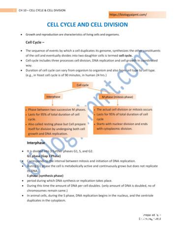
of the cell and eventually divides into two daughter cells is termed cell cycle. Cell cycle includes three processes cell division, DNA replication and cell growth in coordinated way. Duration of cell cycle can vary from organism to organism and also from cell type to cell type. (e.g., in Yeast cell cycle is of 90 minutes, in human 24 hrs.)
by the two central base pairs of the DNA response element, interacting with residues of the first zinc finger helix going across the DNA large groove [6,10]. The ligand-binding domain The LBD is a globular domain that harbors a hormone binding site, a dimerization interface (homo- and hetero-dimerization), and a coactivator and corepressor interac-
DNA-binding domain (residues 1-86) and a dimerization domain (residues 86-180) that allows it to form heterodimers with dimerization partner . E2Fs can be captured as DNA-binding proteins activating the adenoviral E2a promoter, implying that E2Fs are potentially involved in viral replication [25-27]. E2F4 can enter the nucleus with the .
Trastuzumab, Pertuzumab, Lapatinib, T-DM1: Complementary Mechanisms HER2 Dimerization domain Pertuzumab HER1/3/4 Trastuzumab Subdomain IV Trastuzumab: Inhibits ligand-independent HER2 signaling Activates ADCC Prevents HER2 ECD shedding Pertuzumab: Inhibits ligand-dependent HER2 dimerization and signaling Activates ADCC Lapatinib
UNIT-V:CELL STRUCTURE AND FUNCTION: 9. Cell- The Unit of Life: Cell- Cell theory and cell as the basic unit of life- overview of the cell. Prokaryotic and Eukoryotic cells, Ultra Structure of Plant cell (structure in detail and functions in brief), Cell membrane, Cell wall, Cell organelles: Endoplasmic reticulum, Mitochondria, Plastids,
Ground Penetrating Radar Fundamentals by Jeffrey J. Daniels, Department of Geological Sciences, The Ohio State University Prepared as an appendix to a report to the U.S.EPA, Region V Nov. 25, 2000 Introduction Ground penetrating radar (commonly called GPR) is a high resolution electromagnetic
analysis of Hawking [9, 10] showed that black holes are thermodynamic systems that emit black body radiation. Hawking’s discovery resulted in the precise formulation of. Chapter 1. Introduction 4 black hole entropy S BH A d 4G d; (1.3) where A d is the area of the event horizon, G d is the d-dimensional Newton’s constant, and c k B 1. The breakthrough in understanding .


