Exotic Ribosomal Enzymology
Digital Comprehensive Summaries of Uppsala Dissertationsfrom the Faculty of Science and Technology 1770Exotic Ribosomal EnzymologyJOSEFINE SN 1651-6214ISBN 978-91-513-0567-7urn:nbn:se:uu:diva-374965
Dissertation presented at Uppsala University to be publicly examined in A1:111a, BMC,Husargatan 3, Uppsala, Friday, 8 March 2019 at 09:15 for the degree of Doctor of Philosophy.The examination will be conducted in English. Faculty examiner: Christophe Danelon (DelftUniversity of Technology, Kavli Institute of NanoScience, Department of Bionanoscience).AbstractLiljeruhm, J. 2019. Exotic Ribosomal Enzymology. Digital Comprehensive Summaries ofUppsala Dissertations from the Faculty of Science and Technology 1770. 43 pp. Uppsala:Acta Universitatis Upsaliensis. ISBN 978-91-513-0567-7.This thesis clarifies intriguing enzymology of the ribosome, the multiRNA/multiproteincomplex that catalyzes protein synthesis (translation). The large ribosomal RNAs (23S and16S rRNAs in E. coli) are post-transcriptionally modified by many specific modificationenzymes, yet the functions of the modifications remain enigmatic. A deeper insight intotwo of the 23S rRNA S-adenosyl-methionine-requiring methyltransferase enzymes, RlmMand RlmJ, was given by investigating substrate specificity in vitro. Both enzymes were ableto methylate in vitro-transcribed, modification-free, protein-free, 2659-nucleotide-long 23SrRNA. Furthermore, RlmM was able to methylate the 611-nucleotide-long Domain V of the23S rRNA alone and RlmJ could modify the A2030 with only 25 surrounding nucleotides.Translation is evolutionary optimized to incorporate L-amino acids to the exclusion of Damino acids in the cell. To understand how, and how to engineer around this restriction forpharmacological applications, detailed kinetics of ribosomal dipeptide formation with D- versusL-phenylalanine-tRNA were determined. This was done by varying the concentrations of EFTu (which delivers aminoacyl-tRNAs to the ribosome) and the ribosome, as well as changingthe tRNA adaptor. Binding to EF-Tu was shown to be rate limiting for D-Phe-tRNA at alow concentration of EF-Tu. Surprisingly, at a higher (physiological) concentration of EFTu, binding and subsequent dipeptide synthesis became so efficient that D-Phe incorporationbecame competitive with L-Phe, and accommodation/peptide bond formation was unmasked asa new rate-limiting step. This highlighted the importance of D-aminoacyl-tRNA deacylase inrestricting translation with D-amino acids in vivo.Although polypeptides are intrinsically colorless, it is remarkable that evolution hasnevertheless enabled ribosomes to synthesize highly colored proteins (chromoproteins).Such eukaryotic proteins reside in coral reefs and undergo self-catalyzed, intramolecular,chromophore formation by reacting with oxygen in a manner highly similar to that ofgreen fluorescent protein. The potential utility of different colored chromoproteins in E.coli was analyzed via codon-optimized over-expression and quantification of maturationtimes, color intensities and cellular fitness costs. No chromoprotein was found to have thecombined characteristics of fast maturation, intense color and low fitness cost. However, semirational mutagenesis created different colored variants with identical fitness costs suitable forcompetition assays and teaching.Keywords: rRNA modification, Methyltransferase, RlmM, RlmJ, D-amino acid, Unnaturalamino acid, ChromoproteinJosefine Liljeruhm, Department of Cell and Molecular Biology, Molecular Biology, Box 596,Uppsala University, SE-751 24 Uppsala, Sweden. Josefine Liljeruhm 2019ISSN 1651-6214ISBN 978-91-513-0567-7urn:nbn:se:uu:diva-374965 (http://urn.kb.se/resolve?urn urn:nbn:se:uu:diva-374965)
Till Sandra
List of PapersThis thesis is based on the following papers, which are referred to in the textby their Roman numerals.IPunekar AS, Shepherd TR, Liljeruhm J, Forster AC, Selmer M (2012)Crystal structure of RlmM, the 2'O-ribose methyltransferase for C2498of Escherichia coli 23S rRNA. Nucleic Acids Res. 40, 10507-20.II Punekar AS, Liljeruhm J, Shepherd TR, Forster AC, Selmer M (2013)Structural and functional insights into the molecular mechanism ofrRNA m6A methyltransferase RlmJ. Nucleic Acids Res. 41, 9537-48.III Liljeruhm J, Wang J, Kwiatkowski M, Sabari S, Forster, AC (2019)Kinetics of D-amino acid incorporation in translation. ACS Chem. Biol.,DOI: 10.1021/acschembio.8b00952, 2019.IV Liljeruhm J, Funk SK, Tietscher S, Edlund AD, Jamal S, WistrandYuen P, Dyrhage K, Gynnå A, Ivermark K, Lövgren J, Törnblom V,Virtanen A, Lundin ER, Wistrand-Yuen E, Forster AC (2018) Engineering a palette of eukaryotic chromoproteins for bacterial synthetic biology. J. Biol. Eng. 12:8, 1-10.Reprints were made with permission from the respective publishers.
Related Publications by the Author(Not included in this thesis)V Kjellander M, Götz K, Liljeruhm J, Boman M, Johansson G (2013)Steady-state generation of hydrogen peroxide: kinetics and stability ofalcohol oxidase immobilized on nanoporous alumina. Biotechnol. Lett.35, 585-90.VI Liljeruhm J, Gullberg E, Forster AC. Synthetic Biology: a lab manual(2014) World Scientific Press, 204 pp, ISBN: 9789814579551.VII Shepherd TR, Du L, Liljeruhm J, Samudyata, Wang J, Sjödin MOD,Wetterhall M, Yomo T, Forster AC (2017) De novo design and synthesisof a 30-cistron translation-factor module. Nucleic Acids Res. 45, 10895905.
ContentsIntroduction . 11The central dogma . 11Bacterial protein synthesis and the genetic code . 12Dissertation overview . 15The present work . 17Papers I and II: Methylation of E. coli 23S ribosomal RNA . 17rRNA modification enzymes . 17Analysis of modified RNAs . 19Methods summary . 20Results and conclusions . 21Paper III: the kinetics of D-AA incorporation in translation . 22D-amino acids . 22D-AA-tRNA synthesis in vitro . 24Methods summary . 24Results and conclusions . 25Paper IV: Eukaryotic chromoproteins in a prokaryotic host . 26Chromoproteins. 26Methods summary . 28Results and conclusions . 29Future outlook . 32rRNA modifications . 32Incorporation of D-AAs in translation. 32CPs . 33Sammanfattning på svenska . 34Acknowledgements . 37References . 38
Abbreviations3HAAAAARSAA-tRNAA siteAMVATPCcDNACMCCPDDNAE siteE. P sitePTCPAGEPCRtritiumadenineamino acidaminoacyl-tRNA synthetaseaminoacyl-tRNAribosomal minoacyl-tRNA siteAvian Myeloblastosis virusadenosine 5’ triphosphatecytidinecomplementary nucleic acidribosomal exit tRNA siteEscherichia colielongator factor thermo-unstableformylmethioninefluorescent proteinguaninegreen fluorescent proteinguanosine 5’diphosphateguanosine 5’triphosphatehigh-pressure liquid chromatographyinitiation factorinternational Genetically Engineered Machinelevorotatorymatrix-assisted laser desorption ionizationmessenger RNAmass spectrometernucleotide triphosphateribosomal peptidyl-tRNA siteribosomal peptidyltransferase centrepolyacrylamide gel electrophoresispolymerase chain reaction
PheRNARlmrRNARTSAMTtRNAUphenylalanineribonucleic acidrRNA large subunit methtyltransferaseribosomal RNAreverse transcriptaseS-adenosylmethioninethyminetransfer RNAuridine
IntroductionThe central dogmaThe molecular-level mechanism of life, encoded in genes of an organism, isuniversal, likely due to an original common ancestor. Together, genes areassembled to a genome, consisting of polymerized deoxyribonucleotidescontaining the nucleobases adenine (A), guanine (G), cytosine (C) and thymine (T) in a highly organized and specific order held within doublestranded deoxyribonucleic acid (DNA). A genome is duplicated in a processcalled replication, which enables the cell to divide into two and where bothcells then continue to carry the entire genome.In a process called transcription, several ribonucleic acids polymers (RNAs,which contain ribose instead of deoxyribose and uracil instead of thymine)are transcribed from the genome, for example as messenger RNAs (mRNAs;a temporary intermediate form of genetic information), transfer RNAs(tRNAs) and ribosomal RNAs (rRNAs). All three of these classes are required for ribosomal protein synthesis, and many of them must first undergopost-transcriptional chemical alteration, called RNA modification.Organisms are functional predominantly due to their catalytically-activeproteins that carryout life-maintaining reactions and/or mechanisms. Theseproteins are synthesized by first transcribing DNA into single-strandedmRNAs and then mRNAs are translated into proteins (Fig. 1).11
Figure 1. The central dogma. Image taken, with permission, from Liljeruhm et al.(2014), courtesy of E. Wistrand-Yuen.Bacterial protein synthesis and the genetic codeProtein synthesis (translation) is mRNA decoding by the ribosome, a macromolecular complex. The E. coli ribosome consists of two subunits, 30Sand 50S (Schmeing et al. 2009), that incorporate 21 and 33 ribosomal proteins respectively and catalytic centre constitutes three RNA parts, the 23S(2904 nt), 16S (1542 nt) and 5S (120 nt) rRNAs (Cate et al. 1999; Yusupovet al. 2001; Yonath et al. 2002).Bacterial translation initiation requires the initiation factors 1, 2 and 3 (IF1,IF2 and IF3; Antoun et al. 2006) which position the 30S at the start region(ribosome binding site, RBS) of an mRNA. The initiator tRNA, formylMethionine-tRNAformylMethionine (fMet-tRNAfMet) base pairs with the start codon(usually AUG; Liljeruhm et al. 2014, Fig. 7a) at the ribosome’s PeptidyltRNA site (P-site; Shine & Dalgarno 1975). Lastly, the 50S subunit is recruited and the IFs dissociate (Huang et al. 2010).12
Elongator aminoacyl-tRNAs (aa-tRNAs) form a complex with elongatorfactor thermo-unstable (EF-Tu) and guanosine 5’triphosphate (GTP), termedthe ternary complex. The ternary complex delivers the next AA to the ribosome and the anticodon sequence on the AA-tRNA is placed in the decodingcentre of the ribosome but with the EF-Tu:GTP still remained bound (Fig.2). When the codon-anticodon interaction is sufficiently stable, i.e. a codonanticodon match, GTP is hydrolysed to GDP and EF-Tu:GDP is released(Ogle et al. 2003). The AA-tRNA accommodates into the Aminoacyl-site(A-site) of the ribosome. Next, a peptide bond forms (peptidyl-transfer) between the AA on the A-site tRNA and the fMet (or peptide) on the P-sitetRNA through the peptidyl transferase reaction, which is catalysed by theribosome (Wilson & Nierhaus 2003):The Elongator factor G, through the energy from the GTP-to-GDP conversion, will move the ribosome one codon downstream of the mRNA. Thus,the peptidyl-tRNA (former AA-tRNA) at the A-site is translocated to the Psite whereas the tRNA at the P-site move to the Exit site (Fig. 2; Rodnina etal. 1997). This enables a new round of AA incorporation with a new AAtRNA being delivered and accommodated at the A-site. The translationalmechanisms of charging tRNAs, binding to EF-Tu:GTP, delivery to the ribosome and peptide bond formation are all stereospecific to enable the exclusive incorporation of amino acids in the L-isomeric form.Figure 2. A kinetic scheme of the incorporation of a (D-)AA in translation (elongation). The image is adapted from Liljeruhm et al. 2019.Although the genetic code is essentially universal, due to the redundancy incodons per amino acid, there is room for organismal preferences in terms offavoured codons, i.e. there is a codon usage bias (Fig. 3). Codon bias affectswhich codons are more preferred in the genome and, subsequently, whichtRNAs are up-regulated.Finally, when one of 3 stop codons (UAA, UAG and UGA; Fig. 3) arrives atthe A site, it is recognised by a release factor (RF) which catalyses release13
(hydrolysis) of the peptidyl-tRNA from the tRNA, and the nascent proteincompletes its folding into a specific three-dimensional structure (Scolnick etal. 1968).Figure 3. The genetic code and codon usage frequencies in the most abundant proteins in E. coli. Image taken, with permission, from Liljeruhm et al. (2014).14
Dissertation overviewThis thesis encompasses "exotic" enzymology centred on bacterial translation. The enzymology includes(i) very slowly acting, specific, rRNA modification enzymes,(ii) kinetics of unnatural D-AA incorporation in translation, and(iii) expression of self-catalysing chromoproteins, a unique trick enablingribosomes to directly produce coloured products.The overarching theme is to better characterize these exotic processes tofacilitate applications in synthetic biology, which can be defined as “thecomplex engineering of replicating systems.” The applications envisioned ofthe three respective projects above are:(i) synthesis of a ribosome (toward synthesis and directed evolution of selfreplication),(ii) directed evolution of D-AA-containing, protease-resistant drug candidates, and(iii) development of colorimetric cellular biosensors.The experimental results described in the next chapter have been performedby me, unless otherwise stated, and correspond to following parts of PapersI-IV in the Appendix:Paper ICharacterization studies on the rRNA methyltransferase enzyme RlmM. Theminimal substrate for recognition was defined, and time-course assays confirmed the relatively slow enzymatic reaction of methylases.Paper IICharacterization studies on the rRNA methyltransferase enzyme RlmJ. Aminimal substrate for recognition was defined.Paper IIIKinetic studies of D-AA incorporation in translation in vitro. The rate ofpeptide formation was examined with different tRNA bodies, EF-Tu concentrations and ribosome concentrations to elucidate the rate-limiting steps.15
Paper IVStudy of 14 eukaryotic CPs to visualize and compare their properties interms of colour strength, gene stability and growth defects in E. coli.16
The present workPapers I and II: Methylation of E. coli 23S ribosomalRNArRNA modification enzymesThe initial stages of ribosome assembly require ribosomal RNA to be transcribed, and ribosomal protein mRNAs to be transcribed and translated. Anadditional step is nucleotide modification (35 in total for E. coli) of the nascent 23S and 16S rRNA transcripts. Curiously, these modification reactionsare amongst the slowest enzymatic reactions known.Based on knock out studies of individual modification enzymes in E. coli(Baba et al. 2006), it is remarkable that none of them are individually essential. The combination of the modifications is thus presumed to be essential.Although many cluster around functionally active regions, such as the peptidyl transferase centre (PTC; Fig. 4), A, E and P site and the polypeptideexit tunnel, little is known about their specific functions. However becauseof this crowding, one hypothesis is that they are important for the function ofthe ribosome (Brimacombe et al. 1993; Decatur & Fournier 2002). On theother hand, three-dimensional rRNA modification maps (Decatur & Fournier2002) showed that they are not phylogenically conserved between E. coliand Saccharomyces cerevisiae (S. cerevisiae), suggesting other, nontranslational, functions are more likely instead, e.g. structural stabilization.This is further strengthened by reports on different types of modified nucleotides that either serve as aid in subunit assembly to avoid mis-folding(Grosjean et al. 2005), stabilize helices and other secondary structures (Hayrapetyan et al. 2009), prevent hydrolysis of internucleotide bonds, protect thepre-assembled unprotected RNA from ribonucleases (Decatur & Fournier2002; Nierhaus & Lafontaine 2006), or improve base stacking interactions(Hayrapetyan et al. 2009).Groups of modified nucleotides have been tested for E. coli 50S subunit invitro reconstitution/activity (fragment reaction with puromycin) by digestingand matching different lengths of in vitro transcribed and wild-type 23SrRNAs to form a full-length 23S rRNA. The loss of modifications had a low17
effect on the peptidyl-transferase activity, except for an 80-nucleotide region, now called the critical region. This region includes six modified nucleotides within the PTC (Fig. 4) (Green & Noller 1996), which were then necessary for the ribsome’s peptidyl transfer activity.Figure 4. Secondary structure of the PTC in the domain V of 23S rRNA of E. coli.Blue letters indicate the nucleotides that lies within the Critical region from Green &Noller 1996. Image courtesy of T. Shepherd.One of these six "critical" modifications is the 2’O-ribose methylation ofC2498 (Cm2498), positioned in helix no. 89 (nucleotides 2455 – 2496). Inan earlier study, the enzyme rRNA large subunit methyltransferase M(RlmM, YgdE) was experimentally shown to catalyse the methylation ofC2498 on 23S rRNA (Purta et al. 2009). Methyltransferases utilize the cofactor S-adenosylmethionine (SAM) as a methyl donor substrate, which isthen converted to S-adenosyl-homocysteine (SAH):18
They proved, through in vitro methylation assays, that RlmM can methylatepurified rRNA from a !ygdE strain, i.e. protein-free 23S rRNA which stillcontains all its other modifications. On the other hand, the enzyme did notseem to methylate 50S subunits or 70S ribosomes purified from the !ygdEstrain (Purta et al. 2009). This complements another study where Cm2498was categorized among those that get modified in the early stage of the 50Ssubunit assembly (Siibak & Remme 2010).The helix no. 72 (nucleotides 2023 – 2040) folds against helix no. 89 in thetertiary structure of E. coli 23S rRNA (Borovinskaya et al. 2008) and includes the N6-methylated A2030 nucleotide (m6A2030). This methylationwas recently discovered to be linked to the enzyme RlmJ (YhiR). Apart fromidentifying the gene, they also showed that RlmJ could methylate proteinfree 23S rRNA purified from a !yhiR strain, but not the 50S subunit (Golovina et al. 2012).Analysis of modified RNAsReverse transcription and primer extensionReverse transcription is a process that was originally discovered in virusesand is the synthesis of a complementary DNA (cDNA) to an RNA strand(Temin & Mizutani 1970). Avian Myeloblastosis Virus (AMV; Kacian et al.1971) RTase extends from the 3’ end of a [32P]-labelled primer and willpause or terminate upon reaching a methylated nucleotide or rigid secondarystructure (optimization of the dNTP concentration was crucial to achieveefficient specific pausing). The premature halt is either due to unsuccessfulbase pairing between the modified residue and the supposed complementarydNTP or the RTase fails to recognize it. This stop will generate a cDNA thatwill be one nucleotide shorter than the length between the primer’s 5’-endand the modification. The cDNA is then separated by long denaturing polyacrylamide gel electrophoresis (PAGE), next to sequencing lanes for identification of modified DNA through sequence comparison.The AMV RTase is an inexpensive enzyme, which make this method accessible. However, while methylations are readily detected, pseudouridines willrequire a prior chemical treatment with e.g. arbodiimide (CMC). As well, this techniquedoesn’t determine which type of modification the RTase terminated for if itis unknown, but preliminary conclusions could be drawn based on dNTP19
concentration sensitivity (detection of a 2’O ribose methylation needs a primer extension reaction with a significantly lower amount of the complementary dNTP than of a base methylation). However, if the cDNA is co-run withSanger sequencing lanes, the exact position of the modification in the RNAcan directly be determined and this technique is semi-quantifiable throughcomparison of band intensity with wild-type control.Tritium labelling assayRNA methyltransferases typically use S-Adenosylmethionine (SAM) as acofactor for the transfer of the methyl to the specific base. Analysis withtritiated [3H]-SAM allows for the detection of any [3H]-methyl group transfer between SAM (converted to S-adenosylhomocysteine) and the newlymethylated substrate. After an in vitro methylation reaction the RNA is precipitation in 10 % trichloroacetic acid (TCA) and subsequently applied tonitrocellulose filer. Thereafter the filter is rinsed with additional volumes ofcold TCA, in order to remove the excess cofactor. Then the filters are analysed in a scintillation counter. This technique yields quantitative values andis suitable when assaying methylation of small RNA fragments. Howeverthis method relies on in vitro methylation and therefore it cannot comparethe reaction efficiency to in vivo levels of methylation. Also, this method canonly assay one type of modification.Matrix-assisted laser desorption ionization (MALDI) mass spectrometer(MS)This method (Karas et al. 1985) was not used in this thesis, but it is widelyused in the modification field (Purta et al. 2009; Kimura et al. 2011; Golovina et al. 2012). Briefly, fragmentized modified RNA is analysed throughmass-to-charge ratio. This method requires access to expensive machines,but, on the other hand, can be used for exact determination of what type ofmodification it is, for many type of modifications (Kammerer et al. 2005),regardless of the size on the RNA, and (to a certain degree) identify the nucleotide position.Methods summaryThe His-tagged methyltransferase enzymes RlmM and RlmJ were overexpressed and purified on a Ni-Sepharose column (Paper I, method heading“Cloning, protein expression and purification”; Paper II, method heading“Crystallization and crystallographic data collection”; performed by T.Shepherd).23S rRNA was transcribed in vitro from the E. coli rrlB gene (plasmidpCW1). The domain V fragment was cloned through PCR amplification20
from pCW1 and then transcribed in vitro (Paper I, see method heading “Invitro transcription and RNA preparation”).In vitro methylation was carried out at 37 C as described (Paper I, see method heading “In vitro methylation assay”; Paper II, see method heading “Invitro methylation assay and primer extension analysis” and “in vitro modification and tritium labelling analysis”).The analysis of the in vitro methylated rRNA with non-radioactive SAM,was performed through reverse transcription (RT) with subsequent PAGEand exposure (of the dried gel) to a phosphor storage film (Paper I, seemethod heading “Primer extension”; Paper II, see method heading “In vitromodification and primer extension analysis”). RlmJ was also assayed by atritium-labelling assay with [3H]-SAM (Paper II, see method heading “invitro modification and tritium labelling analysis”).Results and conclusionsIn the characterization of the methyltransferase RlmM, I showed that it couldmethylate completely unmodified, ribosomal protein-free, in vitrotranscribed 23S rRNA to a wild-type level (Fig. 2a in Paper I) through invitro methylation with overexpressed and purified enzyme. Furthermore, Icould prove that RlmM can methylate an even smaller substrate, only thedomain V (nucleotides 2016 – 2625) of the 23S rRNA, to a similar extentas the full length rRNA (Paper I, Fig. 2b). Additionally, RlmM was shown tohave a slow turn-over rate under our in vitro condition, where only around80 % of wild-type-level modification was obtained after 45 minutes (Paper I,Fig. 2c).For RlmJ, I showed that the enzyme could methylate completely unmodified, ribosomal protein-free, in vitro-transcribed 23S rRNA to a wild-typelevel within 30 minutes (Paper II, Fig. 6a). As well, a lower but still detectable level of the methylation was achieved within 30 seconds (Paper II, Fig.6a). We also proved, by tritium labelling analysis, that RlmJ only requiresthe helix 72 to recognize and methylate the A2030 (Paper II, Fig. 6b; performed by T. Shepherd and me). It was also elucidated that the enzyme onlycan methylate an adenine residue at 2030 as substrates containing singlepoint mutations of the adenine to guandine, cytosine or uracil were notmethylated (Paper II, Fig 6b; performed by T. Shepherd). Tritium labellingassays were also carried out with RlmJ mutations with the Helix 72 as substrate, which identified key amino acid residues (Y4, H6, K18 and D164)important for the methyl transfer catalysis (Paper II, Fig 6c; performed byA.S. Punekar and T. Shepherd).21
In conclusion, RlmM and RlmJ can methylate in vitro transcribed 23S rRNAand shorter fragments of it at levels which are similar to those in vivo. However, both methyltransferases required unphysiologically long incubationtimes for unknown reasons, perhaps due to alternative RNA structures invitro. These results are nevertheless consistent with prior suggestions that theenzymes act in vivo early in ribosome synthesis (Siibak & Remme 2010)before the rRNA reaches its mature conformation fully bound by ribosomalproteins.Paper III: the kinetics of D-AA incorporation intranslationD-amino acidsAmino acids occur naturally in two enantiomeric forms termed levorotatory(L-) and dextrorotatory (D-) that structurally mirror each other (Fig. 5).D-AAs are used as signalling molecules, carbon sources or building blocksfor peptidoglycan cell wall synthesis, with free D-AA concentrations in vivohaving been measured at up to millimolar amounts (Melnikov et al. 2018).The L-AAs are, on the other hand, the only isomeric form that ultimately getincorporated into polypeptides in the in vivo translation process to form active full-length proteins. This preference renders D-AAs pharmacologicallyinteresting since D-AA peptides are for this reason not recognized in vivo bythe immune system or degraded to the same extent by proteases (Dintzis etal. 1993; Wade et al. 1990). Considering that D-amino acids are found innatural antibiotics, incorporating them into the translation process couldgreatly enhance their use in drug discovery (Wade et al. 1990).Some AARSs (HisRS, LysRS, TrpRS and TyrRS) can synthesize D-AAtRNAs (Achenbach 2015), implying that in vivo misacylation of tRNAs byAARS must occur. This may explain the apparently universal necessity ofcellular D-AA-tRNA deacylases (DADs) which hydrolyse D-AA-tRNAs(Wydau et al. 2009). Knocking out or mutating the DAD gene (yihZ in E.coli) rendered strains D-AA sensitive (Soutourina et al. 1999; Leiman et al.2013; Wydua et al. 2009), implying that the in vivo AA-tRNA pool is undercontroll by the DADs.22
Figure 5. tRNA bodies and chemical structures of the two phenylalanine isomers.The tRNAs are in vitro transcribed and therefore lack their modifications. Changesare in blue and the wild-type nucleotide in black (anticodons in purple) on yellowbackground. The image is adapted from Liljeruhm et al. 2019.Incorporations of D-AAs in in vitro translation systems have historicallybeen seen as low efficiency, variable and at risk for unwanted L-AA contamination. The variability could be explained by the use of different reactionconditions, reaction systems, factor concentrations, D-AAs and tRNA bodies(Englander et al. 2015; Fujino et al. 2013; Goto et al. 2008; Tan et al. 2004).Possible contaminations in the translation system have to be considered seriously due to the natural L-AA preferences of AARSs, EF-Tu and the ribosomal PTC (Yamane et al. 1981).An early paper comparing D- vs L-AA translation kinetics showed Ltyrosine (L-Tyr) to be 30x faster than D-Tyr at 0 C, an unphysiological temperature (Yamane et al. 1981). The effect on D-AA incorporation in EF-Tuomission control experiments was much more severe at 37 C (40 fold,Englander et al. 2015) than at 0 C (1.05 fold, Yamane et al. 1981). At 37 C,the D-AA was incorporated 3 orders of magnitude slower in vitro (Englanderet al. 2015) compared with L-AA in vivo incorporation rates (Liang et al.2000). Yet, despite this unphysiologically slow peptide synthesis from DAAs, one study found that competition between D- vs L-Ser-tRNA gavesurprisingly comparable rates of translation incorporation ( 7.5 vs 5minutes; Fujino et al. 2013).23
D-AA-tRNA synthesis in vitroAn important consideration is the tRNA charging method. Our tRNA charging method in Paper III produced D- and L-Phe-tRNA substrates by chemo
Liljeruhm, J. 2019. Exotic Ribosomal Enzymology. Digital Comprehensive Summaries of Uppsala Dissertations from the Faculty of Science and Technology 1770. 43 pp. Uppsala: Acta Universitatis Upsaliensis. ISBN 978-91-513-0567-7. This thesis clarifies intriguing enzymology of the ribosome, the multiRNA/multiprotein
Invasive Exotic Plant - An aggressive plant that is known to displace native plant species. Invasive exotic species are unwanted plants which are harmful or destructive to man or other organisms (Holmes, 1979; Webster). State Listed Noxious Weeds – Invasive exotic plants prohibited or restricted by Colorado Law.
Exotic Melee Weapons Improvised Weapons Unarmed Melee Weapons Throwing Weapons Ballistic Projectiles Exotic Ranged Weapons Hold-Out Pistols Tasers Light Pistols Heavy Pistols SMGs . Mace 4 1 (STR 5)P -3 4 150 SR4:Ars Victorinox Smart Staff –
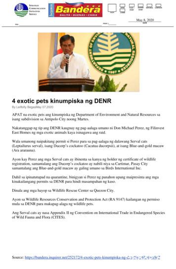
4 exotic pets kinumpiska ng DENR By Leifbilly BegasMay 07,2020 APAT na exotic pets ang kinumpiska ng Department of Environment and Natural Resources sa isang subdivision sa Antipolo City noong Martes. Nakatanggap ng tip ang DENR kaugnay ng pag-aalaga umano ni Don Michael Perez, ng Filinvest East Homes ng mga exotic animals kaya isinagawa ang raid.
4 Eukaryotic genes 1) ribosomal RNA (rRNA) - 4 ribosomal RNA genes from 2 transcripts 2) transfer RNA (tRNA) - carry amino acids that are incorporated into proteins during translation. 3) messenger RNA (mRNA) - translated into proteins 4) heterogeneous nuclear RNA (hnRNA) - an umbrella term that encompases a
be updated and extended to reflect current understanding and finally all the data must be prepared in a coherent, easily accessible manner. These tasks are beyond the scope of the INSDC databases and therefore performed by domain-specific databases. The Ribosomal Database Project (RDP-II) (2,3) and greengenes (4) both cover the
Methods in Enzymology Volume 148 Plant Cell Membranes EDITED BY Lester Packer Roland Douce DEPARTMENT OF PHYSIOLOGY AND ANATOMY DEPARTEMENT DE RECHERCHE FONDAMENTALE UNIVERSITY OF CALIFORNIA CENTRE D'ETUDES NUCLEA1RES ET UNIVERSITE BERKELEY, CALIFORNIA DE GRENOBLE GRENOBLE, FRANCE ACADEMIC PRESS, INC. Harcourt Brace Jovanovich, Publishers
Introduction to Enzymology. Enzymes - Biological catalysts By definition a Catalyst : - Accelerates the rate of chemical reactions . physical methods to solve structure of enzymes Conformation with or without substrate provides functional/biological information Used to identify amino acids involved
Description Logic RWTH Aachen Germany 4. Introduction to DL I A Description Logic - mainly characterised by a set of constructors that allow to build complex concepts and roles from atomic ones, concepts correspond to classes / are interpreted as sets of objects, roles correspond to relations / are interpreted as binary relations on objects, Example: Happy Father in the DL ALC Manu (9has-child .


