Anaesthetic Considerations For Congenital Heart Disease .
3Anaesthetic Considerations forCongenital Heart Disease PatientMohammad HamidAga Khan UniversityPakistan1. IntroductionIncidence of congenital heart disease (CHD) is about 0.8%1 and most of these CHD children(80%) survive to adulthood in developed countries due to early diagnosis and interventionalong with improved surgical and anaesthetic techniques. But the situation is different inmost of the third world countries, where 90% of these children receive suboptimal or nocare2. These patients commonly admitted in the hospital for procedures like cardiaccatheterization, radiological procedures3 4, dental and cardiac surgery.There is increased risk of mortality and morbidity5 under anaesthesia as their anaestheticmanagement in the operating room is challenging in several respect. Few heart defects areso complex that you need to involve paediatric cardiologist and intensivist for completeunderstanding of anatomy and pathophysiology of heart defect.Adult population with congenital heart defects has also increased6 7 over the years andposes more challenges for anaesthesiologist in perioperative period. It is now expected thatsoon there will be more adult with congenital heart defects than children. Grown upcongenital heart (GUCH) is a separate entity, which requires expertise of differentdisciplines to prevent associated morbidity and mortality8 during operations (cardiac or noncardiac) particularly in uncorrected defects and in pregnant patients.When a cardiac defect is recognized in a paediatric patient then the presence of other cardiacand extracardiac lesion is a possibility. The incidence of extra cardiac malformation is as highas 20 – 45% and chromosomal abnormalities in these CHD patients is found to be 5-10%.Perioperative anaesthetic considerations include preoperative evaluation, management ofhypoxaemia, shunt, polycythaemia, pulmonary hypertension and ventricular dysfunction.2. ClassificationSeveral classifications of CHD have been introduced. Two are given below2.1 Cyanotic/Acyanotic CHD2.1.1 Acyanotica. Ventricular Septal Defect (VSD)b. Atrial Septal Defect (ASD)c. Patent Ductus Arteriosus (PDA)d. Atrio ventricular Septal Defect (AVSD)www.intechopen.com
58e.f.g.Perioperative Considerations in Cardiac SurgeryPulmonary Stenosis (PS)Aortic Stenosis (AS)Coarctation of the Aorta2.1.2 Cyanotic CHDa. Tetralogy of Fallotb. Transposition of the Great Arteries (TGA)c. Total Anomalous Pulmonary Venous Return(TAPVR)d. Tricuspid Atresiae. Truncus Arteriosusf. Uncommon, each 1% of CHD, pulmonary atresia, Ebstein’s anomaly.2.2 Classification on the basis of pulmonary and systemic flow1. Excessive Pulmonary Blood Flowa. VSD, ASD, PDA, PAPVR2. Inadequate Pulmonary Blood Flowa. Tetralogy of Fallot, Pulmonary atresia3. Inadequate or obstruction to systemic blood flowa. Coarctation of Aorta4. Abnormal Mixinga. TGA3. Preoperative considerationThree type of paediatric CHD patients are expected to come for evaluation.1. Patients with uncorrected cardiac defect2. Patients who had previous palliative surgerya. ToF with BT shuntb. Atrial septostomy for TGA3. Patients in whom total correction has been done but they may have residual defectsrequiring certain procedures9Preoperative evaluation should include detailed information about cardiac lesion, alteredphysiology and its implications. There are few questions which should be clearly answeredduring preoperative evaluation of these CHD patients. These includesa. Complete understanding of the anatomical changes due to cardiac defect or palliativeprocedureb. Direction and amount of shuntingc. Presence and severity of pulmonary hypertensiond. To what extent pulmonary flow reduced or increased?e. Degree of hypoxaemia, Polycythaemiaf. Coagulation abnormalitiesg. What associated pathophysiological findings likely will influence the management?h. Functional status of the patientFatigue, headache, visual disturbances, depressed mentation and paraesthesia of toes andfingers are presenting symptoms of polycythaemia. History of cyanosis and congestive heartfailure (CHF) are major manifestations of CHD. Fatigue and dyspnoea on feeding andirritability indicate poor functional status. Cyanosis occurs due to decrease pulmonary flowwww.intechopen.com
Anaesthetic Considerations for Congenital Heart Disease Patient59anatomically or functionally (Mixing lesion). Cyanosis may be permanent or appearintermittently. Cyanosis may not be seen in new born due to presence of faetal haemoglobinwhich is highly saturated at a given PaO2.High pulmonary flow leads to pulmonary edema. Failure to thrive and feeding problemsare common in patients with history of repeated pulmonary congestion. Patient maypresents with tachycardia, tachypnoea, irritability, cardiomegaly and hepatomegaly. Theright ventricular function should also be assessed as it is equally important in paediatricCHD patient.Try to avoid dehydration in cyanotic CHD patients by allowing clear liquids two hoursprior to surgery (Table 1). Children also have low glycogen stores which makes themvulnerable to hypoglycemia. If timing of surgery uncertain then start an intravenous lineand give glucose containing solution. Midazolam10 11 is a preferred sedative in the doses of0.5 to 1mg/kg or even higher doses in few studies given orally half hour before surgery(Table 2). If patient is on prostaglandin (PGE1) infusion then it should be continued.2hrsClear liquids (water, apple juice, pedialyte)4hrsBreast milk6hrsFormula & Cow milk6hrsSolidsTable 1. NPO OrdersAgePremedication 6 monthsNone6 months to 8 yearsMidazolamOral 0.5- 1.0mg/kg (Max. 12 mg)Intravenously 0.05 – 0.2mg/kgChloral hydrate 40 – 50mg/kg 8 yearsMidazolam 7.5 mg POMorphine 0.1mg/kg IMKetamine 4mg/kg IMTable 2. Premedication orders4. InvestigationPolycythaemia is very common which increases blood viscosity and leads to thrombosis andinfarction in cerebral, renal and pulmonary region. Although polycythaemia leads tointravascular volume expansion but at the same time reduces plasma volume. Coagulationabnormalities also occur due to hypofibrinogenaemia and factor deficiencies. Platelet count,PT and PTT should be ordered in all patients coming for surgery. Preoperative phlebotomycan be performed in patients with symptomatic hyperviscosity and haematicrit 65%.www.intechopen.com
60Perioperative Considerations in Cardiac SurgeryElectrolyte abnormalities are commonly seen in patients who receive diuretics and parentalnutrition. Hypocalcaemia commonly found in patients with Di George syndrome.ECG may show ventricular strain or hypertrophy pattern. Echocardiography is used fordoppler and color flow mapping while catheterization is used for information aboutpressures in different chambers, magnitude of shunt and coronary anatomy. Examine chestX-Ray for heart position (Dextrocardia) and size, atelectasis, acute respiratory infection,vascular markings and elevated hemidiaphragm. High pulmonary flow will leads toincreased pulmonary marking while reduced flow causes oligaemic lung fields.Neurological assessment and MRI12 may also be needed in these patients. Delayed braindevelopment is associated with certain CHD. Fetal MRI can help in early assessment ofimmature brain.5. Intraoperative considerationsPresence of CHD in paediatric patients poses a great challenge for anaesthetist13 asmorbidity and mortality is quite high. Incidence of cardiac arrest in these paediatric patientsunder anaesthesia is higher14 than non CHD patients and mainly due to pharmacologicalinteraction and over dose5.Intravenous line must be placed in all patients even for minor procedure. All intravenoustubings should be free of air bubble. Polycythaemic patient must be well hydrated beforeinduction either by IV or orally.Sevoflurane15 is preferred over halothane due to better haemodynamic stability in CHDpatients. Most of the CHD patients tolerate inhalation induction with sevoflurane whilepatients with poor cardiac function, may not tolerate inhalation induction. Ionotropesshould be continued if patient is on ionotropes.5.1 MonitoringMonitoring in paediatric CHD is the same as in adult cardiac surgery but there are fewdifferences and considerations during surgery. Monitoring during surgery ranges fromsimple ECG to blood glucose, which is controversial due to non availability of evidence thattight blood sugar control improves outcome16.5.1.1 ElectrocardiogramAlthough ECG can be helpful in the detection of ST changes but is mainly used forarrhythmia detection in paediatric patients. Even arrhythmia detection is difficult due tobaseline tachycardia. Skin should be prepared for electrode by rubbing with alcohol pad orswab. Three leads system is commonly used while in older children five leads system canalso be used.5.1.2 Blood pressure monitoringNon invasive monitoringNon invasive blood pressure should always be monitored even in the presence of arterialline. Cuff should be 20% wider than the diameter of limb where non invasive blood pressureis monitored. Smaller cuff results in erroneously high pressure while larger cuff will givelower pressures.www.intechopen.com
61Anaesthetic Considerations for Congenital Heart Disease PatientInvasive pressure monitoringIt not only provides beat to beat continuous blood pressure monitoring but also provideseasy access for blood sampling. Pressure monitoring tubing and stopcocks should be free ofair to prevent air embolism and damping of system. It is also a major source of fluidoverload as system continuously flushes 2-4 ml/hr per invasive line. In addition a quickflush also pushes about 1-2 ml of fluid per second . Dextrose can be used but usually normalsaline is the flushing solution as bacterial growth is less likely.5.1.3 Central venous pressureCentral venous access not only helpful in monitoring but also provides a reliable route fordrugs, fluid and blood. Right internal jugular vein (Table 3) is commonly used due to itsstraight course to right atrium while left side is avoided due to concerns about its persistentconnection to left SVC (which may be ligated during surgery). Alternatively femoral andsubclavian veins can also be used.WeightCVP 5kg or less than one year4F, 5cm5 – 20 kg4F, 8cm 20 kg5F, 8 or 13cmAdult7FTable 3. Central venous catheter (Internal Jugular Vein) size according to weight5.1.4 Pulse oximetersUsually two oximeters are placed , one in the upper limb and other in the lower extremity.Pulse oximeter uses two light emitting diodes and one photodiode for detection of red andinfra red lights.Accuracy of pulse oximeter is affected by1. Hypotension2. Hypothermia3. Electrocautery4. Artifacts due toa. Thick skinb. Dark colorc. Bright outside lightd. Presence of dyes like indocyanine green and methylene blue5. Abnormal haemoglobinsa. Met Hbb. Carboxy HbNot affected by fetal hemoglobin.www.intechopen.com
62Perioperative Considerations in Cardiac Surgery5.1.5 Cerebral oximeterTranscranial near-infrared spectroscopy (NIRS)17 is a sensitive measure of regionalhypoperfusion. It measures all haemogloblins and useful in non pulsatile cardiopulmonarybypass and circulatory arrest. Cerebral oximeter detects intravascular haemoglobin oxygensaturation of cerebral cortex.5.1.6 EchocardiographyIntraoperative transesophageal echocardiography (TEE) plays a critical role in improvingsurgical outcome in CHD surgeries by confirming diagnosis and identifying residualdefects. It is also helpful in the placement of devices in catheterization lab.Micromultiplane TEE probe and three dimensional technologies are new advances inechocardiography. Epicardial echocardiography is an alternative option in institutionswhere smaller TEE probe is not available18. Adult TEE probe can be used in patientweighing more than 20Kg.Arterial blood gases and blood glucose should also be done frequently. Tight blood glucosecontrol is suggested by certain authors as high blood sugar is toxic to mitochondria.Intraoperative managementAnaesthetic management during surgery depends on presence or absence of shunt,pulmonary hypertension, hypoxaemia, Ventricular dysfunction, pulmonary flow andarrhythmia.5.2 ShuntShunting through these defects depends upon diameter of defect and balance betweensystemic and vascular resistance. Balance between systemic vascular resistance (SVR) andpulmonary vascular resistance (PVR) is essential in the anaesthetic management of patientwith shunts.Normal pulmonary: Systemic ratio (Qp:Qs ratio) is 1:1 which indicate either no shunting orbidirectional shunt of equal magnitude. Qp:Qs ratio of 2:1 indicate left to right shunt whileless than 1:1 ratio (0.8:1) means right to left shunt. The ratio is estimated from oxygensaturation measurements at pulmonary veins, pulmonary artery, systemic arterial andmixed venous blood.5.2.1 Left to right shunt1. Atrial septal defect (ASD)2. Ventricular septal defect (VSD)3. patent ductus arteriosus (PDA)4. Atrio ventricular (AV) canal defects5. Complete anamolous venous return (CAVR)6. Partial anomalous venous return (PAVR)7. Artificial Blalock taussig (BT shunt)L –R shunt reduces greatly with drop in SVR or an increase in PVR. It leads to excesspulmonary blood flow. Patients are usually acynotic but deterioration in gas exchange mayresult from pulmonary congestion. Avoid 100% oxygen and hyperventilation in patientswith L R shunt.www.intechopen.com
Anaesthetic Considerations for Congenital Heart Disease Patient63Patients with PDA are vulnerable to coronary ischaemia11 due to ongoing pulmonaryrunoff during the diastolic phase. Therefore diastolic blood pressure (DBP) should bemonitored during surgery. Diameter of Modified Blalock Taussig shunt is fixed so itsoutput is proportional to SVR and in case of systemic hypotension, the pulmonary bloodflow will be reduced. Blood pressure in the arm will be low due to BT shunt, so usecontralateral limb.5.2.2 Right to leftThese intra cardiac shunts lead to prolong inhalation induction. R L shunt (e.g. tetralogyof fallot (TOF) or shunt reversal12 occur when SVR drops or PVR increases. Hypercyanoticspell under anaesthesia will respond to volume, Increase SVR with alpha agonists such asPhenylephrine.5.3 HypoxaemiaInadequate pulmonary blood flow and/or admixture of deoxygenated with oxygenatedblood in systemic circulation are usually responsible for ischaemia. In addition pulmonarycongestion with inadequate exchange of gases can also leads to hypoxaemia.Persistent hypoxaemia leads to following changesa. Slightly HRb. Hyperventilationc. Polycythaemiad. Chemoreceptor response to hypoxaemia reducede. Cerebral and myocardial oxygenation maintain but Visceral and muscular oxygenationreducedf. Reduced metabolic activity of many organsa. Growth retardationg. Myocardial ischaemia can occurh. Myocardial dysfunctioni. Down regulation of β - receptorsThe anaesthetic management includes adequate hydration, maintenance of systemic bloodpressure, minimizing additional resistance to pulmonary blood flow and avoids suddenincrease in oxygen demand (crying, struggling, and inadequate level of anaesthesia).5.4 Pulmonary hypertension (HTN)During early stages, the pulmonary HTN is reactive and responds to hypothermia, stress,pain, acidosis, hypercarbia, hypoxia and elevated intrathoracic pressure but later pulmonaryHTN becomes fixed. This last stage, where pulmonary vascular resistance exceeds SVR andsymptoms appear due to R L shunt, is the Eisenmenger syndrome13.Anaesthetic risk is quite high including right ventricular failure, bronchospasm, pulmonaryhypertensive crisis and cardiac arrest. Anaesthetic management focus on preventing furtherincrease in R-L shunt by keeping SVR high and PVR low, maintaining myocardialcontractility and prevention of arrhythmia and hypovolemia.5.5 Ventricular dysfunctionChronic volume overload (Large shunts, valvular regurgitation), Obstructive conditions andcardiac muscle diseases leads to reduced ventricular function. Blood gas and X-Ray maywww.intechopen.com
64Perioperative Considerations in Cardiac Surgeryshow metabolic acidosis and pulmonary edema respectively. Patients are usually ondigoxin, diuretics and ionotropes.Anaesthetic considerations includes1. Preoperative optimization of following before surgerya. Ionotopesb. Diureticsc. Digoxind. Antiarrhythmics or ablation in patients with arrhythmia2. Preoperative CBC and electrolytes3. Etomidate and fentanyl provide cardiovascular stability at the time of induction4. Avoid or limit the use of inhalation anaesthetics due to associated myocardialdepression5. Maintain normal sinus rhythm6. Maintain preload during anaesthesia.7. After load reduction in certain situations5.6 Miscellaneous concerns5.6.1 Neurological outcomeThere is growing concern about their quality of life and neurocognitive function, as the longterm survival of these children is now possible. 20 -50% may develop neurologicalimpairment due to chronic hypoxaemia, prolong deep hypothermic circulatory arrest andprolong exposure to anaesthetics. Non pulsatile low flow during cardiopulmonary bypasscausing ischaemia/reperfusion injury may also play a part19.Brain adapts to chronic hypoxia due to presence of NMDA 2B receptors in early life. Corticalneurons may reduce by 30% due to chronic hypoxia causing reduction in brain volume. Butthis reduction is compensated when normoxia develops after surgery. Although most of thearticle have supported the use of high dose narcotics in over all outcome but at present thereis no concrete evidence about best anaesthetic agents for congenital heart surgery.5.6.2 Coagulation disturbancesCoagulation abnormalities are very common in CHD patients particularly in cyanosed andpolycythaemic patient.Coagulation derangement associated with polycythaemia includes:1. Decreased platelet count and function2. Primary fibrinolysis3. Impaired coagulation factors production4. Contracted serum volumeUse of blood products is common in paediatric cardiac surgery due to coagulopathy duringsurgery and several strategies have been instituted to minimize this practice. Preoperativeexchange transfusion of 20 ml/kg FFP to replace same amount of blood is an effectivemethod to counter coagulopathy. Antifibrinolytics like aprotinin and tranexamic acid20 havebeen used for this purpose. Aprotinin is no longer recommended in cardiac surgery due tohigher incidence of renal failure, stroke and myocardial infarction while the use oftranexamic acid has increased.Tranexamic acid as a part of blood saving strategy is given as a bolus of 100mg/kg followedby 10 mg/kg/hr infusion. Whole blood transfusion is quite effective in coagulopathicwww.intechopen.com
Anaesthetic Considerations for Congenital Heart Disease Patient65patients. Factor VII in the dose of 90 microgram/kg is increasingly used in paediatriccongenital heart surgeries.5.6.3 Grown up congenital heart (GUCH)PregnancyIncreased in blood volume during pregnancy may further aggravate the situation andpatient may develop arrhythmias, pulmonary congestion and heart failure. Considerationduring pregnancy ranges from termination of pregnancy to the safe delivery by caesareansection. A multidisciplinary approach involving obstetrician, paediatric cardiac surgeon,paediatric cardiologist, intensivist, anaesthetist and neonatologist is essential in decisionmaking process.Anaesthetic challenges and considerations include1. Invasive line monitoring according to the severity of cardiac defect2. Slow infusion of lowest effective dose of oxytocin as vigorous uterine contraction leadsto high pre load3. Use of inline air filter4. Reduction in SVR should be avoided5. Coagulation abnormalities should also be considered6. Prevention of thromboembolic eventsEisenmengerMost of the patients with Eisenmenger started with simple correctable cardiac defects buteventually leads to severe pulmonary hypertension (PVR 800 dynes/cm-5) which does notrespond to pulmonary vasodilators. Hypoxaemia, myocardial dysfunction and arrhythmiais a common finding.Perioperative risk includes1. Arrhythmia2. Cardiac arrest3. Pulmonary hypertensive crisis4. Bleeding5. ThrombosisAnaesthetic management includes1. Phlebotomy in hyperviscosity syndrome2. Avoid dehydration in preoperative period1. Avoid myocardial depressants2. Keep SVR high3. Try to reduce PVR4. Regional anaesthesia can be used but general anaesthesia is preferred5. Postoperative pain should be adequately managed6. Will require intensive care after surgery6. Common CHD6.1 Ventricular septal defect (VSD)VSD is the most common congenital heart defect. It may be an isolated cardiac defect or maybe associated with other cardiac defects like ASD, PDA or a part of complex defectswww.intechopen.com
66Perioperative Considerations in Cardiac Surgery(tetralogy, AV canal defect). Communication between two ventricles can be of any size andcan occur at any part of septum. Most common type of VSD is peri membranous (also calledsubaortic or infracristal). Other less common defects are subpulmonary (Supra cristal,infundibular or outlet type), Inlet type (canal type) and muscular. Spontaneous closure ispossible in muscular and membranous type of defects.Smaller defects are not associated with large shunting of blood from left ventricle to rightventricle may not diagnose early in life but they are prone to infective endocarditis. Whereaslarger defects cause shunting of blood from left to right ventricle this led to higherpulmonary blood flow and consequently pulmonary congestion. Due to early developmentof symptoms these patients diagnosed earlier. During systole LV ejects blood not only in theaorta but also in the pulmonary artery causing volume overload of pulmonary vessels, atriaand left ventricle. These patients will develop high pulmonary vascular resistance (PVR)and if untreated will leads to Eisemenger.A device like amplatzer can be placed to close few of these defects by interventionalcardiologist. This procedure is performed in the cath lab as a daycare procedure but thereare certain criteria needs to be fulfilled. There should be an adequate rim around the defectswhere amplatzer can be placed. Surgically VSD can be approached through ventricle, aorta,pulmonary artery or right atrium.Anaesthetic considerationsAlways consider high pulmonary vascular resistance in these patients and be ready to treathigh PVR and right ventricular failure by inhaled NO, dobutamine and milrinone. Desirablehaemodynamic goals by anaesthetists are to have slightly higher preload and pulmonaryvascular resistance while keeping the SVR on the lower side and at the same timemaintaining heart rate and contractility. Up to 10% of patients may develop conductionabnormalities after VSD repair which may be transient or permanent.Intraoperative transesophageal echocardiography (TEE) will be beneficial in recognizingresidual defects, intracardiac air and right ventricular function. Smaller VSD are sometimesbecomes apparent after closure of large defect. In uncomplicated VSD closure patient can beextubated in the operating room.6.2 Atrial septal defect (ASD)Normally there is no communication between right and left atria due to presence of aseptum. This atrial septum composed of septum primum and septum secundum whichmerges with endocardial cushion, superior and inferior vena cava.Several types of defects can occur in this septum leading to shunting across. Apart fromsecundum defect other less common are primum, sinus venosus and coronary sinus type.Most common defect is ostium secundum which usually located in the centre (also calledfossa ovalis type) and occurs due to deficient septum primum. It may be single or haveseveral small defects called fenestrated type. Patent foramen ovale commonly seen at thesame site in 25 – 30% of normal patients. Usually PFO do not permit left to right shuntingbut right to left shunting can occurs if right atrial pressure exceeds left atrial pressure(sneezing, valsalva)Sinus venosus defect is usually associated with partial anomalous pulmonary venousdrainage and appears either at the junction of superior vena cava and atrial septum (Highup) or at the junction of inferior vena cava and septum (located lower part of septum).Repair some time may cause injury to SA node.www.intechopen.com
Anaesthetic Considerations for Congenital Heart Disease Patient67Ostium primum defect is due to failure of fusion between endocardial cushion and lowerpart of interatrial septum leading to communication between two atria and usuallyassociated with cleft at anterior mitral leaflet.Coronary sinus type defect is due to absence of wall between left atrium and coronary sinusleading to communication between left and right atrium. It may be associated withpersistent left SVC.Left to right shunting depends on the size of defects and compliance of ventricles asshunting usually occurs during diastole when both mitral and tricuspid valves are open. Ifthe defect is small (less than 5 mm) then it’s called restrictive type while larger defects arenon restrictive and associated with right atrial dilatation, RV volume overload andincreased pulmonary blood flow. Spontaneous closure is possible but most require deviceclosure by cardiologist or surgery.Anaesthetic considerationsInhalation induction in infants and very young and intra venous induction in older childrenis acceptable technique. Intramuscular ketamine can be alternative for induction or intravenous line placement in some children. Pulmonary hypertension is generally not seen inthese patients and their management is usually simple with the goals of higher preload andslightly high PVR to reduce pulmonary flow. Presence of ASD is not usually poses higherrisk for infective endocarditis.TEE is helpful to see the residual ASD, mitral valve repair (primum type), four pulmonaryveins opening in left atrium (Sinus venosus type). Tracheal extubation in the operating roomwill help in minimizing the charges.6.3 Tetralogy of Fallot (ToF)ToF is the most common cyanotic CHD, accounting for 10% of all CHD. It comprises of fouranatomical defects: (i) VSD (ii) RVOT obstruction (iii) RV hypertrophy (iv) Over riding ofaorta.VSD is usually large, non restrictive which led to equalization of RV and LV pressures andshunting through VSD depends primarily on systemic and pulmonary vascular resistance.RVOT obstruction is dynamic due to hypertrophied infundibulum but fixed obstruction canalso occur due to v pulmonary valve stenosis.Due to reduce pulmonary flow, main and branched pulmonary arteries hypoplasia may alsobe seen. Right ventricular hypertrophy is more marked when VSD is restrictive. ToF mayalso be associated with certain defects like anomalous origin of LAD crossing the RVOT,pulmonary atresia, absent pulmonary valve and complete AV canal defect.Palliative surgeryThe classic Blalock Taussig shunt was performed in 1944 to relieve the ToF related cyanosiswhere end to side anastomosis of subclavian artery to pulmonary artery was performed.Today modified BT shunt is the most commonly performed palliative procedure in CHDpatients where a synthetic graft is interpositioned between subclavian artery and ipsilateralpulmonary artery.Complete surgical repairFirst total correction was performed by Lillehei in 1954. Surgical correction involvesinfundibular muscle resection through right ventriculotomy or transpulmonarry approach.www.intechopen.com
68Perioperative Considerations in Cardiac SurgeryPulmonary valve is removed or dilated accordingly and a transannular patch is placed. VSDis also closed at the same setting. Main Pulmonary artery and its branches are also inspectedfor narrowing. Some centres creates small ASD to counteract high right sided pressures.There is trend towards early total correction rather than palliative surgery which is followedby total correction.Anaesthetic managementGoal of anaesthetic management is to avoid low SVR and ionotropes before bypass. Ifpatient is on prostaglandin E1 then it should be continued in pre bypass period. Avoidcatecholamine release in preoperative phase and at the time of induction by providing goodpremedication and adequate analgesia and anaesthesia.Induction can be done with ketamine and fentanyl if intravenous line is in place. Inhalationinduction can also be performed while maintaining SVR. Remember infundibular stenosisincreased by increasing contractility and heart rate, so minimize noxious stimulus avoidcatecholamine release. This is achieved by high dose fentanyl at the maintenance phase.Arterial line and central line should be placed after induction and intubation.Acute desaturation at any time should be considered as tet spell and treated by analgesicsand volume. Phenylephrine should also be available to treat low systemic vascularresistance and hypotension. Steroids given at the time of induction can help in reducingrelease of inflammatory markers during cardio pulmonary bypass.TEE is helpful in assessing residual VSD and infundibular stenosis and degree of pulmonaryregurgitation. In case of tet spell, give 100% O2, Phenyl ephrine, volume, increase depth ofanaesthesia, hyperventilate and give bicarbonate. In addition esmolol or proponol can betried to reduce infundibular spasm.During postbypass period be ready for arrhythmias and heart block, RV dysfunction andcoagulopathy. Ionotrpic support is mandatory in postbypass period along with high fillingpressure particularly if right ventriculotomy was performed. Blood products should beavailable and antifibrinolytics should be started for coagulopathy.6.4 Patent ductus arteriosus (PDA)Ductus arteriosus is a normal communication in fetus, which constrict and closes within 1015 hrs of birth and later closed anatomically by fibrosis in 2 – 3 weeks. Various mechanismhave been described for initial functional closure, which includes increased PaO2, absence ofplacental derived prostaglandins and presence of catecholamines and bradykinins in newborn.Ductus venosus provides a communication between junction of main and
3 Anaesthetic Considerations for Congenital Heart Disease Patient Mohammad Hamid Aga Khan University Pakistan 1. Introduction Incidence of congenital heart disease (CHD) is about 0.8% 1 and most of these CHD children (80%) survive to adulthood in developed countr ies due to
Arrhythmias in congenital heart disease: a position paperof the European Heart Rhythm Association (EHRA), Association for European Paediatric and Congenital Cardiology (AEPC), and the European SocietyofCardiology (ESC) Working Group on Grown-up Congenital Heart Disease, endorsedby HRS, PACES, APHRS, and SOLAECE
Congenital heart disease after childhood: an expanding patient population. 22nd Bethesda Conference, Maryland, October 18-19, 1990. . Repaired congenital heart disease with residual shunts or valvular regurgitation at the site or . Update on Adult Congenital Heart Disease
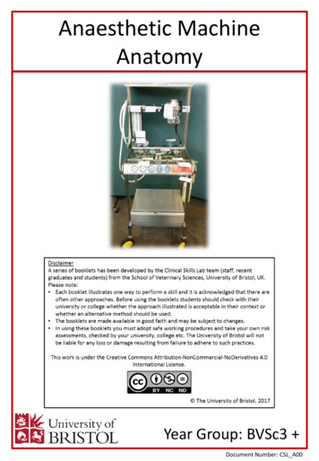
Anaesthetic Machine Anatomy O 2 flow-meter N 2 O flow-meter Link 22. Clinical Skills: 27 28 Vaporisers: This is situated on the back bar of the anaesthetic machine downstream of the flowmeter It contains the volatile liquid anaesthetic agent (e.g. isoflurane, sevoflurane). Gas is passed from the flowmeter through the vaporiser. The gas picks up vapour from the vaporiser to deliver to the .
Bruksanvisning för bilstereo . Bruksanvisning for bilstereo . Instrukcja obsługi samochodowego odtwarzacza stereo . Operating Instructions for Car Stereo . 610-104 . SV . Bruksanvisning i original
Congenital femoral deficiency and fibular hemimelia are rare and complex congenital disorders of the lower limb, with an incidence of approximately one in 50,000 live births for congenital femoral deficiency [12, 19, 30] and between 7.4 to 20 per million live births [6, 11, 33] for fibular hemimelia. Congenital femoral deficiency and fibular
10 tips och tricks för att lyckas med ert sap-projekt 20 SAPSANYTT 2/2015 De flesta projektledare känner säkert till Cobb’s paradox. Martin Cobb verkade som CIO för sekretariatet för Treasury Board of Canada 1995 då han ställde frågan
service i Norge och Finland drivs inom ramen för ett enskilt företag (NRK. 1 och Yleisradio), fin ns det i Sverige tre: Ett för tv (Sveriges Television , SVT ), ett för radio (Sveriges Radio , SR ) och ett för utbildnings program (Sveriges Utbildningsradio, UR, vilket till följd av sin begränsade storlek inte återfinns bland de 25 största
CISC4/681 Introduction to Artificial Intelligence 1 Russell and Norvig: 2 Agents? agent percepts sensors actions environment CISC4/681 Introduction to Artificial Intelligence 2 Agent – perceives the environment through sensors and acts on it through actuators Percept – agent’s perceptual input (the basis for its actions) Percept Sequence – complete history of what has been .


