Part I Sectional Anatomy Of The Body (Chest Abdomen Pelvis .
Outline11.1 Body ImagingPart ISectional Anatomy of the Body(Chest Abdomen(Chest,Abdomen, Pelvis)Carolyn Kaut Roth, RT (R)(MR)(CT)(M)(CV) FSMRTCEO Imaging Education Associatescandi@imaginged.comwww.imaginged.comPart I Planes of the Chest, Abdomen & Pelvis Sectional Anatomy of the Chest & Heart MR imaging of the Chest Sectional Anatomy of the Abdomen & Pelvis MR imaging of the Abdomen & PelvisSlide # 1Imaging PlanesCT ImagesSlide # 2Median LineFrontal orCoronal PlaneMid-sagittal PlaneParasagittal PlanesChest Coronal, Heart & LungsMRI ImagesTransverseOr AxialPlaneMRAAxialAxialCTAHeartCoronalCoronal ReformatLungsSagittal ReformatSagittalSagittalSlide # 3AxialEnhanced Coronal CTCoronalCoronal MRISlide # 4Chest Coronal, VasculatureChest Coronal, Lungs & AirwayUpper Lobe of the right lungUpper Lobe of the left lungMRACTABronchi - carinaMiddle lobe of the right lung*remember there is nomiddle left because of the heart!Pulmonary ArteriesPulmonary VeinsLower lobe of the right lungLower lobe of the left lungDiaphragmCoronal MRICoronal CTEnhanced Coronal CTCoronal MRISlide # 5Slide # 61
Chest Coronal Muscles of the ChestSagittal Chest – “Candy Cane Shot”Pectoralis musclesHeart MuscleAortic ArchAxial MRIAxial CTAscending AortaDescending AortaTrapeziusLatissimusHeartIntercostal MusclesSagittal MRIDiaphragmCoronal MRICoronal CTSagittal CTSlide # 7Axial chestAxial slice #1Axial slice #2Axial slice #3Axial slice #1Axial slice #2Axial slice #3Axial slice #1Axial slice #2Axial slice #3Slide # 8Pectoralis MuscleAortic ArchLatissimus MuscleSpine (vertebral body)Ascending AortaPulmonary arteriesPulmonary veinsDescending AortaHeartSlide # 9Axial slice #1Axial slice #2CT AXIALSlide # 11MRI CORONALMRI AXIALMRI CORONALMRI AXIALSlide # 10Heart AnatomyCTA CORONALCTA CORONALHeart flowIVC & SVC (Inferior & Superior vena cava)RA (right atrium)TriRvP valvePaLungsPvLaBiLvAortic valveAorta – coronaryCT AXIALarchAxial slice #3Heart AnatomyHeart flowSVC (superior vena cava)RA (right atrium)Tri cuspid valveRvP valvePaLungsPvLaBiLvAortic valveAorta – coronaryarchHeart anatomyCTA CORONALHeart flowIVC & SVC (Inferior & Superior vena cava)RA (right atrium)Tri cuspid valveRV (right ventricle)P valvePaLungsP-S vLaBiLvAortic valveAorta – coronaryCT AXIALarchMRI CORONALMRI AXIALSlide # 122
Heart AnatomyCTA CORONALHeart flowIVC & SVC (Inferior & Superior vena cava)RA (right atrium)Tri cuspid valveRV (right ventricle)Pulmonary valvePA (Pulmonary artery)LungsPvLaBiLvAortic valveCT AXIALAorta – coronaryarchMRI CORONALMRI AXIALHeart AnatomyHeart flowIVC & SVC (Inferior & Superior vena cava)RA (right atrium)Tri cuspid valveRV (right ventricle)Pulmonary valvePA (Pulmonary artery)LungsPV (pulmonary veins)LABiLVAortic valveCT AXIALAorta – coronaryArchSlide # 13Heart anatomyCTA CORONALHeart flowIVC & SVC (Inferior & Superior vena cava)RA (right atrium)Tri cuspid valveRV (right ventricle)Pulmonary valvePA (Pulmonary artery)LungsPV (pulmonary veins)LA (Left Atrium)BiLVAortic valveAorta – coronaryCT AXIALArchCTA CORONALHeart flowIVC & SVC (Inferior & Superior vena cava)RA (right atrium)Tri cuspid valveRV (right ventricle)Pulmonary valvePA (Pulmonary artery)LungsPV (pulmonary veins)LA (Left Atrium)Bi cuspid valveLV (Left Ventricle)Aortic valveAorta – coronaryCT AXIALArchSlide # 17MRI CORONALMRI AXIALSlide # 14MRI CORONALMRI AXIALHeart anatomyCTA CORONALHeart flowIVC & SVC (Inferior & Superior vena cava)RA (right atrium)Tri cuspid valveRV (right ventricle)Pulmonary valvePA (Pulmonary artery)LungsPV (pulmonary veins)LA (Left Atrium)Bi cuspid valveLVAortic valveAorta – coronaryCT AXIALArchSlide # 15Heart anatomyCTA CORONALMRI CORONALMRI AXIALSlide # 16MRI CORONALMRI AXIALHeart anatomyCTA CORONALHeart flowIVC & SVC (Inferior & Superior vena cava)RA (right atrium)Tri cuspid valveRV (right ventricle)Pulmonary valvePA (Pulmonary artery)LungsPV (pulmonary veins)LA (Left Atrium)Bi cuspid valveLV (Left Ventricle)Aortic valveAorta – coronaryCT AXIALArchMRI CORONALMRI AXIALSlide # 183
Aortic Arch – the “ A, B, C’s”Aortic Arch – the “ A, B, C’s”Ascending aortaCoronariesBrachiocephalic (aka Innominate)right common carotidright subclavianright vertebralLeft common carotidAscending aortaCoronariesBrachiocephalic (aka Innominate)right common carotidright subclavianright vertebralLeft common carotidLeft SubclavianLeft vertebralLeft SubclavianLeft vertebralMRA CORONALMRA CORONALCTA CORONALCTA CORONALSlide # 19Slide # 20Aortic Arch – the “ A, B, C’s”Aortic Arch – the “ A, B, C’s”Ascending aortaCoronariesBrachiocephalic (aka Innominate)right common carotidright subclavianright vertebralLeft common carotidAscending aortaCoronariesBrachiocephalic (aka Innominate)right common carotidright subclavianright vertebralLeft common carotidLeft SubclavianLeft vertebralLeft SubclavianLeft vertebralMRA CORONALMRA CORONALCTA CORONALCTA CORONALSlide # 21Slide # 22Frontal orCoronal PlaneMedian LineMid-sagittal PlaneImaging PlanesFrontal orCoronal PlaneMedian LineMid-sagittal PlaneImaging PlanesParasagittal PlanesParasagittal PlanesTransverseOr AxialPlaneSagittalAxialCoronalTransverseOr AxialPlaneSagittalCT ImagesAxialCoronalreformatAxialCoronalCT ImagesSagittalreformat Slide # 23AxialCoronalSagittalSlide # 244
Coronal AbdomenCoronal AbdomenApproximateSlice locationApproximateSlice locationApproximateSlice locationApproximateSlice tomachSpleenpLiverBowelSmall BowelStructures of the Small esCoronal MR imageCoronal CT imageCoronal CT imageSlide # 25Coronal MR imageSlide # 26Axial AbdomenCoronal AbdomenDiaphragmApproximateSlice locationLiverSpleenumorbody anatomy Slide 2bDiaphragmApproximateSlice locationStomachStructures of the stomach:Lesser curvatureFundusPylorusGreater curvatureLesser curvatureApproximateSlice locationApproximateSlice locationLiverSpleenSmall BowelStructures of the Small bowelDuodenumJejunemIleumCoronal CT imageLarge bowel (colon)Ascending colonTransverse colonDescending colonSlide # 27Sigmoid colonStomachAortaVertebralbodyAxial CT imageAxial MR imageCoronal MR imageSlide # 28Axial AbdomenAxial AbdomenApproximateSlice locationApproximateSlice locationApproximateSlice locationApproximateSlice Vena cavaAxial CT imageVertebralbodySlide # 29KidneysAxial MR imageAxial MR imageAxial CT imageSlide # 305
Sagittal AbdomenAxial AbdomenApproximateSlice locationApproximateSlice locationThoracicSpineRectus abdominus musclesLiverApproximateSlice locationApproximateSlice locationLiverPancreasLumbar SpineAortaGall l MR imageAxial CT imageSagittal CT imageSlide # 31Symphysis PubisSagittal MR imageSlide # 32Abdominal Vasculature“See Spot Run In.”Abdominal Vasculature“See Spot Run In.”(“C” not see) Celiac, SMA, Renals (right & Left), IMA(“C” not see) Celiac, SMA, Renals (right & Left), IMASMAsuperiormesentericartery“C” CeliacgastrichepaticsplenicArises anteriorlyfrom the aortaat the level ofL2-3Arises theaorta at thelevel of L1Providesblood supplyto thestomach,spleen andliverCoronal CTA ImageProvides bloodsupply to thestomach, smallbowel and partof the colonCoronal MRA imageCoronal CTA ImageSlide # 33Coronal MRA imageSlide # 34Abdominal Vasculature“See Spot Run In.”Abdominal Vasculature“See Spot Run In.”(“C” not see) Celiac, SMA, Renals (right & Left), IMA(“C” not see) Celiac, SMA, Renals (right & Left), IMAIMAInfereriorMesentericArteryRenal arteriesrightleftArisesante io l &anteriorlyinferiorlyfrom theaorta at thelevel of L 4-5ArisesBilaterally andposteriorly fromthe aorta at thelevel of L3-4Coronal CTA ImageProvides bloodsupply to theright & leftkidneySlide # 35Coronal MRA imageCoronal CTA ImageProvidesblood supplyto the inferiorcolon,sigmoid andrectumCoronal MRA imageSlide # 366
Abdominal Vasculature“See Spot Run In.”(“C” not see) Celiac, SMA, Renals (right & Left), IMAAbdominal Veins(IVC)Inferiorvena cavaReview Vasculature Almost ALL Arteries:Portal veinSplenic vein– Carry oxygenated blood away fromthe heart– Carry oxyhemaglobin blood to organs(AAA)AbdominalAorticAneurysmLeft renal ve(SMV)SuperiormesentericVeinAlmost ALL Veins:– Carry deoxygenated blood to theheart– Carry deoxyhemaglobin away fromorgansAbdominalAorta(IVC)Inferiorvena cavaExceptions:Iliacarteries– Portal vein-Right iliacvein Carries deoxyhemaglobin to the liver– Pulmonary veins Carries deoxyhemaglobin to thelungsCoronal abdominal venogram– Pulmonary arteriesCoronal CTA ImageCoronal MRA image Carries oxyhemaglobin to the heartSlide # 37Slide # 38Peripheral Vasculature “Run-off’sFemale Pelvis AnatomyFundusEndometriumAbdominal AortaIliac arteries(at the level of the Ileum)ApproximateSlice locationsFemoral ArteriesSuperficial femoral &common femoral(at the level of the Femur)FundusJunctional ZoneEndometriumPopliteal Arteries(at the level of the Knee)Coronal CTA ImageTrifurcation(Lower Leg)*Anterior Tibeal*Posterior Tibealis*Peroneus BrevisSagittal ultrasound imageUterusCervixVaginaBladderSymphysis pubisCoronal MRA image(at the level of the Foot)Dorsalis PedisMedial MalalearSagittal Reformatted CTSlide # 39Sagittal MRI (T2 image)Slide # 40body anatomy Slide 8aFemale Pelvis AnatomyFemale Pelvis AnatomyApproximateSlice locationsApproximateSlice locationsCT #1CT #2UterusJunctional ZoneEndometriumApproximateSlice locationsMR #1MR #2Rectus abdominus musclesUterusendometriumGleuteal musclesOvaryFallopian tuberectumBl ddBladderAxial CT #1Coronal Reformatted CTMusclesGleuteal MusclesIleumAcetabulumAxial MR #1BladderOvaryFemoral headObturator Internus musclesCoronal MRI (T2 image)CervixVaginaObturator internus musclesObturator extermus musclesAxial CT #2Slide # 41Axial MR #2Slide # 427
Male Pelvis AnatomyMale Pelvis AnatomyApproximateSlice locationsApproximateSlice locationsApproximateSlice locationsApproximateSlice locationsNAVELSymphysis PubisPsoas MusclesBladderProstateCentral glandPeripheral zone(normal)Peripheral zone(cancer)Prostate (base)Seminal vessiclesVas defferensUrethraApex of the ProstateAxial CTPubic boneCoronal Oblique MRI (T2 image)High resolution (small FOV)Coronal Reformatted CTObtuaturatorInternusMusclesSlide # 43Seminal vessiclesUrinaryy BladderProstate BasePeripheral zoneProstate ApexSagittal Reformatted CTAxial MRHigh resolution (small FOV)OutlineApproximateSlice locationsRectumRectumSlide # 44Male Pelvis AnatomyRectus abdominusMusclesNeuro vascularbundleGleutealMusclesSagittal MRI (T2 image)High resolution (small FOV)Part I Planes of the Chest, Abdomen & Pelvis Sectional Anatomy of the Chest & Heart MR imaging of the Chest Sectional Anatomy of the Abdomen & Pelvis MR imaging of the Abdomen & PelvisSymphysis PubisSlide # 45Slide # 4611.1 Body ImagingPart I – Anatomy of the Chest, Abdomen & PelvisThank yyou for yyour attention!Click to take your post test and get your creditsCarolyn Kaut Roth, RT (R)(MR)(CT)(M)(CV) FSMRTCEO Imaging Education e # 478
Sectional Anatomy of the Chest & Heart Outline Slide # 46 MR imaging of the Chest Sectional Anatomy of the Abdomen & Pelvis MR imaging of the Abdomen & Pelvis 11.1 Body Imaging Part I – Anatomy of the Chest, Abdomen & Pelvis Thank you for your attention! Slide # 47 Carolyn Kaut Roth, RT (R)(MR)(CT)(M)(CV) FSMRT
Texts of Wow Rosh Hashana II 5780 - Congregation Shearith Israel, Atlanta Georgia Wow ׳ג ׳א:׳א תישארב (א) ׃ץרֶָֽאָּהָּ תאֵֵ֥וְּ םִימִַׁ֖שַָּה תאֵֵ֥ םיקִִ֑לֹאֱ ארָָּ֣ Îָּ תישִִׁ֖ארֵ Îְּ(ב) חַורְָּ֣ו ם
7', 8', 9' and 10' Sofa 52" and 66" Armless Sofa MAXWELL LEATHER COLLECTION Corner Sectional Left-Arm L Sectional Right-Arm L Sectional U-Sofa Sectional Left-Arm Sofa Chaise Sectional Right-Arm Sofa Chaise Sectional . Leather is waxed and oiled in a meticulous 12-step process for superior suppleness Develops a rich, burnished patina .
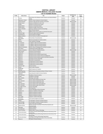
Clinical Anatomy RK Zargar, Sushil Kumar 8. Human Embryology Daksha Dixit 9. Manipal Manual of Anatomy Sampath Madhyastha 10. Exam-Oriented Anatomy Shoukat N Kazi 11. Anatomy and Physiology of Eye AK Khurana, Indu Khurana 12. Surface and Radiological Anatomy A. Halim 13. MCQ in Human Anatomy DK Chopade 14. Exam-Oriented Anatomy for Dental .
39 poddar Handbook of osteology Anatomy Textbook 10 40 Ross ,Pawlina Histology a text & atlas Anatomy Textbook 10 41 Halim A. Human anatomy Abdomen & lower limb Anatomy Referencebook 10 42 B.D. Chaurasia Human anatomy Head & Neck, Brain Anatomy Referencebook 10 43 Halim A. Human anatomy Head & Neck, Brain Anatomy Referencebook 10
teaching anatomy in conventional curricula, clinicians are exposed to more and more cross-sectional anatomy with the sophisticationof the medical imaging []. Certainly the concept of using cross-sectional imaging as an adjunct for teaching anatomy is not new. As early as , % of medical schools in theUnitedStates were using
Descriptive anatomy, anatomy limited to the verbal description of the parts of an organism, usually applied only to human anatomy. Gross anatomy/Macroscopic anatomy, anatomy dealing with the study of structures so far as it can be seen with the naked eye. Microscopic
HUMAN ANATOMY AND PHYSIOLOGY Anatomy: Anatomy is a branch of science in which deals with the internal organ structure is called Anatomy. The word “Anatomy” comes from the Greek word “ana” meaning “up” and “tome” meaning “a cutting”. Father of Anatomy is referred as “Andreas Vesalius”. Ph
1000 days during pregnancy and the first 2 years of life, as called for in the 2008 Series. One of the main drivers of this new international commitment is the Scaling Up Nutrition (SUN) movement.18,19 National commitment in LMICs is growing, donor funding is rising, and civil society and the private sector are increasingly engaged. However, this progress has not yet translated into .


