Surgical Pathology
www.cap.orgSurgical PathologyAnalytes/procedures in bold type are regulated for proficiency testing by the Centers for Medicare & Medicaid ServicesPerformance Improvement Programin Surgical Pathology PIP (PIP1)Category 1 CMEProgramPIPChallenges per ShipmentSurgical pathology case review for one pathologist 10Surgical Pathology Option: PIP1 (For each additional pathologist within the same institution)Note: The PIP1 Program may be ordered only in conjunction with PIP.Product InformationThe Performance Improvement Program in Surgical Pathology (PIP) is designed by pathologists for the education ofpathologists in general surgical pathology. This program provides a practical approach to continuing education insurgical pathology and gives pathologists a method to assess their diagnostic skills and compare their performancewith that of their peers.Each quarterly shipment will contain ten unknown cases with patient histories. PIP case selections represent a varietyof neoplastic and non-neoplastic lesions, including inflammatory and infectious diseases, and will be made fromvarious sites, encompassing essentially all organ sites.The participant selects the appropriate diagnosis from a master list provided with each case. Also included in eachmailing is a review of the case features with diagnostic highlights and educational questions. Participants are asked toreturn to the CAP the completed questionnaire with their diagnoses and answers to the educational questions. Acertificate for CME credit is mailed to the pathologist, and a report tabulated with peer group responses will follow.The slides become the property of the PIP subscribers.PIP is designed for educational purposes only. The program is unsuitable for proficiency testing or grading becauseof the large number of blocks used for each case and their inherent variability and because rare or newly describedlesions are often included.Pathologists can earn a maximum of 40 CME credits (Category 1) per pathologist for completion of an entire year(see Chapter 3, Continuing Education). For those institutions with multiple pathologists interested in participating inPIP and obtaining their own CME credits, the PIP1 option is available.Product Fulfillment Group PIP2008 Surveys & Anatomic Pathology Education ProgramsSurgical Pathology 183
800-323-4040 Option 1 for Customer Contact CenterOnline Virtual Biopsy ProgramProgramOnline virtual biopsy case reviewVBP, VBP1VBP Challenges per Shipment5Category 1 CMEOnline Virtual Biopsy Program Option: VBP1 (For each additional pathologist within the same institution)Note: The VBP1 may be ordered only in conjunction with VBP.Product InformationThe Online Virtual Biopsy Program (VBP) is designed as an educational program for participants to assess andimprove their diagnostic skills in surgical pathology. Using digital image technology to simulate the use of a microscope in evaluating slides enables the use of a wide variety of case materials and provides all participants with identical diagnostic challenges. This VBP is designed as an educational activity and is not designed for proficiency testing.Four online activities will be available in 2008, each containing five diagnostic challenges. Each challenge willconsist of one or more digital images derived from a single case, plus clinical history and other pertinentinformation. Participants will be able to manipulate the digital slide images by scanning throughout the slide fieldand changing the magnification. Some cases may also include gross, radiographic, or endoscopic images.Participants will receive immediate feedback as they select diagnoses from a master list and answer educationalquestions.Case selections will be made from selected organ systems and may include a variety of specimen types (e.g., corebiopsies, endoscopic biopsies, curettings, aspirate smears). The 2008 online VBP activities will focus on thefollowing organ sites:Activity2008-A2008-B2008-C2008-DCourseLiver biopsyGynecologic biopsyProstate biopsySurgical pathology biopsyPathologists can earn a maximum of 10 CME credits (Category 1) for completion of an entire year. For thoseinstitutions with multiple pathologists interested in participating and obtaining their own CME credits, the VBP1option is available.For information on computer requirements and continuing medical education, please see Chapter 3, ContinuingEducation.Online Dates (Participants will be notified by mail when each online activity is available.)College of American Pathologists184Surgical Pathology
www.cap.orgNSH/CAP HistoQIPStain/TissueH&E–Skin (excision, non-neoplastic tissue)H&E–Uterus, to include endometrium and myometriumGrocott methenamine silver (GMS) – Control tissueVerhoeff–van Gieson (VVG) - Lung (excision, non-neoplastic tissue)Cytokeratin 20–Colorectal adenocarcinomaH&E–Breast (to include non-neoplastic parenchyma)H&E–SpleenReticulin–Liver wedge or needle biopsyMucicarmine–Mucin-producing adenocarcinomaHMB45 (or comparable melanoma marker,excluding S100 protein)–MelanomaHQIPHQIP Challenges per ShipmentAB111111111CE1Product InformationThe HistoQIP Program is designed as an educational program to improve the preparation of histologic slides.Participants will receive an evaluation specific to their laboratory, an educational critique, and a participant summaryreport that includes peer comparison data, evaluators’ comments, and performance benchmarking data.Twice each year, participating laboratories will submit one stained and coverslipped glass slide from five differentcases (2 H&E-stained slides, 2 special stains, and 1 immunohistochemical stain). All submitted slides will be recutsof specific surgical tissue types or positive control tissue that will vary from one challenge to the next. Submittedslides will be evaluated for histologic technique by an expert panel of histotechnologists, histotechnicians, andpathologists, using uniform grading criteria. The following areas will be evaluated: fixation, tissue processing andembedding, microtomy, staining, and coverslipping.This Survey provides an education activity that includes reading material found in the Final Critique and onlinelearning assessment questions. All laboratory staff can participate individually and earn free CE credit without leavingthe laboratory.2008 Surveys & Anatomic Pathology Education ProgramsSurgical Pathology 185
800-323-4040 Option 1 for Customer Contact CenterPredictive MarkersAnalytes/procedures in bold type are regulated for proficiency testing by the Centers for Medicare & Medicaid Services (CMS).ImmunohistochemistryCategory 1 CMEMK (MK1)ProcedureMKChallenges per ShipmentImmunohistochemistry 4Immunohistochemistry Option: MK1 (For each additional pathologist within the same institution)Note: The MK1 Program may be ordered only in conjunction with Survey MK.Product InformationSurvey MK is designed as an educational program for laboratories performing immunohistochemistry procedures fordiagnostic support. Participants are asked to stain slides with antibodies to suggested “markers,” using the remainingslides for an H&E stain and appropriate negative controls, and provide interpretation.Each shipment will contain unstained, treated glass slides with brief clinical histories.Pathologists can earn a maximum of four CME credits (Category 1) for completion of an entire year (see Chapter 3,Continuing Education). For those institutions with multiple pathologists interested in participating in Survey MK andobtaining their own CME credits, the MK1 option is available.Product Fulfillment Group MKHER2 Immunohistochemistry, Tissue MicroarrayAnalyteHER2HER2 HER2Challenges per Shipment40Product InformationEach shipment will contain four ten-core tissue microarray slides, which participants are asked to stain for HER2using immunohistochemistry procedures and to provide interpretations.College of American Pathologists186Predictive Markers
www.cap.orgCD117, ER, CD20, EGFR Immunohistochemistry, Tissue MicroarrayPM1, PM2, PM3, PM4AnalyteCD117Estrogen Receptor (ER)CD20Epidermal Growth Factor Receptor (EGFR)PM1PM2PM3 PM4 Challenges per Shipment101010 10Product InformationSurveys PM1, PM2, PM3, and PM4 are designed for laboratories performing immunohistochemistry procedures forpredictive markers. Participants are asked to stain one ten-core tissue microarray slide for the assigned “marker”using immunohistochemistry procedures and provide interpretation.2008 Surveys & Anatomic Pathology Education ProgramsPredictive Markers 187
800-323-4040 Option 1 for Customer Contact CenterCytopathologyAnalytes/procedures in bold type are regulated for proficiency testing by the Centers for Medicare & Medicaid Services (CMS).Interlaboratory Comparison Program inNon-Gynecologic Cytopathology NGC (NGC1)Category 1 CMEProgramNGCChallenges per ShipmentNon-gynecologic cytopathology case review—glass slides 5Non-gynecologic cytopathology case review—online 4 per yearCNGC includes one Laboratory response and two additional individual participants – pathologists orcytotechnologistsCytopathology Options: NGC1 (For each additional pathologist or cytotechnologist within the same institution)Note: The NGC1 Program may be ordered only in conjunction with NGC.CEProduct InformationThe Interlaboratory Comparison Program in Non-Gynecologic Cytopathology (NGC) is designed as an educationalopportunity for participants to assess their screening and interpretive skills. Because NGC cases are chosen for theireducational value, the NGC Program is unsuitable for proficiency testing.Each quarterly shipment will include five specimens on glass slides with patient histories. Cases include fine needleaspirations and exfoliative specimens representing a variety of conditions, both benign and malignant. Referenceinterpretations and laboratory peer performance, along with concise cytologic features and pertinent references, areavailable within 20 minutes by fax, providing rapid educational feedback, peer comparison, and time to furtherreview the material before returning the slides to the CAP.NGC Shipments will contain instructions for accessing online virtual microscopy cases (four per year), which usedigital image technology to simulate the use of a microscope. Online images will consist of rare and unusual nongynecologic cases with ancillary information when appropriate. Participants will be able to manipulate the images byscanning across the slide, moving between planes (where appropriate), and changing the magnification. Participantswill receive immediate feedback as they select interpretations from a master list and answer educational questions.Pathologists can earn a maximum of 22 CME credits (Category 1) and cytotechnologists can earn a maximum of 22CE credits/hours for completion of an entire year: twenty credits for glass slide review and two credits for online casereview (see Chapter 3, Continuing Education).The NGC Program includes a laboratory response and two individual response forms. For institutions with multiplepathologists or cytotechnologists interested in participating in the glass slide portion of the NGC Program andobtaining his/her own credits/hours, the NGC1 option is available. The four online cases may be accessed andcompleted for two CME/CE credits by all staff in a participating institution independent of the number of NGC1options ordered.Product Fulfillment Group NGCSlide sets must be returned by a trackable method within the time period stated in the kit instructions.Laboratories not returning their slides on time will forfeit subsequent shipments and may be ineligible to enroll infuture cytopathology programs.College of American Pathologists188Cytopathology
www.cap.orgLaboratories subject to CLIA should enroll in one of the PAP PT programs (see page 190). For laboratories not subjectto CLIA, the PAPCE1, PAPKE1, PAPME1, and PAPJE1 programs meet the CAP Laboratory Accreditation Programrequirement for participation in a peer educational program.Interlaboratory (PAP Education) Comparison Program inGynecologic Cytopathology PAPCE1, PAPKE1, PAPME1, PAPJE1ProcedureEach module includes the following slide types:ConventionalThinPrep SurePath PAPCE1 PAPKE1PAPME1 PAPJE1 Challenges per Shipment Category 1 CMECE5 slides 2 online cases5 slides 2 online cases5 slides 2 online casesAPAPCE1, APAPKE1, APAPME1, APAPJE1 (for each pathologist or cytotechnologist within the same institution)Product InformationThe Interlaboratory Comparison Program in Gynecologic Cytopathology (PAP) is designed as a peer educationalopportunity for participants to assess their screening and interpretive skills. Each semi-annual shipment will includefive ungraded specimens on glass slides with patient histories. For PAPCE1, the ungraded slides will be conventionalpreparations. For PAPKE1 and PAPME1, the slidesets will contain pure liquid-based slides reflective of the modulechosen at enrollment. For PAPJE1, the slidesets will be a combination of conventional, ThinPrep , and SurePath slides. Reference interpretations and laboratory performance profiles are available within 20 minutes by fax,providing rapid educational feedback, peer comparison, and time to further review the material before returning theslides to the CAP.Each shipment will also contain instructions to access two online virtual microscopy cases (four per year), which usedigital image technology to simulate the use of a microscope. Online images will present diagnostic challengesincorporating Bethesda terminology. Participants will be able to manipulate the images by scanning across the slide,moving between planes, and changing the magnification. Participants will receive immediate feedback as they selectinterpretations from a master list and answer case-related educational questions.Pathologists can earn a maximum of 12 CME credits (Category 1) and cytotechnologists can earn a maximum of 12CE credits/hours for completion of an entire year: ten credits for glass slide review and two credits for online casereview (see Chapter 3).The PAP Education base program includes one laboratory response form. For each pathologist or cytotechnologist toparticipate in the 10-case glass slide portion of the PAP Program and obtain his/her own credits/hours, an APAPoption (APAPCE1/APAPKE1/APAPME1/APAPJE1) should be ordered. The four online cases may be accessed andcompleted for two CME/CE credits by all staff in a participating institution independent of the number of APAP optionsordered.Slide sets must be returned by a trackable method within the time period stated in the kit instructions.Laboratories not returning their slides on time will forfeit subsequent shipments and may be ineligible to enroll infuture cytopathology programs.2008 Surveys & Anatomic Pathology Education ProgramsCytopathology 189
800-323-4040 Option 1 for Customer Contact CenterCMS APPROVED CYTOLOGY PROFICIENCY TESTING PROGRAM2008 Gynecologic Cytology PT ProgramCategory 1 CMECEProcedureConventionalThinPrep SurePath PAP PT Laboratory EnrollmentPAP PTChallenges per ShipmentProficiency Education EducationPAPCPT PAPKPT* PAPMPT* PAPJPT PPTENR TestingAB 5 slides 2 online5 slides 5 slides 5 slides2 online2 online2 online10 slidescasescases APAPCPT, APAPKPT, APAPMPT, APAPJPT (for each pathologist or cytotechnologist on the same testing date within thesame institution)* The PAPKPT and PAPMPT slidesets will contain ten liquid-based slides only.** Per CLIA ’88 regulations, all laboratories must be enrolled in an approved cytology proficiency-testing programand all individuals must be tested by an approved program.The PAP PT Laboratory Enrollment Only is designed for laboratories that possess a CLIA license to perform gyneclogic cytology but have personnel that are testing at another location. The laboratory must be enrolled in aproficiency-testing program in order to be compliant with CLIA ’88, even though personnel are not testing at thatlocation.Product InformationPAP PT builds on the PAP Program that you trust and includes two components: a CMS-approved CytologyProficiency Testing (PT) Program and an educational Interlaboratory Comparison Program in GynecologicCytopathology. To order the PAP PT Program, you must obtain an enrollment packet from the CAP bycalling 800-323-4040 option 1 or by downloading the packet from www.cap.org.The PAP PT Program mailing will occur on one of 24 test sessions based on participant preference and spaceavailability. Successful completion of the PAP PT Program satisfies an individual’s 2008 cytology PT requirementper CLIA.Participation in the two education mailings (the Interlaboratory Comparison Program in Gynecologic Cytopathologyor PAP Education) meets the CAP Laboratory Accreditation Program requirement for participation in a peereducational program. Each of the two education mailings will include five glass slides. Reference interpretations andlaboratory performance profiles for the five glass slides are available within 20 minutes by fax, providing rapideducational feedback, peer comparison, and time to further review the material before returning the slides to theCAP. Laboratories may choose either Education Series 1 or Education Series 2 ship dates.Each education mailing will also contain instructions to access two online virtual microscopy cases (four per year),which use digital image technology to simulate the use of a microscope. Online images will consist of diagnosticchallenges incorporating Bethesda terminology. Participants will be able to manipulate the images by scanning acrossthe slide, moving between planes, and changing the magnification. Participants will receive immediate feedback asthey select interpretations and answer case-related educational questions.Pathologists can earn a maximum of 12 CME credits (Category 1) and cytotechnologists can earn a maximum of 12CE credits/hours for completion of an entire year: ten credits for glass slide review and two credits for online casereview (see Chapter 3).PT Event Date Choices: see shipping calendar at end of catalogCollege of American Pathologists190Cytopathology
www.cap.orgOnline Digital Slide Program in Fine Needle AspirationFNA (FNA1)ProgramOnline digital slide fine needle aspiration case reviewFNACategory 1 CMEChallenges per Shipment5 2Online Digital Slide Program in FNA Option: FNA1 (For each additional pathologist within the same institution)Note: The FNA1 Program may be ordered only in conjunction with FNA.Product InformationThe Online Digital Slide Program in Fine Needle Aspiration (FNA) is an educational program for pathologists andexperienced cytotechnologists to assess and improve their diagnostic skills in non-gynecologic fine needle aspirations. Using digital image technology to simulate the use of a microscope in evaluating slides enables the use of awide variety of case materials and provides all participants with identical diagnostic challenges. This FNA programwill focus on diagnostic dilemmas encountered by pathologists in practice and is designed as an educational activity.It is not suitable for proficiency testing.Two online activities will be available, each activity containing five diagnostic challenges. Each challenge will consistof one or more digital images derived from a single case, plus clinical history and other pertinent information.Participants will be able to manipulate the digital slide images by scanning throughout the slide field, moving betweenplanes (where appropriate), and changing the magnification. Participants will receive immediate feedback as theyselect interpretations from a master list and answer educational questions.Selections of ancillary studies such as immunohistochemical stains, molecular tests, and/or flow cytometry will beavailable as appropriate.Pathologists can earn a maximum of 5 CME credits (Category 1) and cytotechnologists can earn a maximum of 5 CEcredits/hours for completion of an entire year. For those institutions with multiple pathologists and/or cytotechnologists interested in participating and obtaining their own CME/CE credits, the FNA1 option is available.Product Fulfillment Group FNAHuman Papillomavirus for CytologyCHPVD, CHPVM, CHPVK, CHPVJAnalyteHPVCHPVD CHPVM CHPVK CHPVJ NewChallenges per Shipment5Note: These Surveys are intended for cytopathology labs that have their own CLIA number and are performing HPVtesting. Each product will meet regulatory requirements for vial identification.Product InformationSurveys CHPVD, CHPVM, CHPVK, and CHPVJ are designed for laboratories performing nucleic acid amplification forHPV. Each shipment will include simulated cervical specimens.Survey CHPVD Digene Specimen Transport Medium (STM)Survey CHPVM ThinPrep Preservcyt transport mediaSurvey CHPVK SurePath Preservative Fluid transport mediumSurvey CHPVJ Digene, Preservcyt, and/or SurePath transport mediumProduct Fulfillment Group CHPV2008 Surveys & Anatomic Pathology Education ProgramsCytopathology 191
800-323-4040 Option 1 for Customer Contact CenterSpecialty Anatomic PathologyAnalytes/procedures in bold type are regulated for proficiency testing by the Centers for Medicare & Medicaid Services (CMS).Neuropathology ProgramCategory 1 CMEProgramNeuropathology case review Now on CD-ROMNP (NP1)NP Challenges per Shipment8Neuropathology Option: NP1 (For each additional pathologist within the same institution)Note: The NP1 Program may be ordered only in conjunction with NP.Product InformationThe Neuropathology Program is designed as an educational program for anatomic pathologists, neuropathologists,and trainees to assess and improve their diagnostic skills and to learn of new developments in neuropathology.Each shipment contains a CD-ROM with eight cases that cover the spectrum of neoplastic and non-neoplasticdisorders affecting the central and peripheral nervous systems, including infectious, degenerative, developmental,demyelinating, traumatic, toxic-metabolic, vascular, and neuromuscular diseases. Four of the eight cases in eachshipment comprise a mini-symposium focused on a specific problem area in neuropathology. The other four casescover a variety of nervous system diseases.For each case, pertinent clinical and laboratory information and several microscopic images are provided. Somecases also include radiographic and/or gross images. Following review of the case materials, participants answer aseries of questions directed at diagnosis, interpretation, and pathophysiology.All participants receive on the CD-ROM a critique including diagnosis, focused discussion and review, and selectedreferences for each case. Participants can also download case materials from the CD-ROM for educational purposes.The NP cases are selected for their educational value; the case selection and evaluation process make the programunsuitable for proficiency testing or grading.Pathologists can earn a maximum of eight CME credits (Category 1) for completion of an entire year (see Chapter 3,Continuing Education). For those institutions with multiple pathologists interested in participating and obtaining theirown CME credits, the NP1 option is available. Survey NP1 will include only a result form and CME form for anadditional pathologist within the same institution.Product Fulfillment Group NPCollege of American Pathologists192Specialty Anatomic Pathology
www.cap.orgAutopsy PathologyAU (AU1), AUCD (AUCD1)ProgramAUAUCDAutopsy case analysis Autopsy case analysis (CD-ROM) Autopsy Pathology Option:AU1 (For each additional pathologist within the same institution)Autopsy Pathology CD-ROM Option:AUCD1 (For each additional pathologist within the same institution)Category 1 CMEChallenges per Shipment66Note: The AU1 Program may be ordered only in conjunction with Survey AU. The AUCD1 Program may be orderedonly in conjunction with Survey AUCD.Product InformationParticipants in the program will receive detailed discussions of challenging autopsy cases. Each shipment of SurveyAU will include six cases, each consisting of a case description and illustrative 35mm gross and/or microscopicslides, with questions directed at aspects of interpretive analysis, differential diagnosis, pathophysiology, cause ofdeath, and quality assurance. In addition, each case will include a PowerPoint summary slide highlighting the keyteaching points of each case. Survey AU1 will include only a Result Form and CME form for an additional pathologistwithin the same institution. To provide participants with immediate feedback, detailed discussions with currentreferences will be included in the original mailing.Each shipment of Survey AUCD will include the same six cases as above, but instead of 35mm slides, the images willbe provided on a CD-ROM. Survey AUCD1 will include only a Result Form and CME form for an additional pathologistwithin the same institution. To provide participants with immediate feedback, detailed discussions with current references will be included in the original mailing.Pathologists can earn a maximum of 12 CME credits (Category 1) for completion of an entire year (see Chapter 3,Continuing Education). For those institutions with multiple pathologists interested in participating and obtaining CMEcredit, the AU1 and AUCD1 options are available. Surveys AU and AUCD are designed for educational purposes andare not suitable for proficiency testing.Discover This Unique PublicationAn Introduction to Autopsy Technique, 2nd EditionKim A. Collins, MD, and Grover M. Hutchins, MD, editorsThe 2nd edition of this “how-to” manual depicts both general and specialized techniques forperforming autopsies in postmortem examinations in a hospital setting. 2-volume set, softcoverplus laminated diagrams.2008 Surveys & Anatomic Pathology Education ProgramsSpecialty Anatomic Pathology 193
800-323-4040 Option 1 for Customer Contact CenterCytogenetics and Molecular PathologyAnalytes/procedures in bold type are regulated for proficiency testing by the Centers for Medicare & Medicaid Services (CMS).ACMG/CAP Fluorescence In Situ Hybridization – Breast Cancer(HER2 Gene Amplification) CYHProcedureHER2 gene amplification for breast cancerCYH Challenges per Shipment10Product InformationSurvey CYH is designed for clinical laboratories that perform fluorescence in situ hybridization (FISH) usingchromosome-specific HER2 DNA probes. Participants will use their own chromosome-specific DNA probes andFISH methodology to process and score a series of interphase nuclei for each specimen.Each mailing will consist of two five-core tissue microarray slides that offer a combined total of ten paraffin-embedded breast tissues specimens. A duplicate set of H&E stained tissue microarray slides will be provided with theshipment.College of American Pathologists194Cytogenetics and Molecular Pathology
www.cap.orgACMG/CAP FISH For Paraffin-Embedded Tissue – CYJ, CYK, CYLChallengesper ShipmentGene/Region of InterestCYJ(Glioma tissue)1p/19qMYCNALKCHOPMYCCYK(Sarcoma tissue orpediatric neoplasm)CYL(Lymphoma tissue) A111B111Product InformationThese Surveys are designed for clinical laboratories that perform fluorescence in situ hybridization (FISH) usingchromosome-specific DNA probes in paraffin-embedded tissue (see CYF for FISH in cell suspension specimens).Participants will use their own chromosome-specific DNA probes and FISH methodology to process and score aseries of interphase nuclei for each specimen. All specimens will be 4-micron tissue sections mounted on positivelycharged glass slides. One hematoxylin-eosin stained slide will also be provided with each challenge for reference.The first mailing (CYP-A) will include one challenge each for CYJ, CYK, and CYL. CYJ-A will consist of four unstainedslides from paraffin-embedded tissue representing a neurological cancer specimen (glioma). Participants are to useFISH probes for detection of deletions within 1p36 and 19q13 band regions. CYK-A will consist of two unstainedslides from paraffin-embedded tissue representing a pediatric neoplasm. Participants are to use FISH probes to detectaberrations of the MYCN gene (2p24.3). CYL-A will consist of two unstained slides from paraffin-embedded tissuerepresenting a lymphoma specimen. Participants are to use FISH probes to detect rearrangement of the ALK gene(2p23).The second mailing (CYP-B) will include one challenge each for CYJ, CYK and CYL. CYJ-B will consist of fourunstained slides from paraffin-embedded tissue representing a neurological cancer specimen (glioma). Participantsare to use FISH probes for detection of deletions within 1p36 and 19q13 band regions. CYK-B will consist of twounstained slides from paraffin-embedded tissue representing a sarcoma tissue. Participants are to use FISH probes todetect aberrations of the CHOP (DDIT3) gene (12q13). CYL-B will consist of two unstained slides from paraffinembedded tissue representing a lymphoma specimen. Participants are to use FISH probes to detect rearrangement ofthe MYC gene (8q24.1).Product Fulfillment Group CYP2008 Surveys & Anatomic Pathology Education ProgramsCytogenetics and Molecular Pathologyy 195
800-323-4040 Option 1 for Customer Contact CenterIn Situ HybridizationProcedureIn situ hybridizationKappa lambdaISHISH Challenges per Shipment11Note: Brightfield in situ hybridization for HER2 is now offered in ISH2Product InformationSurvey ISH offers laboratories performing clinical in situ hybridization tests the opportunity to objectively evaluatetheir performance for targets including human papillomavirus (HPV) and Epstein-Barr virus (EBV). Informationincluding the conditions of slide pretreatment, probe type, hybridization conditions, and detection systems will beused to facilitate interlaboratory comparison of methods and standardization. Laboratories performing FISH forinterphase chromosomal targets in paraffin sections should refer to CY Surveys.Each shipment for in situ hybridization analysis will include a set of treated glass sli
Surgical Pathology 183 2008 Surveys & Anatomic Pathology Education Programs.cap.org Program PIP Challenges per Shipment Surgical pathology case review for one pathologist 10 Product Information The Performance Improvement Program in Surgical Pathology (PIP) is designed by pathologists for the
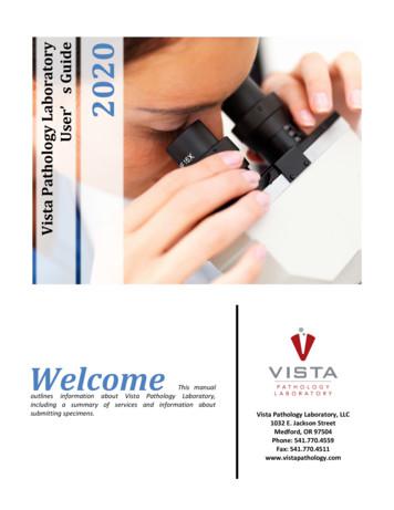
Vista Pathology Laboratory – User’s Guide 1 Who We Are Reedy, Michael MD Pathology Nixon, Randal MD, PhD Pathology Neuropathology Loudermilk, Allison MD Pathology Hematopathology Wu, Bryan MD Pathology Breast Pathology Dermatopathology Pike, Robin MD Pathology Cy
Pathology: Molecular Pathology Page updated: August 2020 This section contains information to help providers bill for clinical laboratory tests or examinations related to molecular pathology and diagnostic services. Molecular Pathology Code Chart The chart included later in this section correlates molecular pathology CPT and HCPCS
Subspecialty: Hepatobiliary Pathology, Gastrointestinal Pathology Assistant Professor of Pathology Dr. Kiyoko Oshima is the Director of Clinical Hepatic Pathology and Assistant Professor in the Department of Pathology at the Johns Hopkins Hospital University School of Medicine. She joined the Hopkins faculty in 2017.
Chicago Pathology Society CLINICAL INTERESTS: Neuropathology, Cytopathology, Autopsy, Surgical Pathology pathology.osu.edu Saman SeyedAhmadian, MD is an Assistant Professor - Clinical for Ohio State’s Department of Pathology. Insert photo here THE OHIO STATE UNIVERSITY DEPARTMENT OF PATHOLOGY
unexpected diagnoses in surgical pathology and cytopathology from the College of American Pathologists and Association of Directors of Anatomic and Surgical Pathology. Arch Pathol Lab Med. 2012; 136(2): 148-154. Rosai J, Bonfiglio TA, Carson JM, et. al. Standardization of the surgical pathology report. Mod Pathol. 1992; 5(2): 197-199.
Gynecologic pathology 5% *3 most common non-surgical pathology fellowships completed by trainees. (Gratzinger et al. Arch Pathol Lab Med. 2018 Apr;142(4):490-495.) Zynger DL, Pernick N. Understanding the Pathology Job Market: An Analysis of 2330 Pathology Job Advertisements From 2013 Through 2017. Ar
Surgical Pathology Dissection An Illustrated Guide Second Edition William H. Westra, M.D. Ralph H. Hruban, M.D. Department of Pathology Department of Pathology The Johns Hopkins University The Johns Hopkins University School of Medicine School of Medicine Baltimore, Maryland Baltimore, Maryland Timothy H. Phelps, M.S. Christina Isacson, M.D.
Unit 14: Advanced Management Accounting Unit code Y/508/0537 Unit level 5 Credit value 15 Introduction The overall aim of this unit is to develop students’ understanding of management accounting. The focus of this unit is on critiquing management accounting techniques and using management accounting to evaluate company performance. Students will explore how the decisions taken through the .


