Specifying Neural Crest Cells: From Chromatin To .
Received: 1 May 2017Revised: 26 March 2018Accepted: 27 March 2018DOI: 10.1002/wdev.322ADVANCED REVIEWSpecifying neural crest cells: From chromatin to morphogensand factors in betweenCrystal D. Rogers1 Shuyi Nie21Department of Biology, College of Science andMathematics, California State UniversityNorthridge, Northridge, California2School of Biological Sciences and Petit Institutefor Bioengineering and Bioscience, GeorgiaInstitute of Technology, Atlanta, GeorgiaCorrespondenceCrystal D. Rogers, Department of Biology,College of Science and Mathematics, CaliforniaState University Northridge, 18111 NordhoffStreet, Northridge, CA 91330-8238.Email: crystal.rogers@csun.eduandShuyi Nie, School of Biological Sciences and PetitInstitute for Bioengineering and Bioscience,Georgia Institute of Technology, Atlanta, GA30332.Email: shuyi.nie@biology.gatech.eduNeural crest (NC) cells are a stem-like multipotent population of progenitor cellsthat are present in vertebrate embryos, traveling to various regions in the developing organism. Known as the “fourth germ layer”, these cells originate in the ectoderm between the neural plate (NP), which will become the brain and spinal cord,and nonneural tissues that will become the skin and the sensory organs. NC cellscan differentiate into more than 30 different derivatives in response to the appropriate signals including, but not limited to, craniofacial bone and cartilage, sensorynerves and ganglia, pigment cells, and connective tissue. The molecular and cellular mechanisms that control the induction and specification of NC cells include epigenetic control, multiple interactive and redundant transcriptional pathways,secreted signaling molecules, and adhesion molecules. NC cells are important notonly because they transform into a wide variety of tissue types, but also becausetheir ability to detach from their epithelial neighbors and migrate throughout developing embryos utilizes mechanisms similar to those used by metastatic cancer cells.In this review, we discuss the mechanisms required for the induction and specification of NC cells in various vertebrate species, focusing on the roles of earlymorphogenesis, cell adhesion, signaling from adjacent tissues, and the massivetranscriptional network that controls the formation of these amazing cells.This article is categorized under:Nervous System Development Vertebrates: General PrinciplesGene Expression and Transcriptional Hierarchies Regulatory MechanismsGene Expression and Transcriptional Hierarchies Gene Networks andGenomicsSignaling Pathways Cell Fate SignalingKEYWORDSBMP, epigenetic, FGF, morphogen, neural crest, specification, Wnt1 INTRODUCTIONThe complexity of the vertebrate form requires gene and protein regulation at many levels, as well as tissue interactions, cellmovements and perfect timing (Martik & Bronner, 2017). Although we have yet to discover all of these controls, the development of one cell type in particular, neural crest (NC) cells, has been studied at length. Ectodermal cells respond to many instructive signals very early in development to form neural tissue (Gaur et al., 2016; Lamb et al., 1993; Rogers, Archer, Cunningham,Grammer, & Casey, 2008), epidermal/placodal tissue (Nordin & LaBonne, 2014; Schlosser, 2014), or NC tissue (Duband,Dady, & Fleury, 2015). The epigenetic and molecular specification of each of these tissue-types is followed by morphogeneticevents such as neural tube closure and NC cell migration. The neural plate begins as a flat epithelial layer (Figure 1a, blue), butrises to meet in the center of the embryo and create the neural tube (Wilde, Petersen, & Niswander, 2014) (Figure 1b,d), whichWIREs Dev Biol. ley.com/devbio 2018 Wiley Periodicals, Inc.1 of 23
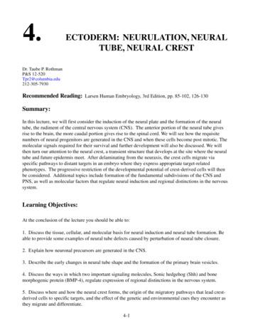
2 of 23ROGERS AND NIEMorphogenesis and NC specification. (a, a0 ) In chicken embryos, the NPB (green) is specified by Hamburger Hamilton stage 5 (HH5) prior tothe onset of definitive neural crest (NC) markers and expresses transcription factors such as MSX1, ZIC1, and PAX7. (b, b0 ) As neurulation proceeds at HH7HH8, the neural folds rise and bend toward the midline. At this stage the neural folds are being specified as definitive NC cells. (c, c0 ) By HH8, definitive NCmarkers are expressed in the dorsal neural tube (SOX9, SOX10, SNAI2, and FOXD3), the neural tube is closed, and the ectodermal cells are converging onthe midline to cover the neural tube. (d, d0 ) By HH9, the NC cells are beginning to undergo EMT and start detaching from the neural tube. ECT, ectoderm;NC, neural crest; NF, neural folds; NPB, neural plate border; NP, neural plateFIGURE 1gives rise to the central nervous system. At the same time, the nonneural ectodermal (NNE) cells meet and separate from the neural tube, covering the embryo (Figure 1d, orange), eventually differentiating into epidermis and placodes (Groves & LaBonne,2014). In chicken embryos, NC cells, which begin as neuroepithelial cells (Figure 1c, green), undergo an epithelial to mesenchymal transition (EMT) after the neural tube closes, and leave their former neighbors, the neural and NNE, and migrate throughoutthe embryo creating diverse derivatives (Figure 1d, green) (Rogers, Jayasena, Nie, & Bronner, 2012). However, in different species, NC cells are specified adjacent to the neural plate (NP) and migrate while the neural tube is closing (mouse and frog)(R. T. Lee et al., 2013; Linker, Bronner-Fraser, & Mayor, 2000). The molecular and morphogenetic mechanisms that regulate thespecification of each of these tissues act in concert with symphonic precision, and in this review we will highlight some of themolecular mechanisms that drive NC induction and specification.NC cells, in their current form are thought to be a unique vertebrate trait; however, recent evidence has suggested that the NChas its origins in multiple migratory and/or pigmented cell types in closely related chordates (Abitua, Wagner, Navarrete, & Levine,2012; Oonuma et al., 2016; Stolfi, Ryan, Meinertzhagen, & Christiaen, 2015). Analysis of both Ciona and Amphioxus embryosdemonstrated that NC-related proteins seem to have conserved functions with vertebrate NC proteins, and that these pigmentedand/or migratory cell types are controlled by conserved NC-specific transcription factors (Abitua et al., 2012; Tai et al., 2016). However, in the less-derived species, NC cells do not form the traditional derivatives such as craniofacial structures (Green, SimoesCosta, & Bronner, 2015). Formation of the NC is mediated by a series of regulatory interactions including epigenetic changes and atightly regulated transcriptional gene regulatory network (GRN) that is largely conserved across vertebrates (Green et al., 2015). TheNP border (NPB), induced during gastrulation, includes the tissues that will give rise to the NC. However, NC cells only becomemorphologically recognizable as neurulation proceeds, where they manifest in an anterior to posterior fashion arising in(e.g., chicken), or adjacent to (e.g., frog), the dorsal neural tube as neural tube closure occurs. These cells are first specified in thehead (cranial NC) and proceeding caudally to form cardiac and vagal NC, then trunk and finally sacral NC cells (graphical abstract).Although premigratory NC cells are neuroepithelial as they are specified, they eventually alter the expression of their cell–celladhesion molecules, and undergo cytoskeletal changes that result in an EMT, allowing them to delaminate from the epithelialsheet and start migrating both collectively and individually in the developing embryo (Theveneau et al., 2013). Normal formationand migration of NC cells is crucial for the development of craniofacial structures, pigment cells, and the peripheral nervous system among a multitude of derivatives. Additionally, the abilities of NC cells to migrate extensively and to differentiate intodiverse cell types, are reminiscent of stem cells and metastatic cancer cells in that they utilize similar molecular pathways to selfrenew (Kerosuo, Nie, Bajpai, & Bronner, 2015), migrate, invade tissues, and proliferate (Gallik et al., 2017). These unique characteristics have made NC cells an interesting and well-studied topic for many years. This review will focus on the molecularevents controlling the specification of NC cells in vertebrate embryos, specifically characterizing the events in amphibians (frog)and avians (chick). Here, we give an updated view of early patterning of the NPB and the segregation of NC cells from neuralectoderm with a focus on morphogenetic events, gene regulation and the signaling involved in the process.
ROGERS AND NIE3 of 232 M O R P H O G E N E S I S , T IS S U E I N T E R A C T I O N S , A N D N C I N D U C T I O N2.1 Morphogenetic movements during NC specificationThe process of gastrulation allows for the creation of the three germ layers, endoderm, mesoderm, and ectoderm. The mostsuperficial germ layer, the ectoderm, divides into neural, nonneural, and NPB cells soon after the ectoderm is specified. Inmany species, the formation of the NC from the unspecified ectoderm relies on the concomitant formation of the adjacent NP,and a specific transcriptome is activated in response to signaling pathways in the early embryo. In frog embryos, this earlytranscriptome has been described by an EctoMap, which details the spatiotemporal localization of ectoderm specification cascades (Plouhinec et al., 2017; Simoes-Costa, Tan-Cabugao, Antoshechkin, Sauka-Spengler, & Bronner, 2014). Multiple studies have detailed the direct and indirect transcriptional interactions during chicken NC specification and induction, and newdetails are ever emerging (Prasad, Sauka-Spengler, & LaBonne, 2012; Simoes-Costa & Bronner, 2015; Simoes-Costa,Stone, & Bronner, 2015). Due to the abundance of genes and proteins involved in NC specification, we will use mouse genenomenclature throughout the review, highlighting specific species when necessary. As the three ectodermal derivatives areseparating, neural tube and NPB cells share the expression of some transcription factors like SOX2 and PAX3/7, suggestingthat their fate is not yet fixed and is ultimately determined by the instructive signals they receive (Figure 1a,a’) (Roellig, TanCabugao, Esaian, & Bronner, 2017). Figure 1 depicts a developing chicken embryo from mid-gastrula stage through earlyEMT and shows the location of different tissues as well as some of the factors that are expressed in these developing tissuesduring neurulation. Although morphogenesis and NC cell formation varies between organisms due to different embryonicanatomy, the regulatory networks and major factors that control specification are conserved between organisms (Green et al.,2015). The NPB, which flanks the NP bilaterally, expresses a host of transcription factors that are distinct from both the neuraltube and the nonneural ectoderm. These factors, known as NPB specifier proteins, include MSX1, PAX3 (frog)/PAX7 (chick),and ZIC1 (both), and the genes that code for these proteins are expressed in the NPB in both amphibian and amniote embryos(Figure 1b,b’ Msx1 and Zic1 would overlap with Pax3/7 at this stage) (Basch, Bronner-Fraser, & Garcia-Castro, 2006; McMahon & Merzdorf, 2010; Monsoro-Burq, Wang, & Harland, 2005; Plouhinec et al., 2014). As development proceeds from gastrulation to neurulation and the neural folds rise to meet at the midline (Figure 1c,c’), the NPB proteins begin to activate theexpression of bona fide NC transcription factors that become restricted to the dorsal neural tube, although there is still someoverlap with neural tube markers such as Sox2 and Sox3 in both frog and chick embryos at these stages (Figure 1d,d’)(Roellig et al., 2017). As the neural tube invaginates, the neural folds meet at the dorsal midline and bona fide NC markersincluding the SoxE genes, Sox8, Sox9, and Sox10 (Aoki et al., 2003; K. M. Bell, Western, & Sinclair, 2000; Cheng, Cheung,Abu-Elmagd, Orme, & Scotting, 2000; O'Donnell, Hong, Huang, Delnicki, & Saint-Jeannet, 2006; Spokony, Aoki, Saint-Germain, Magner-Fink, & Saint-Jeannet, 2002; Wakamatsu, Nomura, Osumi, & Suzuki, 2014), as well as Snai2 (Aybar, Nieto, &Mayor, 2003; Taneyhill, Coles, & Bronner-Fraser, 2007), FoxD3 (Sasai, Mizuseki, & Sasai, 2001; Simoes-Costa, McKeown,Tan-Cabugao, Sauka-Spengler, & Bronner, 2012), and cMyc (Bellmeyer, Krase, Lindgren, & LaBonne, 2003; Kerosuo &Bronner, 2016) are expressed in the most dorsal regions (Figure 1c,c’). Subsequently, after neural tube closure, NC cellsundergo an EMT, allowing them to delaminate from the neuroepithelium and to migrate throughout the embryo, starting at thelevel of the midbrain and then proceeding in an anteroposterior wave (Figure 1d,d’). As the NC cells leave the neural tube,they alter the expression of many adhesion molecules, but maintain the expression of most bona fide NC specifiers(Figure 1d,d’).2.2 Tissue interactions and NC inductionThe NPB is not only flanked by NP and nonneural ectoderm, but also overlays the paraxial mesoderm in cranial and trunkregions (Figures 1a and 2) (Trainor, Tan, & Tam, 1994). The proximity of these different tissues to the prospective NPB andNC-forming region allows the cells to communicate with each other via paracrine, autocrine, and direct cell–cell signaling.Each of these adjacent tissues has been proposed to act as an NC inducer after perturbation studies have deemed them necessary and/or sufficient for expression of NC markers. The original experiments in frog embryos established that interactionsbetween the NP and nonneural ectoderm are involved in NC formation (Moury & Jacobson, 1990), and that mesoderm wassufficient to induce the expression of NC specifiers (Bonstein, Elias, & Frank, 1998; Marchant, Linker, Ruiz, Guerrero, &Mayor, 1998; Mayor, Morgan, & Sargent, 1995). Since that time, much work has been done to establish the network of intraand extracellular factors that control NPB and NC formation from adjacent tissues (reviewed in Rogers et al., 2012; Schille,Heller, & Schambony, 2016; Shyamala, Yanduri, Girish, & Murgod, 2015), and the interaction between nonneural ectodermas well as mesoderm and neural ectoderm is crucial for the development of presumptive NC cells.Multiple signaling pathways have been implicated in patterning the NPB and formation of NC in different species(Table 1). In this review, we will focus on the four most well-studied pathways that function during this process in amphibian
4 of 23ROGERS AND NIEComparative gene expression of morphogens and NC transcription factors in frog and chick. Diagrams depicting the expression of NCtranscription factors Pax3/7 and Snai2 compared to various genes coding for morphogens that regulate their early expression (Bmp2/4, Wnt1/8A, andFgf2/4/8). (a, b) In the frog embryo, Pax3 is expressed by late gastrula stage (stage 12.5) in the presumptive NC region adjacent to the NP. Its expression isbroader than Snai2, which is also in the presumptive NC region. At this stage, Bmp4, Fgf2, and Wnt8 transcripts are expressed posteriorly near the blastopore.At neurula stage (stage 17), the NC cells are preparing to migrate ventrolaterally away from the midline, and the expression of both Pax7 and Snai2 remainsadjacent to the rising neural folds. At this stage, BMPs remain in the nonneural ectoderm and mesoderm while Fgf8 is expressed in the anterior region, in thebrain, and Fgf2 and Fgf4 are in the posterior mesoderm. By stage 17, Wnt8 is expressed in the developing neural folds. (c, d) At late gastrula stage (HH5) inthe chicken embryo, Pax7 is expressed in the NPB, but Snai2 is limited to the primitive streak/mesoderm. Bmp4 is expressed in the ectoderm and NPB, Fgf8is expressed in the mesoderm, and Wnt8 is expressed in the NPB and the mesoderm. At neurula stage (HH8), Pax7 is expressed in the definitive NC as well asthe dorsal neural tube, and the NPB in the posterior region. Snai2 is expressed in the definitive NC cells in the head. Bmp4 is expressed in the neural folds andthe mesoderm, and Wnt1 is expressed in the dorsal neural folds and Wnt8A is expressed in the paraxial mesoderm. The differences in expression of thesemorphogens may explain some of the differences in NC induction between organisms. Embryos are depicted as follows: (a, c) Anterior to the top, posterior tothe bottom, dorsal out. (b, d) Dorsal up, ventral down. All expression patterns (a–d) were found in Xenbase and GeishaFIGURE 2and avian embryos, Wingless/Int (WNT), fibroblast growth factor (FGF), and bone morphogenetic protein (BMP). NC cellsare ectodermally derived, but without instructive information, the ectodermal cells do not autonomously develop into NCcells, rather, they become either neural or epidermal tissue depending on the species studied, and whether or not the tissuesare dissociated (Hurtado & De Robertis, 2007; Lamb et al., 1993; Rogers, Moody, & Casey, 2009; Streit et al., 1998). Therefore, the unique location of the presumptive NPB and NC cells between multiple inducing tissues suggests that NC cells areformed concurrent or subsequent to the other ectodermal derivatives. However, evidence suggests that signals secreted fromthe adjacent, surrounding, and underlying tissues drive the formation of the NC from ectoderm (Figure 2). The tissues that areinvolved include the nonneural ectoderm as well as the mesoderm, which secrete specific members of the BMP pathway(Andree, Duprez, Vorbusch, Arnold, & Brand, 1998; Chapman, Schubert, Schoenwolf, & Lumsden, 2002; Joubin & Stern,2001). The role of BMP signaling in NC induction has been studied in multiple species, but not all BMP-family proteins areinvolved in this process (Takahashi et al., 1996; Varley & Maxwell, 1996). Additionally, both the canonical and noncanonicalWNT pathways have been identified as negative (Carmona-Fontaine, Acuna, Ellwanger, Niehrs, & Mayor, 2007) and positive(Ikeya, Lee, Johnson, McMahon, & Takada, 1997; Schmidt, McGonnell, Allen, Otto, & Patel, 2007; Simoes-Costa et al.,2015) regulators of NC specification, migration, and development at multiple embryonic stages. Wnt genes are expressed inthe nonneural ectoderm and mesoderm similarly to Bmps at the stages of NC induction and specification, but are generallylocalized in the posterior side of the embryo (Figure 2a,c) (Christian, McMahon, McMahon, & Moon, 1991; Mikawa, Poh,Kelly, Ishii, & Reese, 2004), while their targets are more widespread. The FGF pathway is also a player in NC inductionand specification as well as a factor that imbues general competence to ectodermal cells, allowing them to respond to theinstructive signals from other tissues (LaBonne & Bronner-Fraser, 1998; Mayor, Guerrero, & Martinez, 1997). FGF
ROGERS AND NIETABLE 15 of 23Recent papers detailing involvement of morphogens or their effectors in NC rentiation FGF ChickMartinez-Morales et al., 2011ChickSasai, Kutejova, & Briscoe, 2014ZebrafishCiarlo et al., 2017MouseAnderson, Schimmang, & Lewandoski, 2016Martinez-Morales et al., 2011ZebrafishJimenez et al., 2016 FrogShi, Severson, Yang, Wedlich, & Klymkowsky, 2011 ChickSasai et al., 2014 MouseAnderson et al., 2016 FrogSchille, Bayerlova, Bleckmann, & Schambony,2016; Schille, Heller, & Schambony, 2016 WNTReferenceChickRABMP OrganismChickMcLennan et al., 2017 Frog, chickGarcia-Castro, Marcelle, & Bronner-Fraser, 2002 Frog, chickSato, Sasai, & Sasai, 2005 FrogShi et al., 2011FrogPodleschny, Grund, Berger, Rollwitz, & Borchers, 2015FrogSchille, Bayerlova, et al., 2016;Schille, Heller, & Schambony, 2016 FrogMaj et al., 2016Mouse*Masek, Machon, Korinek, Taketo, & Kozmik, 2016 Frog, chickRabadán et al., 2016 FrogVega-Lopez, et al., 2015ChickSasai et al., 2014 Notch SHH *[Correction added on 15 May 2018, after first online publication: Organism was corrected from ‘Frog’ to ‘Mouse.’]proteins are secreted from the paraxia
markers are expressed in the dorsal neural tube (SOX9, SOX10, SNAI2, and FOXD3), the neural tube is closed, and the ectodermal cells are converging on the midline to cover the neural tube. (d, d 0 ) By HH9, the NC cells are beginning to undergo EMT and start detaching from the neural tube.
and might thus represent murine neural crest stem or progenitor cells. How neural crest cells become committed to the melanocytic . In addition, cells derived from single-cell clones were able to differentiate into multiple lineages including melanocytes. In a three-dimensional skin equivalent model, sphere-forming cells differentiated into .
stress stimuli depending on their developmental and maturation stage [1-3]. In vertebrates, neural crest (NC) cells constitute a stem cell population which originates at the dorsal neural tube and gives rise to multiple cell types throughout the body [4]. NC cells are highly migratory and travel extensively to reach their final destination in .
Neuroblast: an immature neuron. Neuroepithelium: a single layer of rapidly dividing neural stem cells situated adjacent to the lumen of the neural tube (ventricular zone). Neuropore: open portions of the neural tube. The unclosed cephalic and caudal parts of the neural tube are called anterior (cranial) and posterior (caudal) neuropores .
Kristi Glassford; kglassford@clsd.k12.pa.us On Facebook Groups: Cedar Crest Middle School PSP Cedar Crest High School - Falcon Booster Club Email: cedarcrestboosterclub@gmail.com Cedar Crest High School - Cedar Crest Music Aides Steve Moyer, (717) 926-9753; smoyer@clsd.k12.pa.us On Facebook Groups: Cedar Crest Music Aides CORNWALL-LEBANON
3H-Thymidine, and the new cells express neuronal or glial markers. 10 Subventricular Zone (SVZ) x Six types of cells in the SVZ: ependymal cells neural stem cells (B cells) transit amplifying cells (C cells) neuroblasts & glioblasts (A cells) .
with NCCs being primarily a progenitor cell population as opposed to a true stem cell population in the strictest sense. The true stem cells may well be the neural stem cells (NSCs) that constitute the neural ectoderm from which NCCs are ultimately derived as suggested from single neuroepithelial cell in vivo labeling experiments.
expressed markers of embryonic neural crest stem cells including nestin, Pax3, Snail, and Slug suggestive of a neural crest origin (Toma et al., 2005 and Table 1). Sin-gly seeded cells gave rise to clones that were able to generate neurons, glia, smooth muscle, and adipocytes indicating these cell types were all derived from a com-mon progenitor.
BAR and BAN List – Topeka Housing Authority – March 8, 2021 A. Abbey, Shanetta Allen, Sherri A. Ackward, Antonio D. Alejos, Evan Ackward, Word D. Jr. Adams .


