Simultaneous Ipsilateral Floating Hip And Knee: A Complex . - Hindawi
HindawiCase Reports in OrthopedicsVolume 2020, Article ID 9197872, 5 pageshttps://doi.org/10.1155/2020/9197872Case ReportSimultaneous Ipsilateral Floating Hip and Knee: A ComplexCombination and Difficult Surgical ChallengeAbdellatif Benabbouha ,1 Mostapha Boussouga,1 Salaheddine Fjouji,2 Adil Lamkhanter,1and Abdeloihab Jaafar11Department of Orthopedic Surgery and Traumatology, Military Hospital Mohammed V (HMIMV), University Mohammed V,BP 10100 Rabat, Morocco2Department of Anaesthesiology, Military Hospital Mohammed V (HMIMV), University Mohammed V, BP 10100 Rabat, MoroccoCorrespondence should be addressed to Abdellatif Benabbouha; benbouha.abdel@gmail.comReceived 6 October 2019; Revised 16 December 2019; Accepted 10 January 2020; Published 10 February 2020Academic Editor: Elke R. AhlmannCopyright 2020 Abdellatif Benabbouha et al. This is an open access article distributed under the Creative Commons AttributionLicense, which permits unrestricted use, distribution, and reproduction in any medium, provided the original work isproperly cited.Simultaneous ipsilateral floating hip and floating knee are extremely rare. To the best of our knowledge, only four cases have beendescribed in the literature. This uncommon injury is mostly caused by high-velocity impact and associated with life-threateninglesions. We report a unique case of concomitant ipsilateral floating hip and floating knee following road traffic accident. Thepatient presented ipsilateral hip dislocation and acetabular, femoral, and tibial fractures associated with chest trauma. The aimof this report is to highlight the severity and rarity of this combination and to describe the therapeutic recommendations.1. IntroductionSimultaneous ipsilateral floating hip and floating knee areextremely rare. To the best of our knowledge, only four caseshave been published. These special injuries generally occurfollowing high-energy trauma. Hence, they are often associated with life-threatening conditions. In addition, the combination of an ipsilateral floating hip and floating knee remainsa surgical challenge in orthopedics. However, there are nosufficient reports regarding the management of such cases.The authors present a unique case of concomitant ipsilateralfloating hip and knee injuries, in order to underline the severity of this entity and highlight the importance of early surgical management.2. Case ReportA 56-year-old female, who had been followed for diabetessince 2004, was admitted to the emergency service as pedestrian who was hit by a car traveling about 60 miles per hour.On physical examination, she was conscious and hemodynamically stable with a blood pressure of 135/85. Her rightlower limb was short and deformed in abduction and external rotation, and there was open wound in the distal 1/3 anterior of the tibia measuring 7 cm with moderate soft tissueinjury, corresponding to grade II of the Gustilo classification.The neurovascular examination was uneventful. Radiographsindicated right hip dislocation, displaced posterior acetabularwall fracture (Figure 1(a)), femoral shaft fracture at distalthird, and concomitant fracture in the distal third of the tibia(Figures 1(b) and 1(c)). Chest radiograph revealed a smallpneumothorax. A pelvis computed tomography confirmedthe posterior fracture dislocation and showed the presenceof intra-articular fragments (Figure 2). Urgently, the patientwas taken to the operation room following resuscitation.Under general anesthesia, an intercostal tube was insertedfor the pneumothorax. Closed reduction of the hip dislocation was performed. Then, the open fracture of the tibiawas stabilized with an external fixator after debridement ofall devitalized tissues (Figure 3(a)).Two days later, the patient was taken back to the theatre.Under general anesthesia, she was positioned in the left lateral decubitus. First, the diaphyseal femoral fracture wasfixed with an antegrade intramedullary nail with 2 distal
2Case Reports in Orthopedics(a)(b)(c)Figure 1: Initial radiographs of the fracture sites. (a, b) Radiographs showing dislocation of the right hip, displaced posterior wall ofacetabular fracture, and concomitant femur shaft fracture. (c) Radiograph showing tibia fracture.months, the follow-up radiographs showed bony consolidation of all fractures except the tibia which was completelyconsolidated in 10 months (Figures 4(a)–4(c)). We optedto remove the external fixator. We authorized the fullweight-bearing, and we asked the patient to use a walkingstick. At 1 year after trauma, the patient returned to normalactivities. The hip and knee functions were recovered withlimited flexion of the knee at 110 degrees.3. DiscussionFigure 2: Three-dimensional computed tomography imagesshowing a dislocation fracture of the right acetabulum.locking bolts following open reduction (Figure 3(b)). Then,the acetabular fixation was performed through a KocherLangenbeck approach. The posterior wall fracture was stabilized using 2 screws due to the presence of large fragment andgood bone quality. Finally, the intraoperative explorationfound the existence of a detached fragment of the femoralhead, which was fixed by direct screw (Figure 3(c)).Postoperatively, there was no acute complication suchas infection, venous thrombosis. The chest drain wasremoved on the fifth day. The tibial wound was well cicatrized, and the wound cultures were sterile. After twoweeks, the patient was discharged to go home. Initially,she was mobilized non-weight-bearing on crutches for tenweeks. Then, partial weight-bearing was allowed. The rehabilitation protocol included muscular exercises especiallyquadriceps and early mobilization of the hip and knee forprompt recovery of the joint range of motion. After sevenCombined ipsilateral fractures above and below articulationsare known as floating joints that disconnect the joint fromthe rest of the limb [1]. Generally, they are indicative ofhigh-velocity trauma especially road traffic accidents as seenin our case. Although floating hip and floating knee are notuncommon, simultaneous floating hip and floating knee onthe same limb are very rare combination, and only fourreports have been published before [2–4]. The managementof such injuries remains a surgical dilemma in orthopedicsdue to their low prevalence and paucity in literature concerning their management.Most reports regarding management of the floating hipdemonstrated that the surgical fixation of all fractures is thebest option with excellent clinical outcome. But, the maintreatment dilemma is the optimal sequence of stabilizationof these fractures. Indeed, this surgical order has been widelydiscussed in literature and there are no guidelines concerningwhich injury will be fixed first. While Liebergall et al. suggested that the femur should be fixed first as this can facilitatereduction and traction of the acetabulum [1], Kregor andTempleman [5] advocated fixing the acetabular fracture atfirst. On the other hand, Müller et al. [6] reported 42 patientswith floating hip and they fixed the femur first in 38% of thecases. In addition, they found a high rate of postoperativecomplications: iatrogenic injury of the sciatic nerve (24%)
Case Reports in Orthopedics3(a)(b)(c)Figure 3: Postoperative radiographs showing fixation of fractures of (a) tibia, (b) shaft of femur, and (c) acetabulum.and shortening of the femur (21%). Although there is no consensus on management of the floating hip, most authorsagree on early stabilization of unstable pelvic injuries as ameasure of adequate resuscitation according to the principlesof damage control orthopedics [7, 8]. We believe that the surgical order has to be discussed case to case.In our case, the hip dislocation was urgently treated witha closed reduction. The femoral shaft fracture was fixed firstwith an antegrade intramedullary nail in the lateral position.Then, the acetabular and femoral head fractures are stabi-lized at the same time by using the posterior approach. Therewas no iatrogenic nerve injury, but there was a small shortening of the lower limb than 1 cm.Floating knee injuries are almost always caused by highenergy trauma. Hence, they are often associated with lifethreatening injuries. Rios et al. [9] reported that 42% ofpatients had head injury, 16% abdominal injuries, and 28%chest trauma. Moreover, it has been reported that the mortality rate of this severe injury range between 5 and 15% [10].The soft tissues damage is often extensive, and the prevalence
4Case Reports in Orthopedics(a)(b)(c)Figure 4: Radiographs of the (a) pelvis, (b) femur, and (c) tibia at 7-month follow-up.of open fractures was approximately 50–70%, especially atthe tibia. As early as the first description of floating knee byBlake and McBryde in 1975 [11], various classifications havebeen reported in the literature and the most commonly usedis Fraser’s classification. In their report of 222 patients, theauthors identified five patterns of floating knee [12].Currently, immediate surgical fixation of both fractures isgenerally the treatment of choice with excellent functionalresults in over 80% patients, but there is no unanimity aboutthe perfect technique of fixation. The latter depends on different parameters: fracture pattern, soft tissue trauma,patient’s general condition, and surgeon preference. Severalauthors [8–10] recommended intramedullary nailing of thefemur and the tibia that allows prompt mobilization, permitsearly weight-bearing, and prevents knee stiffness. It has beenreported that the use of retrograde femoral nail permits tostabilize both fractures with a single incision [9, 13] andreduces surgery time with less blood loss. However, the plating is recommended for metaphyseal fractures with articularextension [14]. It is known that internal stabilization has certain benefits over external fixation, but in cases where the softtissue is significantly involved, external fixation remains thebest treatment option. Indeed, Theodoratos et al. advocatedinternal fixation except for open fractures type IIIB and C[15]. In our case, the floating knee corresponds to type A ofthe Fraser classification with extra-articular fractures. Therewas an open fracture in the distal tibia, corresponding tograde II of the Gustilo classification. The treating surgeonsopted not to nail both fractures with the same incision fortwo reasons: the first is that open tibial fracture was treatedwith external fixation, the second is that the acetabular andfemoral fractures were stabilized at the same time with thesame approach.4. ConclusionA simultaneous ipsilateral acetabular, femoral, and tibialinjury is a very uncommon combination that is often associated with other vital organ lesions. It seems that aggressive and immediate fixation of all fractures is the currentrecommendation for these injuries. But this orthopedicmanagement should be performed after stabilization andresuscitation of the polytraumatized patient.Conflicts of InterestThe authors declare that there is no conflict of interestsregarding the publication of this paper.References[1] M. Liebergall, R. Mosheiff, O. Safran, A. Peyser, and D. Segal,“The floating hip injury: patterns of injury,” Injury, vol. 33,no. 8, pp. 717–722, 2002.
Case Reports in Orthopedics[2] A. K. Siotia, J. Gunn, R. Muthusamy, and S. Campbell, “Acutemyocardial infarction in young patients: the culprit is notalways a ruptured atherosclerotic plaque,” International Journal of Clinical Practice, vol. 61, no. 9, pp. 1580–1582, 2007.[3] G. Okcu and H. S. Yercan, “A case of ipsilateral floating hipand knee with concomitant arterial injury,” Joint Diseases &Related Surgery, vol. 18, no. 3, pp. 134–138, 2007.[4] Y. Kumar, Dept of Orthopaedics, M S Ramiah Medical College, Bangalore, India -560054, K. B. Nalini, P. Nagaraj, andA. Jawali, “Ipsilateral floating hip and floating knee, a rareentity,” Journal of Orthopaedic Case Reports, vol. 3, no. 3,pp. 3–6, 2013.[5] P. J. Kregor and D. Templeman, “Associated injuries complicating the management of acetabular fractures: review andcase studies,” Orthopedic Clinics of North America, vol. 33,no. 1, pp. 73–95, viii, 2002.[6] E. J. Müller, K. Siebenrock, A. Ekkernkamp, R. Ganz, andG. Muhr, “Ipsilateral fractures of the pelvis and the femur–floating hip? A retrospective analysis of 42 cases,” Archives ofOrthopaedic and Trauma Surgery, vol. 119, no. 3-4, pp. 179–182, 1999.[7] R. K. Sen and L. Jha, “Floating hip,” Journal of Clinical Orthopaedics, vol. 2, no. 1, pp. 43–48, 2017.[8] S. O. Mohamed, W. Ju, Y. Qin, and B. Qi, “The term “floating”used in traumatic orthopedics,” Medicine, vol. 98, no. 7,p. e14497, 2019.[9] J. A. Ríos, V. Ho-Fung, N. Ramírez, and R. A. Hernández,“Floating knee injuries treated with single-incision techniqueversus traditional antegrade femur fixation: a comparativestudy,” American Journal of Orthopedics, vol. 33, no. 9,pp. 468–472, 2004.[10] C. W. Oh, J. K. Oh, W. K. Min et al., “Management of ipsilateral femoral and tibial fractures,” International Orthopaedics,vol. 29, no. 4, pp. 245–250, 2005.[11] R. Blake and A. McBryde Jr., “The floating knee: ipsilateralfractures of the tibia and femur,” Southern Medical Journal,vol. 68, no. 1, pp. 13–16, 1975.[12] R. Fraser, G. Hunter, and J. Waddell, “Ipsilateral fracture of thefemur and tibia,” Journal of Bone and Joint Surgery, vol. 60-B,no. 4, pp. 510–515, 1978.[13] W. Chen, D. Z. Tang, Z. M. Guo et al., “Use a simple lowerlimb outrigger frame in intramedullary nailing fixation of afloating knee,” Orthopaedics and Traumatology: Surgery andResearch, vol. 100, no. 5, pp. 561–564, 2014.[14] S. H. Hung, T. B. Chen, Y. M. Cheng, N. J. Cheng, and S. Y.Lin, “Concomitant fractures of the ipsilateral femur and tibiawith intra-articular extension into the knee joint,” Journal ofTrauma, vol. 48, no. 3, pp. 547–551, 2000.[15] G. Theodoratos, A. Papanikolaou, E. Apergis, and J. Maris,“Simultaneous ipsilateral diaphyseal fractures of the femurand tibia: treatment and complications,” Injury, vol. 32,no. 4, pp. 313–315, 2001.5
Figure 1: Initial radiographs of the fracture sites. (a, b) Radiographs showing dislocation of the right hip, displaced posterior wall of acetabular fracture, and concomitant femur shaft fracture. (c) Radiograph showing tibia fracture. Figure 2: Three-dimensional computed tomography images showing a dislocation fracture of the right acetabulum.
What causes a hip fracture? Falls are the most common cause of a hip fracture. As we get older, our strength and balance can reduce and our bones become thinner due to conditions like osteoporosis. What is a hip fracture? The hip is a ball and socket joint where the pelvis and thigh bone (femur) meet. A hip fracture is
Hip Fracture: Patient Education Handbook AdventHealth - Orthopedic Institute Orlando Understanding the Hip & Hip Fractures Anatomy and Function The hip is a ball and socket joint. The pelvic bone contains the cup shaped “socket” (acetabulum) that holds the “ball” (femoral head). Together they form your hip, and allow
each hip action and the primary muscle group it targets. Figure 8 is based on three hip movements: hip sway, hip tuck, and hip roll are the foundation for the Figure 8 training. With the hip roll, you rotate your hips a complete 360 degrees. This fires and stretches the back, sides, a
brace to limit hip flexion. Hip flexion limit to 45 degrees Quad sets, active-assisted and passive hip and knee flexion, ankle pumps Hip flexion ROM limit 60 flexion None None Weight bearing TDWB crutches Post-op hip brace Limit hip flexion to 45 Phase Two 2 to 6 weeks after surgery PWB 50%
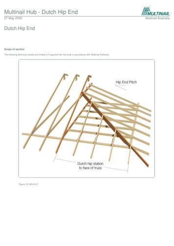
The “Hip Rafters” are positioned from the corner of the span / area and to the centre of the Dutch Hip Girder. Jack Top Chord to Hip Top Chord: Use ‘effective’ flat-head 3/75mm x 3.05Ø nails. Hip Top Chord to be mitre cut. Jack and Hip Top Chord to Waling Plate: Use 30 x 0.8mm looped
A hip-hop dance routine incorporates the look, music, . The most real routines showcase a variety of hip-hop dance styles, signature moves and choreography conveying the character and energy of the street. Hip Hop International (HHI) Hip Hop International founded in 2000 is based in Los Angeles, California. Hip Hop International is
Readers Theatre Scripts Family Tutoring 584 Come Hippopotamus Roles: Reader 1, All, Reader 2, Reader 3 Reader 1 Come hippopotamus All HIP HIP HIP! HIP HIP HIP! Reader 2 What an enormous face you have! Reader 3 What an enormous lip! Reader 1 Can't you come and play a bit? All Dance! Dance! Reader 2 And hop!
Academic writing styles can vary from journal to journal, so you have to check each publication’s guide for writers and follow it carefully and/ or copy other papers in it. Academic writing titles cultural differences and useful phrases Academic papers often have a title with two parts. If the title of an academic paper has two parts, the two parts are usually separated by a colon .


