Multiple Methods For Detecting Apoptosis On The BD Accuri C6 Flow .
BD BiosciencesTechnical BulletinMarch 2012Multiple Methods for Detecting Apoptosis on the BD Accuri C6 Flow CytometerMultiple Methods for Detecting Apoptosison the BD Accuri C6 Flow CytometerTechnical BulletinContents1Introduction2Annexin V4JC-15Caspase-36APO-BrdU and APO-DirectIntroductionApoptosis (programmed cell death) is an important biological process for bothdevelopment and normal tissue homeostasis. Dysregulation of apoptoticpathways can lead to disease.Methods for detecting apoptosis include Western blot, immunofluorescence,enzymatic assays, and flow cytometry. Flow cytometry is especially powerfulbecause researchers can gain quantitative data on both apoptotic and dead cellswithin whole populations and cell subsets.The BD Accuri C6 personal flow cytometer is well suited to the study ofapoptosis. With the ability to detect four fluorochromes in addition to forwardand side scatter, the instrument can perform most flow cytometric apoptosisassays, including Annexin V, caspase activation, PARP cleavage, mitochondrialchange, and DNA fragmentation. Powerful yet easy to use, the BD Accuri C6allows both new and experienced flow cytometry users to perform theseestablished assays.This technical bulletin presents examples of several popular BD Biosciencesapoptosis kits (Table 1) run on the BD Accuri C6. It discusses the backgroundof the assays and includes suggestions to optimize results.
BD BiosciencesTechnical BulletinMarch 2012Multiple Methods for Detecting Apoptosis on the BD Accuri C6 Flow CytometerTable 1. Selected methods for detecting apoptosis using flow cytometry.Apoptosis IndicatorAssayExamplesCat. No.Plasma membrane alterations(Phosphatidylserine exposure)Annexin V binding assays Single conjugates Annexin V kitsAnnexin V FITC Apoptosis Detection Kit556570Annexin V PE Apoptosis Detection Kit559763Mitochondrial changesJC-1 assaysBD MitoScreen (JC-1) Kit551302Caspase activationCaspase activity assay kitsand reagentsCaspase-3 Active Form PE Apoptosis Kit550914APO-BrdU Apoptosis Detection Kit556405DNA fragmentationTUNEL assaysAPO-Direct Apoptosis Detection Kit556381Annexin VChanges in the plasma membrane are one of the first detectable characteristicsof the apoptotic process. In normal cells, phosphatidylserine (PS) molecules areconfined to the inner leaflet of the plasma membrane (Figure 1). Duringapoptosis, these molecules externalize and can be bound to labeled Annexin V(FITC or PE) in the presence of calcium. Dead cells can be excluded withmembrane-impermeant dyes such as propidium iodide (PI) or 7-AAD.Annexin V-PE ConjugateCa Plasma MembranePhospholipid FlippingCa ApoptosisNormal CellCytoplasmExternalization ofPhosphatidylserineApoptotic CellCytoplasmFigure 1. Changes in the plasma membrane are an early sign of apoptosis.Ca
Technical BulletinMarch 2012Multiple Methods for Detecting Apoptosis on the BD Accuri C6 Flow CytometerPage 3DMSO FITC PIQ2-UL0.1%Q2-UR11.1%Q2-LL86.0%Q2-LR2.8%FL3 Propidium Iodide-A3456101010SSC-A1,000,000107DMSO FITC PI1.810500,000P287.4%100Figure 2. Flow cytometric analysis of FITCAnnexin V staining.Jurkat cells (human T-cell leukemia; ATCCTIB-152) were treated with 6 µM ofcamptothecin or 0.1% DMSO (negative control)for 4 hours to induce apoptosis. Cells werestained with FITC Annexin V and PI accordingto the BD Pharmingen Annexin V FITCApoptosis Detection Kit staining protocol (Cat.No. 556570). Results: Camptothecin treatment(lower plots) resulted in an increase in earlyapoptotic cells (PI – Annexin V , shown in green)compared to the DMSO control (upper plots).Dead cell (PI , red) and live cell (PI – Annexin V– ,black) populations were easily distinguished.Data was acquired on a BD Accuri C6 flowcytometer using BD Accuri C6 software.1,704,234BD 610710710Q2-UL0.0%Q2-UR9.1%Q2-LL63.2%Q2-LR27.7%FL3 Propidium FL1 Annexin V FITC-AFSC-ADMSO PE 7AAD106DMSO PE 7AADQ4-UL0.5%Q4-UR8.5%Q4-LL88.4%Q4-LR2.6%FL3 7-AAD-A341010SSC-A1,000,0001051,634,686510Campto FITC PI1,704,234Campto FITC PI5,000,000FSC-A8,606,1712.310451010FL2 Annexin V PE-A6.310Campto 6 um PE 7AAD106Campto 6 um PE 7AAD310Q4-UL0.2%Q4-UR9.3%Q4-LL64.4%Q4-LR26.1%FL3 00P248.5%0Figure 3. Flow cytometric analysis of PEAnnexin V staining.Jurkat cells (human T-cell leukemia; ATCCTIB-152) were treated with 6 µM ofcamptothecin or 0.1% DMSO (negativecontrol) for 4 hours to induce apoptosis. Cellswere stained with PE Annexin V and 7-AADaccording to the BD Pharmingen Annexin V PEApoptosis Detection Kit staining protocol (Cat.No. 559763). Results: Camptothecin treatment(lower plots) resulted in an increase in earlyapoptotic cells (7-AAD –Annexin V , shown inorange), compared to the DMSO control (upperplots). Dead cell (7-AAD , red) and live cell(7-AAD – Annexin V– , black) populations wereeasily distinguished. Data was acquired on aBD Accuri C6 flow cytometer using BD AccuriC6 software.410FL1 Annexin V FITC-A2.310310451010FL2 Annexin V PE-A6.310
BD BiosciencesTechnical BulletinMarch 2012Multiple Methods for Detecting Apoptosis on the BD Accuri C6 Flow CytometerJC-1Changes in mitochondrial membrane potential (ΔΨm) are another early markerfor apoptosis. Lypophilic cationic fluorochromes such as JC-1 penetrate cells. Inhealthy cells, JC-1 accumulates in the mitochondria and forms aggregates. Inapoptotic cells, however, JC-1 does not accumulate in the mitochondria andremains in the cytoplasm as monomers.10 4.610 510 6JC-1 FL-1-A10 6.810 6JC-1 FL-2-AP1010.6%10 5.110,000,000 14,540,969FSC-AP912.9%10 6JC-1 FL-2-A5,000,00010 5.1010 710 7A03 K562 CCCP JC-1Gate: (P1 in all)P987.6%P190.0%0SSC-AA02 K562 untreated JC-1Gate: (P1 in all)A02 K562 untreated JC-1Gate: [No Gating]2,000,0005,093,085Because monomers and aggregates of JC-1 have different emission spectra,changes in ΔΨm can be determined by comparing the ratio of fluorescence betweenthe FL1 and FL2 channels.10 4.6P1084.0%10 510 610 6.8JC-1 FL-1-AFigure 4. Flow cytometric analysis of BD MitoScreen staining.K562 cells (human chronic myelogenous leukemia; ATCC CCL-243) were treated with 100 µMof CCCP (in DMSO) for 5 minutes at 37ºC to induce apoptosis. The cells were stained with JC-1(1:2,500 dilution in assay buffer) for 15 minutes at 37ºC, according to the BD MitoScreen protocol(Cat. No. 551302). The cells were washed with assay buffer as described in the kit insert andcollected on the BD Accuri C6 for 30 seconds on fast speed. Results: CCCP treatment resulted ina shift in mitochondrial membrane potential (red to green). Data was acquired on a BD Accuri C6flow cytometer using BD Accuri C6 software.
BD BiosciencesTechnical BulletinMarch 2012Multiple Methods for Detecting Apoptosis on the BD Accuri C6 Flow CytometerPage 5Caspase-3Changes in ΔΨm as a result of apoptotic triggers make the mitochondrialmembrane more permeable and release soluble proteins such as cytochrome cand procaspases. Procaspases are activated by protein cleavage and, in turn,cleave other proteins. This leads to a loss of cellular function and %2,000,000FSC-A5,218,844310410PE active othecin2.410410510PE active 00SSC-A500,000SSC-A500,0006001,022,601Several caspases are important for apoptosis, including caspase-3, -8, and -9.Methods of detecting caspase cleavage include fluorogenic substrates as well asantibodies specific to the cleaved (activated) forms of caspases.2.410310410PE active caspase-3-A5105.4102.710410510PE active caspase-3-A5.910Figure 5. Flow cytometric analysis of active Caspase-3 staining.Jurkat cells (human T-cell leukemia; ATCC TIB-152) were treated with 6 µM of camptothecin or0.1% DMSO (negative control) for 4 hours to induce apoptosis. Cells were permeabilized, fixed,and stained according to the BD Pharmingen Caspase-3 Assay Kit staining protocol (Cat. No.550914). Results: Camptothecin treatment (lower plots) resulted in an increase in active Caspase-3expression (red) compared to the DMSO control (upper plots), which was primarily negative(green). Data was acquired on a BD Accuri C6 flow cytometer using BD Accuri C6 software.
BD BiosciencesTechnical BulletinMarch 2012Multiple Methods for Detecting Apoptosis on the BD Accuri C6 Flow CytometerAPO-BrdU and APO-DirectDNA fragmentation is one of the final stages of apoptosis. BD Biosciencesprovides two kits to detect DNA fragmentation using flow cytometry.The BD APO-BrdU Kit uses a reaction catalyzed by terminal deoxynucleotidyltransferase (TdT) to detect fragmentation. The method is often called end labelingor TUNEL (TdT dUTP nick end labeling). In this assay, TdT catalyzes a templateindependent addition of bromolated deoxyuridine triphosphates (Br-dUTP) tothe 3'-hydroxyl (OH) termini of double- and single-stranded DNA. After theBr-dUTP is incorporated, the cells are stained with labeled anti-BrdU, and theDNA terminal sites are identified using flow .TTdT Br-dUTPG.CC.GFITC-LabeledAnti-BrdU mAbG.CC.GA.TDNA Strand BreaksC.CA.TA.TG.CC.GA.TAdd Br-dUPT to 3’ - OHDNA EndsAntibody LabeledBreak SitesFigure 6. Schematic representation of APO-BrdU labeling.The enzyme TdT catalyzes a template-independent addition of Br-dUTP to the 3'-hydroxyl ends ofdouble- and single-stranded DNA. After Br-dUTP incorporation, DNA break sites are identified by aFITC-labeled anti-BrdU monoclonal antibody.The BD APO-Direct Kit uses the same catalyst (TdT) in a single-step methodthat labels DNA breaks with a dUTP antibody.GGG.CG.CG.GG.GA.TA.TC.CC.CA.TG.CC.GA.TDNA Strand BreaksTdT FITC-dUTPA.TG.CC.GA.TFITC LabeledBreak SitesFigure 7. Schematic representation of APO-Direct labeling.The enzyme TdT catalyzes a template-independent addition of FITC-labeled deoxyuridine triphosphates (FITC-dUTP) to the 3'-hydroxyl ends of double- and single-stranded DNA. When theFITC-labeled dUTPs are incorporated, the DNA break sites can be identified.
Technical BulletinMarch 2012Multiple Methods for Detecting Apoptosis on the BD Accuri C6 Flow Cytometer05,000,000FSC-A13,182,09910 610 510 510 410200,000400,000FL-2 DNA-A600,000 777,654A04 Kit neg control DirectGate: (P2 in all)10 510 4FL-1 FITC dUTP-A10 3.1936,56210 6.9A05 Kit pos control DirectGate: (P1 in all)10200,000400,000FL-2 DNA-A600,000 777,654A05 Kit pos control DirectGate: (P2 in all)FL-1 FITC dUTP-A0500,000FL-2 DNA-A936,56210 510 6P281.1%R11.2%R154.4%10 4633,860500,000FL-2 DNA-A00FL-2 DNA-HP163.0%600,000 777,65410 6P282.9%013,182,099A05 Kit pos control DirectGate: [No Gating]400,000FL-2 DNA -AR170.0%10 6.9A04 Kit neg control DirectGate: (P1 in all)400,000FSC-A200,000D05 control BRDUGate: (P2 in all)10 6.9936,562200,0005,000,0001010 3.1633,860500,000FL-2 DNA-A0FL-2 DNA-HP166.2%10 410 3.1FL-1 FITC BrdU-A010 3.1FL-1 FITC BrdU-A400,000013,182,099400,000FSC-AR12.7%10 6P268.4%200,0005,000,000A04 Kit neg control DirectGate: [No Gating]2,000,0004,000,000 5,392,677936,562200,000FL-2 DNA-HP139.1%2,000,0004,000,000 5,392,677500,000FL-2 DNA -AD05 control BRDUGate: (P1 in all)633,860D05 control BRDUGate: [No Gating]0SSC-A013,182,099D04 - control BRDUGate: (P2 in all)10 6.9633,860400,000FSC-A0SSC-AP287.0%200,000FL-2 DNA-H5,000,0000SSC-A4,000,000 5,392,67700Figure 9. Flow cytometric analysis ofAPO-Direct staining.Positive control cells (human lymphoma cell linestimulated to undergo apoptosis, bottom row)and negative control cells (untreated lymphomacell line, bottom row), both included in theBD APO-Direct Kit (Cat. No. 556381), werestained according to the kit insert. Sampleswere collected for 30 seconds on fast speedon a BD Accuri C6 flow cytometer, and datawas acquired using BD Accuri C6 software.Clumped cells were excluded by gating onthe DNA Area vs Height plot (middle plots).Results: The positive control cells showeda significant increase in apoptotic cells (R1)compared to the negative control.D04 - control BRDUGate: (P1 in all)02,000,000P158.6%0SSC-APage 7D04 - control BRDUGate: [No Gating]2,000,000Figure 8. Flow cytometric analysis ofAPO-BrdU staining.Positive control cells (human lymphoma cellline stimulated to undergo apoptosis, bottomrow) and negative control cells (untreatedlymphoma cell line, top row), both included inthe BD APO-BrdU Kit (Cat. No. 556405), werestained according to the kit insert. Sampleswere collected for 30 seconds on fast speedon a BD Accuri C6 flow cytometer, and datawas acquired using BD Accuri C6 software.Clumped cells were excluded by gating onthe DNA Area vs Height plot (middle plots).Results: The positive control cells showeda significant increase in apoptotic cells (R1)compared to the negative control.4,000,000 5,392,677BD Biosciences10200,000400,000FL-2 DNA-A600,000 777,654
BD BiosciencesTechnical BulletinMarch 2012Multiple Methods for Detecting Apoptosis on the BD Accuri C6 Flow CytometerFor Research Use Only. Not for use in diagnostic or therapeutic procedures.BD, BD Logo and all other trademarks are property of Becton, Dickinson and Company. 2012 BD23-14155-00BD Biosciences2350 Qume DriveSan Jose, CA 95131US Orders: 855.236.2772BD Accuri Technical Support: ciences.com
collected on the BD Accuri C6 for 30 seconds on fast speed. Results: CCCP treatment resulted in a shift in mitochondrial membrane potential (red to green). Data was acquired on a BD Accuri C6 flow cytometer using BD Accuri C6 software. 0 5,000,000 10,000,000 14,540,969 05,093,085 SSC-A 2,000,000 FSC-A A02 K562 untreated JC-1 Gate: [No Gating .
Bruksanvisning för bilstereo . Bruksanvisning for bilstereo . Instrukcja obsługi samochodowego odtwarzacza stereo . Operating Instructions for Car Stereo . 610-104 . SV . Bruksanvisning i original
RESEARCH Open Access Efficacy of ImageJ in the assessment of apoptosis Iman M Helmy* and Adel M Abdel Azim Abstract Objective: To verify the efficacy of ImageJ 1.43 n in determining the extent of apoptosis which is a complex and multistep process. Study Design: Cisplatin in different concentrations was used to induce apoptosis in cultured Hep2 .
10 tips och tricks för att lyckas med ert sap-projekt 20 SAPSANYTT 2/2015 De flesta projektledare känner säkert till Cobb’s paradox. Martin Cobb verkade som CIO för sekretariatet för Treasury Board of Canada 1995 då han ställde frågan
service i Norge och Finland drivs inom ramen för ett enskilt företag (NRK. 1 och Yleisradio), fin ns det i Sverige tre: Ett för tv (Sveriges Television , SVT ), ett för radio (Sveriges Radio , SR ) och ett för utbildnings program (Sveriges Utbildningsradio, UR, vilket till följd av sin begränsade storlek inte återfinns bland de 25 största
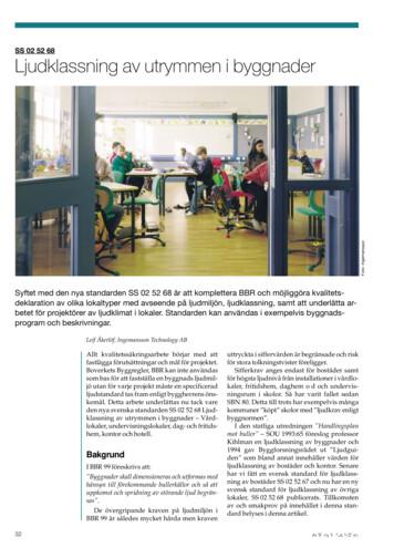
Hotell För hotell anges de tre klasserna A/B, C och D. Det betyder att den "normala" standarden C är acceptabel men att motiven för en högre standard är starka. Ljudklass C motsvarar de tidigare normkraven för hotell, ljudklass A/B motsvarar kraven för moderna hotell med hög standard och ljudklass D kan användas vid
LÄS NOGGRANT FÖLJANDE VILLKOR FÖR APPLE DEVELOPER PROGRAM LICENCE . Apple Developer Program License Agreement Syfte Du vill använda Apple-mjukvara (enligt definitionen nedan) för att utveckla en eller flera Applikationer (enligt definitionen nedan) för Apple-märkta produkter. . Applikationer som utvecklas för iOS-produkter, Apple .
och krav. Maskinerna skriver ut upp till fyra tum breda etiketter med direkt termoteknik och termotransferteknik och är lämpliga för en lång rad användningsområden på vertikala marknader. TD-seriens professionella etikettskrivare för . skrivbordet. Brothers nya avancerade 4-tums etikettskrivare för skrivbordet är effektiva och enkla att
Den kanadensiska språkvetaren Jim Cummins har visat i sin forskning från år 1979 att det kan ta 1 till 3 år för att lära sig ett vardagsspråk och mellan 5 till 7 år för att behärska ett akademiskt språk.4 Han införde två begrepp för att beskriva elevernas språkliga kompetens: BI


