AOHS Foundations Of Anatomy And Physiology II Lesson 20 The .
AOHS Foundations of Anatomy and Physiology IILesson 20The Reproductive SystemStudent ResourcesResourceDescriptionStudent Resource 20.1Terms: The Reproductive SystemStudent Resource 20.2Notes: Reproductive Anatomy and PhysiologyStudent Resource 20.3Reading: Anatomy and Physiology of the Male Reproductive SystemStudent Resource 20.4Reading: Anatomy and Physiology of the Female ReproductiveSystemStudent Resource 20.5Reading: The Menstrual CycleStudent Resource 20.6Graphs: The Menstrual CycleStudent Resource 20.7Reading: ContraceptivesStudent Resource 20.8Posters Summary: Reproductive Health and ConditionsStudent Resource 20.9Lab: ELISA TestStudent Resource 20.10Notes: Genes and ReproductionStudent Resource 20.11Reading: Genes and ReproductionCopyright 2014‒2016 NAF. All rights reserved.
AOHS Foundations of Anatomy and Physiology IILesson 20 The Reproductive SystemStudent Resource 20.12Reading: Stages of Human Growth and DevelopmentStudent Resource 20.13Glossary: The Reproductive System (separate Word file)Student Resource 20.14Guide: Reproduction Expert Blog PostStudent Resource 20.15Peer Review Chart: Reproduction Blog PostCopyright 2014‒2016 NAF. All rights reserved.
AOHS Foundations of Anatomy and Physiology IILesson 20 The Reproductive SystemStudent Resource 20.1Terms: The Reproductive SystemStudent Name: Date:Directions: Write down three medical or common terms for parts of the reproductive system. When yourteacher tells you to begin, mingle with your classmates and collect as many other medical and commonterms as you can.Terms I thought ofTerms I collected from classmatesCopyright 2014‒2016 NAF. All rights reserved.
AOHS Foundations of Anatomy and Physiology IILesson 20 The Reproductive SystemStudent Resource 20.2Notes: Reproductive Anatomy and PhysiologyStudent Name: Date:Directions: Complete the charts and label the diagrams as you watch the presentations on the anatomyand physiology of the male and female reproductive systems.Part 1: The Male Reproductive System1. What are the male and female gametes and what is special about them?2. Label the diagram (lateral view):Lateral viewCopyright 2014‒2016 NAF. All rights reserved.
AOHS Foundations of Anatomy and Physiology IILesson 20 The Reproductive System3. Describe the path sperm follow through the male reproductive organs.4. What organs contribute secretions that become part of semen?5. What is the purpose of semen?6. Why doesn’t a man have to worry about urinating while he is having sex?7. Complete the chart describing hormonal changes a male goes through during puberty.HormoneWhat it does.8. List four secondary male sex characteristicsa)b)c)d)Copyright 2014‒2016 NAF. All rights reserved.
AOHS Foundations of Anatomy and Physiology IILesson 20 The Reproductive SystemPart 2: The Female Reproductive System9. What female organ produces gametes?10. What are these gametes called when they mature?11. Label the diagram:Anterior viewCopyright 2014‒2016 NAF. All rights reserved.
AOHS Foundations of Anatomy and Physiology IILesson 20 The Reproductive System12. Describe the path each gamete follows from the point at which the gamete is made to fertilization.Describe the paths for both the female and male gametes. Include where and when the gamete cell ismade, the organs it passes through, and where the gametes meet.13. Why do oocytes sometimes miss the fallopian tube?14. What is notable about the pH of the vagina and why is it beneficial?15. How does the clitoris resemble the penis?16. Where is milk produced in the breast?17. What is a zygote?Copyright 2014‒2016 NAF. All rights reserved.
AOHS Foundations of Anatomy and Physiology IILesson 20 The Reproductive System18. Complete the following chart about hormones involved in pregnancy and childbirth:Hormone/substanceBy what organProlactinhCGOxytocin (first)Oxytocin (second)Copyright 2014‒2016 NAF. All rights reserved.When secretedEffect
AOHS Foundations of Anatomy and Physiology IILesson 20 The Reproductive SystemStudent Resource 20.3Reading: Anatomy and Physiology of the MaleReproductive SystemCopyright 2014‒2016 NAF. All rights reserved.
AOHS Foundations of Anatomy and Physiology IILesson 20 The Reproductive SystemCopyright 2014‒2016 NAF. All rights reserved.
AOHS Foundations of Anatomy and Physiology IILesson 20 The Reproductive SystemEach of the body systems you’ve studied so far plays a role in keeping you alive. As you know, ifsomething goes wrong with any of these systems, the whole body can be affected. In stark contrast, youcan live without your reproductive system. In fact, the reproductive system can even put some women inlife-threatening situations. In colonial America, for example, each time a woman was pregnant, she hadabout a 1.5% chance of dying from giving birth. Many women had five or six children, raising theirchances of dying to nearly 10%. That is a higher rate of death than we see now from many forms ofcancer.While the reproductive system might not keep you alive, it keeps our species alive. Without reproduction,it wouldn’t take long before humans disappeared from the earth. On a smaller scale, your personalreproductive system exists for the purpose of making sure that part of you—your genes—continues toexist after you do.That’s a task that takes a lot of energy (especially if you’re female), and so your body devotes itself toreproduction on a more punctuated basis than your other body systems operate.Copyright 2014‒2016 NAF. All rights reserved.
AOHS Foundations of Anatomy and Physiology IILesson 20 The Reproductive SystemAnother unique factor of the reproductive system is that it’s the only system in your body that producescells with the mission of being part of the creation of another member of your species. Such cells arecalled gametes. They contain only half the DNA of an ordinary cell. Female gametes are eggs, also calledova (singular, ovum). You may recall that a chicken egg is a single cell. A human egg isn’t so different,except for its smaller size. Still, the egg cell is the largest cell (in diameter) in the human body, measuringabout a tenth of a millimeter. They’re so big that you don’t even need a microscope to see them. Sperm,the male gametes, are about 25 times smaller. They have a powerful tail that propels them through thefemale reproductive tract. Sperm are expert swimmers―tiny cells carrying little baggage. They have feworganelles and don’t store much energy.Copyright 2014‒2016 NAF. All rights reserved.
AOHS Foundations of Anatomy and Physiology IILesson 20 The Reproductive SystemGametes need only half as much DNA as a normal cell because of the task they are called to: to becomeone with another gamete of the opposite sex. When a sperm meets an egg, the meeting sets in motion asequence of events that ultimately results in the nucleus of the sperm making its way into the egg. Afterseveral hours, the two nuclei fuse and become one, with a full complement of DNA. From there, thefertilized cell begins dividing, eventually producing all the different types of cells needed for a neworganism—skin cells, nerves, muscle, brain, and the rest.Copyright 2014‒2016 NAF. All rights reserved.
AOHS Foundations of Anatomy and Physiology IILesson 20 The Reproductive SystemTestosterone plays many roles for both sexes but is particularly important for men. The initial productionof testosterone is the driver behind the growth spurt that many boys experience during puberty. Itaccounts for the way a boy’s voice deepens, his bones and muscles fill out, and he finds himself needingto shave. It also directs the development of his reproductive organs to their adult size and function.Testosterone fuels sex drive throughout a man’s life. Levels of the hormone decrease as a man ages, butthey remain present into old age.Copyright 2014‒2016 NAF. All rights reserved.
AOHS Foundations of Anatomy and Physiology IILesson 20 The Reproductive SystemBoth boys and girls are born with reproductive systems that aren’t mature, which means they aren’t ableto reproduce. Puberty begins as the reproductive organs start to mature. Men are most fertile in theirteens and 20s, when they produce the most sperm, and sperm that are strong swimmers. At around age30, a man’s testosterone production begins to decline, and therefore so does his sperm production. In hislater years, a man may produce sperm that aren’t shaped as well or aren’t as strong as those heproduced when he was younger. Older men can usually still reproduce, and many men have childrenwhen they are older. Several recent studies have shown, however, that children of older men are morelikely to have a range of disorders such as autism and Down syndrome.A very common condition among older men is an enlarged prostate. Many men have mild enough casesthat there’s no need to treatment. But because the prostate surrounds the urethra, when the prostatebecomes too enlarged, it can make it hard for the man to urinate. There are many kinds of treatment foran enlarged prostate, and most men are able to resolve the problem. Keeping your weight under controland getting physical activity can help prevent an enlarged prostate.Copyright 2014‒2016 NAF. All rights reserved.
AOHS Foundations of Anatomy and Physiology IILesson 20 The Reproductive SystemStudent Resource 20.4Reading: Anatomy and Physiology of the FemaleReproductive SystemCopyright 2014‒2016 NAF. All rights reserved.
AOHS Foundations of Anatomy and Physiology IILesson 20 The Reproductive SystemCopyright 2014‒2016 NAF. All rights reserved.
AOHS Foundations of Anatomy and Physiology IILesson 20 The Reproductive SystemThe only task the male reproductive system is faced with is delivering sperm successfully to the femalereproductive tract. The female reproductive system has much more work to do: it provides space for afetus to grow and receive nourishment (a process called gestation). This system enables the female bodyto give birth and feed the newborn. Because these functions are much more complicated than those ofthe male reproductive system, the female hormonal cycles and physiology are much more complicated aswell.Copyright 2014‒2016 NAF. All rights reserved.
AOHS Foundations of Anatomy and Physiology IILesson 20 The Reproductive SystemThe ovaries are considered the primary sexual organ of the female because they produce the gametes.They’re about 2 inches across and anchored to the pelvis and the uterus with ligaments. The cells thatwill eventually become egg cells develop in the ovaries during fetal development. When a girl beginspuberty, her brain begins sending hormonal signals to her ovaries once a month. Those signals promptan egg cell precursor to mature into a cell called an oocyte and enter the reproductive tract.Copyright 2014‒2016 NAF. All rights reserved.
AOHS Foundations of Anatomy and Physiology IILesson 20 The Reproductive SystemThe ovaries aren’t connected directly to the reproductive tract. Instead, when a mature oocyte is releasedfrom the ovary, it floats through a small space and into the nearby fallopian tube. As you might expect, notevery egg makes it; plenty of them miss the mark. When the oocyte does make it to the fallopian tube(which happens frequently enough that our species has had little difficulty reproducing), it has about 4inches of tube to make it through to reach the uterus. This journey takes three to four days, but the oocyteis only fresh enough for fertilization for about 24 hours. This limited time window means that the bestchance for fertilization is when the oocyte is still in the fallopian tube, before it’s reached the uterus.Therefore, sperm need to make their way not only into the female reproductive tract but almost all theway through it, to reach the egg while it’s in the distant end of the fallopian tube.Copyright 2014‒2016 NAF. All rights reserved.
AOHS Foundations of Anatomy and Physiology IILesson 20 The Reproductive SystemThe uterus—also sometimes called the womb—is about the size of a fist, shaped like an upside-downpear, except during pregnancy. The fallopian tubes are lined with cilia that help usher the fertilized egg (orunfertilized oocyte) into the uterus. If the cell is fertilized, it will implant on the inner lining of the uterus andremain there through all of its fetal development. If the cell isn’t fertilized, it just passes out of the woman’sreproductive tract through menstruation.The walls of the uterus are composed of three layers that can stretch to a great degree. Imagine howlarge a pear is. Not so big, right? Now imagine that you put that pear in, say, a pair of nylons. Thenimagine that, in the same nylons, you have to fit a newborn baby. That would have to be some stretchynylon! You can see how stretchy the walls of the uterus must be.Along with being stretchy, the uterine walls also need to be muscular, because the most rigorous task inreproduction—giving birth—requires some real force. During birth, hormones make the uterus go throughintense contractions that ultimately push the baby out of the womb and into the world.Copyright 2014‒2016 NAF. All rights reserved.
AOHS Foundations of Anatomy and Physiology IILesson 20 The Reproductive SystemAt the base of the uterus, where the top of the upside-down pear would be, is a cylinder of muscle calledthe cervix. Normally, there’s a small space in the center of the cervix that allows body fluids out of theuterus and lets sperm in. But when a pregnant woman is about to give birth, the cervix dilates, making theopening much wider. Often the person assisting a birth will look at how far the cervix has dilated toestimate how close the woman is to delivering the baby. Many times, the top of the baby’s head can beseen pushing against the opening of the cervix before it’s born.Cancer of the cervix, and precancerous cells, aren’t uncommon. It’s recommended that once a woman isabout 21, she should start being screened for cervical cancer using a test called a pap smear. Manycervical cancers in young, sexually active women arise from infection with the human papilloma virus(HPV). There is now a vaccine available that protects against several forms of the virus. Many schoolhealth programs suggest that both boys and girls 11 to 12 years old get vaccinated.Copyright 2014‒2016 NAF. All rights reserved.
AOHS Foundations of Anatomy and Physiology IILesson 20 The Reproductive SystemThe vagina, also sometimes called the birth canal, is a tube about 3‒4 inches long. The vagina is wherethe penis enters the female reproductive system during intercourse and is the canal a fetus must passthrough to exit the mother’s body. The walls of the vagina have lots of folds. Think back to the pear in thenylons, followed by the baby in the nylons. The same need for stretchiness is true for the vagina. A tubethat’s usually only slightly open must expand to accommodate the passage of a whole infant, head,shoulders and all. The vagina possesses the ability to stretch this much because of the many folds thatallow it to unfold and expand.As in the digestive tract, there are also microorganisms in the vagina, and they help maintain balance inthe environment. The inside of the vagina is slightly acidic, which is an environment that many harmfulbacteria can’t tolerate. A common beneficial bacterium in the vagina is lactobacillus, which is also foundin fermented milk products like yogurt.Toward the distal end of the vagina is a thin fold of tissue called the hymen. The hymen contains a lot ofblood vessels, and in many women, the hymen is ruptured the first time they have intercourse, releasingsome blood. For many women in the United States, the hymen is ruptured while inserting a tampon,playing sports, or getting a first pelvic exam.Copyright 2014‒2016 NAF. All rights reserved.
AOHS Foundations of Anatomy and Physiology IILesson 20 The Reproductive SystemWhile most of the reproductive plumbing in females is inside the body, organs outside serve to protect thevaginal opening and to make sex physically pleasurable. Outside the vagina are two separate folds offlesh, the labia majora and the labia minora. The larger outer folds, the majora, have hair on them. Theirfleshiness helps cushion and protect the bony parts of the pelvis underneath. The more delicate innerfolds, the labia minora, cover the vaginal opening, the urethra, and the clitoris. Near the posterior end ofthe labia majora are small glands that secrete a fluid that helps provide lubrication during intercourse.The clitoris is similar to a penis in that it has a shaft and a tip that is highly sensitive to sexual stimulation.The clitoris also contains erectile tissues that swell when filled with blood. A woman can achieve orgasmthrough stimulating the clitoris, just as a man can reach orgasm through stimulating the penis. Thoughwomen don’t emit sperm, they do have waves of muscular contractions in the vagina and sometimes inthe uterus as well. These contractions may help usher sperm through the cervix and into the reproductivetract.Copyright 2014‒2016 NAF. All rights reserved.
AOHS Foundations of Anatomy and Physiology IILesson 20 The Reproductive SystemAlthough males and females have the same urinary organs, they are situated in the pelvic cavity indifferent ways to make room for the differing reproductive organs. The male bladder is located posterior tothe vas differens and superior to the prostate and other glands. In females, the bladder is right up frontand against the inferior wall of the cavity, allowing the larger uterus to fit behind it. The male urethra, asyou’ve learned, carries both sperm and urine and is quite a bit longer than the female urethra, whichcarries only urine. In both sexes, the anus and rectum are posterior to the reproductive organs.Copyright 2014‒2016 NAF. All rights reserved.
AOHS Foundations of Anatomy and Physiology IILesson 20 The Reproductive SystemBreasts, also called mammary glands, provide an infant with nourishment until it’s old enough to digestsolid food. Breasts begin to develop during puberty. Girls’ breasts contain special cells that, when mature,can produce milk after pregnancy. At the start of puberty, hormones signal to these cells that it’s time tomature. As the milk-producing cells develop, so do fatty tissues around them, to protect the future infantfood supply. It’s these protective fatty tissues that give breasts their size and shape.After a woman has a baby, her pituitary gland secretes a hormone called prolactin. Prolactin stimulatesmilk production in the breast. A baby’s suckling the breast to get milk stimulates the mother’s pituitary torelease oxytocin. The hormone causes contractions in the breast lobules that help them expel milk,similar to how the hormone causes uterine contractions during birth.Copyright 2014‒2016 NAF. All rights reserved.
AOHS Foundations of Anatomy and Physiology IILesson 20 The Reproductive SystemIt’s harder than you might think for a sperm to get a crack at fertilizing an egg cell. And the privilege goesto only a single sperm out of the millions that will be ejaculated into the woman’s vagina. The oocyte hasa casing of other cells around it, and this casing has to be broken down by enzymes carried on thesperm. It actually takes the work of many sperm to break this casing down enough so a sperm mightmake its way through the cell membrane. The sperm that is the first to make this connection wins. Theoocyte absorbs the sperm’s nucleus and its half-set of DNA. In several hours, the cell will begin to divide.The cell is no longer an egg; it is now called a zygote. Its cells continue to divide until it has gone througha certain number of divisions and is considered an embryo. All the while, it is traveling through thefallopian tube to the uterus.Copyright 2014‒2016 NAF. All rights reserved.
AOHS Foundations of Anatomy and Physiology IILesson 20 The Reproductive SystemWhen the embryo reaches the uterus, around three days after fertilization, it attaches to the inner lining ofthe uterus. This attachment sets a whole cycle of hormone-driven changes in motion as the mother’sbody takes on the task of pregnancy. The ovaries shut down and won’t release any eggs until the motheris no longer pregnant. In fact, the menstrual cycle is put completely on hold, and the lining that wasbuilding up in the uterus remains. That’s thanks to a hormone called human chorionic gonadotropin, orhCG. The embryo produces hCG, and the hormone is detectable in the mother’s blood as early as 10 to14 days after the embryo implants in the uterus. It’s those early levels of hCG that allow a woman to do ahome pregnancy test not long after the first time she misses a period.Copyright 2014‒2016 NAF. All rights reserved.
AOHS Foundations of Anatomy and Physiology IILesson 20 The Reproductive SystemThe amniotic sac forms around the embryo (and fetus). It is filled with fluid that protects and bathes thefetus during development. The fluid cushions the fetus and keeps it lubricated so that its developingfingers and limbs don’t grow together.The placenta is a layer of tissue that forms from the walls of the uterus and separates the fetus from theuterine wall. In the first trimester of pregnancy, the placenta actually becomes an endocrine organ,producing lots of estrogen and progesterone. The umbilical cord is the fetus’ lifeline and connects to oneside of the placenta. The circulatory and respiratory systems aren’t functional. The lungs, even oncethey’ve developed, are completely collapsed and contain no oxygen. Because the fetus is in a fluidenvironment, there is no air to breathe into the lungs. All nutrient and gas exchange takes place acrossthe placenta, but the blood supplies do not mix, which allows the fetus to have a different blood type fromthe mother. The mother does have to send quite a bit of blood to the placenta. A pregnant woman getswinded more easily than usual because she’s sending blood to bring oxygen to someone else’s cellsbesides her own.Copyright 2014‒2016 NAF. All rights reserved.
AOHS Foundations of Anatomy and Physiology IILesson 20 The Reproductive SystemYou may have heard mothers joke that the baby makes the decision about when it will be born. Althougha pregnant woman doesn’t go into labor because her baby thought, “It’s time!,” recent research suggeststhat labor is brought about by hormones produced by the fetus, namely the hormone oxytocin. Oxytocinfrom the fetus triggers a cycle of oxytocin release from the mother’s pituitary gland that result in the oftenpainful uterine contractions known as labor. When the uterus contracts, it signals the mother’s pituitary toproduce additional oxytocin. More oxytocin means more frequent and stronger contractions.Copyright 2014‒2016 NAF. All rights reserved.
AOHS Foundations of Anatomy and Physiology IILesson 20 The Reproductive SystemThe more often the uterus contracts, the more oxytocin is released. The result is a positive feedback loopin which the levels of oxytocin continue to rise and uterine contractions come more often and with moreforce. During this time, the mother’s cervix is also dilating. Eventually, the contractions are strong enoughto push the fetus out of the uterus, through the dilated cervical opening and through the birth canal. Thisis no small effort—remember that sock analogy? Getting the fetus along its exit path requiresconsiderable force. Sometimes the force isn’t enough and labor continues so long that it endangers eitherthe fetus or the mother. In these cases, doctors will deliver the baby surgically by a cesarean section, ofC-section, which involves cutting the abdominal and uterine walls.Copyright 2014‒2016 NAF. All rights reserved.
AOHS Foundations of Anatomy and Physiology IILesson 20 The Reproductive SystemOxytocin has lots of effects in the body beyond those during childbirth. Men have oxytocin too, thoughoften lower levels of it. Oxytocin brings on feelings of care, affection, and bonding, particularly for amother who just gave birth or who is nursing. Oxytocin is also released by the brains of both men andwomen during sex, playing a role in romantic attachment.Oxytocin affects many other behaviors, such as generosity toward others, trust, and empathy.Researchers are studying these many behavioral effects of the hormone, seeking possible ways toaddress certain kinds of antisocial behaviors. In the meantime, what is known for certain is that, from birthonward, the hormone plays an important and varied role in the relationship between mother and child.Copyright 2014‒2016 NAF. All rights reserved.
AOHS Foundations of Anatomy and Physiology IILesson 20 The Reproductive SystemOver the course of her lifetime, a woman will ovulate between 300 and 400 eggs. As she approaches herlate 30s or early 40s, the cycles of estrogen and progesterone start to fluctuate. In turn, that means levelsof LH and FSH also fluctuate in ways they hadn’t before. This hormonal roller coaster can produce somedisruptive systems, like night sweats, hot flashes, insomnia, and mood swings. It also produces irregularperiods, and eventually the woman stops ovulating altogether. This process of reproductive winding downis called menopause. Once a woman has gone through menopause, she’s not able to conceive anymore.The way women experience menopause varies widely. For some, it happens over the course of a fewmonths, and for others it can take five years or more. Some women have symptoms bad enough todisrupt their lives; for others it’s much easier.The lack of hormones, particularly estrogen, also causes changes in the body. The skin, muscles, andbones of both men and women weaken with age, but the effect can be more pronounced in women.(Remember, men also produce estrogen, but much less, so hormonal changes as men age are not asdrastic.) Estrogen aids bone function, which means that postmenopausal women are at risk forosteoporosis. Older women can protect their bones by making sure to get enough calcium and do weightbearing exercise, but the best insurance is to strengthen bones while you’re young, because there will bemore to make up for the bone loss that happens when you’re older.Copyright 2014‒2016 NAF. All rights reserved.
AOHS Foundations of Anatomy and Physiology IILesson 20 The Reproductive SystemStudent Resource 20.5Reading: The Menstrual CycleStudent Name: Date:Directions: Complete the reading and answer the questions at the end.Everyone has heard of “that time of the month.” That particular time is part of a cycle that repeats itselfabout every 28 days, and the cycle is the continual rise and fall of a woman’s fertility.The 28-day menstrual cycle occurs because two separate cycles affect each other, and these two cycleshave to occur in a synchronized way in order for a woman to be fertile. The ovarian cycle involves hormones secreted by the anterior pituitary that affect the developmentof the egg in the ovary. The uterine cycle involves hormones released by the ovary that affect the state of the uterinelining and of the maturing oocyte.The first day of a woman’s period is considered to be the first day of the menstrual cycle.The ovarian cycleAt the start of menstruation, the ovaries are fairly quiet. Oocytes await the signal from pituitary hormonesthat it’s time to move on to the next stage of maturity. Usually, only one oocyte at a time gets the signal,which comes in the form of the follicle stimulating hormone (FSH).In the ovary, each egg cell sits in a tiny sac called a follicle. As the pituitary continues to send FSH overthe course of about 10 days, the egg matures inside the follicle. At around day 14, the anterior pituitarysends out a surge of luteinizing hormone (LH) that makes the oocyte mature further and then burst out ofthe follicle. This process is called ovulation. After its escape, the oocyte is swept into the nearest fallopiantube, where it will remain alive for a day at most, unless it is fertilized. The levels of both LH and FSHplummet, and the empty follicle becomes a temporary structure called the corpus luteum (literally “yellowbody”). FSH levels begin to rise again during the last few days of the cycle.Below is a graph showing FSH and LH levels during the 28-day cycle:Copyright 2014‒2016 NAF. All rights reserved.
AOHS Foundations of Anatomy and Physiology IILesson 20 The Reproductive SystemDuring the ovarian cycle, the follicle (and the resulting corpus luteum) act as an endocrine gland,secreting hormones that set the uterine cycle in motion.The uterine cycleEstrogen and progesterone are two important sex hormones that play a significant role in the fertilitycycle. In the days prior to the first day of menstruation, their amounts in the system are declining, whichcan cause mood swings, bloating, and appetite surges in some women. These symptoms, and others,are collectively known as premenstrual syndrome, or PMS. During the first days of menstruation, theovaries remain quiet not only in terms of egg production but also in terms of hormone production. RisingFSH levels in the follicle prompt it to begin secreting estrogen around day 6 of the cycle, whenmenstruation is almost over. Estrogen makes the inner wall of the uterus, called the endometrium, growand develop a rich blood supply. This thickening is the uterus getting prepared to provide a nourishingsite for an embryo should the oocyte that will be released into the fallopian tube become fertilized. Whenblood levels of estrogen are high, the pituitary senses it and slows the production of FSH.After ovulation occurs, levels of estrogen begin to fall, and the corpus luteum secretes progesterone.Progesterone causes the lining that’s developed in the uterus to remain
AOHS Foundations of Anatomy and Physiology II Lesson 20 The Reproductive System Student Resources Resource Description Student Resource 20.1 Terms: The Reproductive System Student Resource 20.2 Notes: Reproductive Anatomy and Physiology Student Resource 20.3 Reading: Anatomy and Physiology of the Male Reproductive System
Health Sci Anatomy & Physio, 8417100 Health Sci Anatomy & Physio, 8417100 Health Sci Foundations, 8417110 Care & Prevention of Athletic Injuries, 1502490 -1 semester EKG Technician, 8427130 First Aid, 0800320 -1 semester (These athletic courses are not an official part of the AoHS, but many AoHS students take them as electives.)
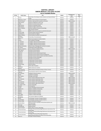
Clinical Anatomy RK Zargar, Sushil Kumar 8. Human Embryology Daksha Dixit 9. Manipal Manual of Anatomy Sampath Madhyastha 10. Exam-Oriented Anatomy Shoukat N Kazi 11. Anatomy and Physiology of Eye AK Khurana, Indu Khurana 12. Surface and Radiological Anatomy A. Halim 13. MCQ in Human Anatomy DK Chopade 14. Exam-Oriented Anatomy for Dental .
39 poddar Handbook of osteology Anatomy Textbook 10 40 Ross ,Pawlina Histology a text & atlas Anatomy Textbook 10 41 Halim A. Human anatomy Abdomen & lower limb Anatomy Referencebook 10 42 B.D. Chaurasia Human anatomy Head & Neck, Brain Anatomy Referencebook 10 43 Halim A. Human anatomy Head & Neck, Brain Anatomy Referencebook 10
HUMAN ANATOMY AND PHYSIOLOGY Anatomy: Anatomy is a branch of science in which deals with the internal organ structure is called Anatomy. The word “Anatomy” comes from the Greek word “ana” meaning “up” and “tome” meaning “a cutting”. Father of Anatomy is referred as “Andreas Vesalius”. Ph
Pearson Benjamin Cummings Anatomy and Physiology Integrated Anatomy – Gross anatomy, or macroscopic anatomy, examines large, visible structures Surface anatomy: exterior features Regional anatomy: body areas Systemic anatomy: groups of organs working
Anatomy titles: Atlas of Anatomy (Gilroy) Anatomy for Dental Medicine (Baker) Anatomy: An Essential Textbook (Gilroy) Anatomy: Internal Organs (Schuenke) Anatomy: Head, Neck, and Neuroanatomy (Schuenke) General Anatomy and Musculoskeletal System (Schuenke) Fo
Descriptive anatomy, anatomy limited to the verbal description of the parts of an organism, usually applied only to human anatomy. Gross anatomy/Macroscopic anatomy, anatomy dealing with the study of structures so far as it can be seen with the naked eye. Microscopic
as advanced engineering mathematics and applied numerical methods. The greatest beneÞt to the reader will probably be derived through study of the programs relat-' 2003 by CRC Press LLC. ing mainly to physics and engineering applications. Furthermore, we believe that several of the MATLAB functions are useful as general utilities. Typical examples include routines for spline interpolation .


