Experiments In Human Anatomy And Physiology
Experiments in HumanAnatomy and PhysiologyDepartment of BiologyEight Edition
ForwardWelcome to Human Anatomy and Physiology II (Bio 269). This laboratory manual isdesigned to act as a guide through experiments in human physiology. Laboratory inHuman Anatomy and Physiology II has three aims:1. To learn physiological concepts,2. To develop an understanding of the scientific approach (i.e., how scientists approacha problem and attempt to answer their questions using the scientific method), and3. To engage in creative and critical thinking.Some laboratory exercises in this manual are more structured (i.e., give more directionto you) than others. This has been done on purpose so that once you have an idea ofhow to perform the experiment, you will be asked (challenged) to come up with somenovel experiments to test on your own (with the instructors guidance of course). In thismanner you will become more involved in the decision making process, and hopefullywill become creative and critical in your thinking towards physiology.I hope that you find this laboratory course both challenging and interesting. Above allelse have fun. I created this lab manual and as such any errors that you may find aremine alone. If you do find an error, or if in your opinion something is unclear, pleasedo not hesitate to point it out to me, so that I may correct it for future students.Again, welcome to the course and good luck.Dr. Giovanni Casotti2
Table of ContentsTable of Contents . 3Timetable and Assessment . 4Introduction to inquiry-based labs and oral presentations . 5Introduction to LabChart . 9Graphing Assignment . 24Psychophysiology I . 26Psychophysiology II: Independent investigation. 37Hematology . 38Electrocardiography I and Heart Sounds . 48Electrocardiography II . 64Respiratory Physiology I. 65Respiratory Physiology II . 80Renal Physiology. 101Digestive Physiology . 111All experiments using PowerLab are Copyright ADInstruments 2016.Reprinted and modified with permission.3
Timetable and AssessmentWeek **TopicAssessment1Introduction to inquiry-based learning2Psychophysiology I3Psychophysiology II4Hematology5Electrocardiography I6Electrocardiography II7Lab exam 1 **8Respiratory Physiology9Respiratory physiology I10Renal Physiology11Digestive Physiology (spring only)12Lab exam 2 **SmartBoard presentationSmartBoard presentation** For an accurate display of lab dates and exam dates please consult the Human Anatomyand Physiology II web site.Laboratory assessment will be as follows:1. Introductory exercise2. Lab quizzes3. Lab exams4. Oral presentationsTotal103012040200(5 pts each)(2 @ 60 pts)(2 @ 20 pts)4
Introduction to inquiry-based labs and oralpresentationsIn lab, you will have several opportunities to design and conduct independent investigationswith your lab group. Each group project lasts for two weeks and is worth 20 points (40 points for 2projects). In the first week, you will gain experience in using the equipment, test specific variables,decide what question you will ask, what methods you will use, and possibly gather some preliminarydata. In the second week, you need to come to lab prepared with some background material thatrelates to your question, finish collecting data, organize those data into graphs or tables, makeconclusions, and prepare and make your presentation. You will have about 1 hour to complete yourexperiment and then your group will have 5 min to present the results to the class. We feel that it isimportant for you to report the results of your work orally because you will 1) improve yourcommunication skills, 2) learn from the other groups, and 3) experience the ways in which scientistsexplain, and sometimes defend, their work.Because inquiry and problem-solving labs run more than one week, you will need tocommunicate with each other between lab sessions. In the space below, exchange full names,telephone numbers, and e-mail addresses with your group members.5
Oral Presentation Rubric (20 points total):Team Members:Project title:Date:1) Background material (2 points)Does the background material (at least 2 prior experiments) relate to the topic of the investigation?Highly relevantRelevantSomewhatModeratelyNot relevant2 pts1.5 ptsrelevant 1 ptrelevant 0.5 pts0 ptsHow could this be improved upon?2) Variable tested (1 point)Were the dependent and independent variables tested, clearly articulated to the audience?Clearly statedVaguely statedNot stated1 pt0.5 pts0 ptsHow could this be improved upon?3) Hypothesis and prediction (2 points)Was a hypothesis presented and a prediction made, based on the background information?Highly relevantSomewhat relevantNot relevant/stated2 pts1 pt0 ptsHow could this be improved upon?4) Method (3 points)The methods were clearly outlined and appropriate for the proposed question:Clearly statedVaguely statedNot stated2 pt1 pts0 ptsThe experiment was properly controlled with replicates:YesNo1 pt0 ptsHow could this be improved upon?5) Results (4 points)Figures or tables were clear, appropriately labeled, and well prepared:ExcellentVery goodGoodFair2 pts1.5 pts1pt0.5 ptsThis presentation style was an effective way to summarize the results:YesNeeds improvementNo2 pts1 pt0 ptsHow could this be improved upon?6Poor0 pts
6) Discussion (4 points)Results were explained well using physiological principles:ExcellentVery goodGoodFair2 pts1.5 pts1pt0.5 ptsPoor0 ptsReflection on the original hypothesis was made and discussed:ExcellentVery goodGoodFair2 pts1.5 pts1pt0.5 ptsHow could this be improved upon?Poor0 pts7) Future Directions (1 point)Future directions the research should go were presented and appropriate based on the study:Clearly statedVaguely statedNot stated1 pt0.5 pts0 ptsHow could this be improved upon?8) References (1 point)At least 3 books or journal articles were presented using the correct formatting style:CompleteIncompleteNot done1 pt0.5 pts0 ptsHow could this be improved upon?9) Evidence of teamwork (see Supplemental rubric) (1 point)The group worked cooperatively and shared responsibility:YesNeeds improvement1 pt0.5 ptsHow could this be improved upon?10) Presentation style (1 point)The presentation was clear, well organized, and well presented:Well doneNeeds improvement1 pt0.5 ptsHow could this be improved upon?7No0 ptsPoor0 pts
Oral Presentation Supplemental RubricLab members (full first/last names)DateTitle of lab project:In the table below, sign your initials in the boxes on the top row and check off the areas for the oralpresentation that you performed.Lab ationExperimentaldesignData collectionData analysisPreparation oftables or graphsSummary ofresultsMakingconnections tolecture materialIdeas for futureexperimentsIdentify at least 3 things that your group learned from this project:1.2.3.8
Introduction to LabChartIn this experiment, you will learn how to acquire data with the PowerLab Data Acquisition Unit and analyzethe data using the LabChart software. You will make simple recordings and measurements using the FingerPulse Transducer.BackgroundThe purpose of the PowerLab system is to acquire, store, and analyze data. The raw input signal is in theform of an analog voltage whose amplitude varies continuously over time. This voltage is monitored by thehardware, which can modify it by amplification and filtering processes called signal conditioning. Signalconditioning may also include zeroing, the removal of an unwanted steady offset voltage from a transducer’soutput. After signal conditioning, the analog voltage is sampled at regular intervals. The signal is thenconverted from analog to digital form before transmission to the attached computer. Figure 1 shows asummary of the acquisition.The LabChart software usually displays the data directly; it plots the sampled and digitized data points andreconstructs the original waveform by drawing lines between the points. Digital data can be stored for laterretrieval. The software can also easily manipulate and analyze the data in a variety of ways.Figure 1. A Summary of Data Acquisition Using a PowerLab SystemThe basic hardware is a PowerLab, a recording instrument that measures electrical signals, usually throughthe inputs on its front panel. It can also generate output signals. Added hardware, such as the Finger PulseTransducer, can extend its capabilities. There are various PowerLab models, but PowerLab 26T, which isrecommended for this experiment, is designed especially for teaching experiments. It is a four-channelrecording instrument with built-in front-ends that allow optimal recording of biological signals (through BioAmplifiers) and provide safe stimuli for humans (through the Isolated Stimulator). Students are able torecord physiological measurements such as finger pulse, blood pressure, respiration, and even morecomplicated measurements like an EMG, or electromyography, recording. The front of PowerLab 26T isshown in Figure 2. The back is shown in Figure 3.9
Figure 2. The Front Panel of PowerLab 26T1.2.3.4.5.6.7.8.Power indicator light: illuminates when the PowerLab is turned onAnalog output connections: provide a voltage output in the 10 V rangeThis is NOT safe for direct connection to humansIsolated Stimulator status light: indicates if the Isolated Stimulator is working properly (green) or out of compliance(yellow)Dual Bio Amp input: connects a 5 lead Bio Amp cable to the PowerLab; reads as inputs 3 and 4Isolated Stimulator outputs: for connecting stimulating electrodes to the Isolated StimulatorIsolated Stimulator switch: turns on/off the Isolated StimulatorPod ports: 8-pin connectors for attaching pods and certain transducers to Input; these supply a DC Power to thepods and transducersTrigger input: can be used to start or stop a recording eventFigure 3. The Back Panel of PowerLab 26T9.10.11.12.13.14.15.16.Audio output connector: standard 1/8'' (3mm) phono jack for sound output of recordings from the Bio AmpDigital Output ConnectorEarthing post: used to ground the PowerLab, if grounded power supply if unavailablePower switchPower cord connectorDigital Input ConnectorUSB connector: connects a computer to the PowerLabI2C connector: connects the PowerLab to special ADInstruments signal conditioners called front-endsThe PowerLab should be connected to your computer and turned on. The hardware is controlled through thesoftware, so there are no knobs or dials on the PowerLab. As the LabChart software controls the PowerLabhardware, it displays the electrical signals measured by the PowerLab on the computer screen. The displayformat resembles a traditional chart recorder with a scrolling area of the window acting as the paper.10
Required Equipment LabChart softwarePowerLab Data Acquisition UnitFinger Pulse TransducerProcedureWords appearing in bold are items to click in LabChart. If the word appears in bold and a color, it isreferred to in the Student Quick Reference Guide. Use the color dividers in the guide to find the appropriatesection for your topic. All blue text appears in Part One: Acquisition, all green text appears in Part Two:Data Analysis, and all red text appears in Part Three: Troubleshooting. An introduction to the PowerLab andLabChart appears in the purple section.Exercise 1: Equipment Setup and Starting the SoftwareConnecting the Finger Pulse Transducer1. Connect the Finger Pulse Transducer to Input 1 on the front panel of the PowerLab (Figure 4).2. Place the pressure pad of the Finger Pulse Transducer on the tip of the index or middle finger ofeither hand of the volunteer. Use the Velcro strap to attach it firmly but without cutting off circulation. If the strap is too loose, the signal will be weak, intermittent, or noisy. If the strap is tootight, blood flow to the finger will be reduced causing a weak signal and discomfort. Youmay need to adjust the strap in the next stage of the exercise.3. Have the volunteer face away from the monitor. In most experiments, you do not want thevolunteer to see the data while it is being recorded. Make sure the Finger Pulse Transducer cable canstill reach the PowerLab while allowing the volunteer to sit comfortably. The volunteer should sit in acomfortable position and relax. The Finger Pulse Transducer cannot rest on any surface. The volunteershould support your wrist with your leg and have your fingers hang freely.Figure 4. How to Connect the Finger PulseTransducer with the PowerLab and Finger11
Starting the LabChart Software1. Start the LabChart software as you would any other computer program. LabChart will appear witheither an empty Chart Window (Figure 5) or the Experiments Gallery dialog. If the Welcome Centerappears, close it to see the Chart Window.2. The Chart Window is divided up into a number of recording channels shown as horizontal stripsacross the screen. Various controls are located around the window. Take time to locate the ones shownand learn their functions.Menu BarStart / StopbuttonLabChartToolbarSampling Ratepop-up menuChannelsRange pop-upmenuChannel Functionpop-up menuScalingbuttonsMarkertoolStart / StopbuttonView buttons(time scale / compression)Figure 5. LabChart Interface with Default Windows and SettingsOpening a Settings File1. Close the Chart Window. Do not save the file if asked. Under the File menu go to Welcome Center.Open An Introduction, Settings File, then choose Pulse Settings.2. The Chart Window you originally saw when you first opened LabChart should now be replaced by amodified version of the window with only one channel visible.12
Figure 6. Welcome CenterUsing the Input Amplifier DialogThrough the Input Amplifier dialog, you can modify signals so they are displayed optimally when you startrecording.1. Select Input Amplifier from the Channel 1 Channel Function pop-up menu (Figure 7). The InputAmplifier dialog will appear with a scrolling signal in the display area on the left side of the dialog (Figure8).Figure 7. Channel Function Pop-up Menu2. The signal from the Finger Pulse Transducer is much smaller than 10 V, so you have to adjust therange to view the signal. To adjust the sensitivity of the channel, choose an appropriate range settingfrom the Range pop-up menu in the Input Amplifier dialog. The number displayed in the range menuindicates the maximum input voltage currently selected.Note: Make sure the Finger Pulse Transducer is not touching any surface and is still attached to thevolunteer. Refer to “Connecting the Finger Pulse Transducer” above.3. Select the 500 mV range. You will notice the vertical scale changes and the small rhythmic deflectionsthat appear on the signal trace. Select 200 mV and note the signal trace now has a much largerdeflection. Continue to adjust the range setting until the deflection fills about one half to two-thirds ofthe data display area (as shown in Figure 8) and press OK.13
4. The signal from the Finger Pulse Transducer has not changed, only the sensitivity of the recordingsystem has. If the rhythmic signal is a series of downward deflections, click in the Invert checkbox toreverse the direction.Figure 8. Input Amplifier DialogCreating Digital Voltmeter Mini-windows (DVMs)It is possible to create mini-windows of the Rate/Time and Range/Amplitude. This allows you to see thenumbers clearly if you are recording data away from the monitor.3. Position the cursor over the Rate/Time. Click-and-drag this area to create the DVM. You canthen do the same for Range. Figure 9 shows where to position the cursor. Once created, you canmove the DVMs anywhere on the screen. When the cursor in is the data channels, the DVMs will display the time and amplitude.When the cursor is elsewhere in the window, the DVMs will be blank. An example ofthese DVMs is shown in Figure 10.Figure 9. The Circles Indicate Where toPosition the Cursor to Create DVMsFigure 10. DVMsSaving the FileIt is wise to save work frequently when working with any computer. Saving files in LabChart is the same assaving any file you would on your personal computer. If you choose to save your files, the Save As dialogwill appear so you can save the file under a suitable name and location.14
Closing the FileIn this experiment, you can leave the file open between exercises. In other experiments, when you arefinished with a file, you can close it the same way you would files in other programs. If you have anyunsaved changes, an alert box will appear asking if you want to save them. If your instructor requires you tosave the file, or if you wish to analyze the results at a later time, click Yes to save the recording as aLabChart data file. If you accidentally close the file or program, click Cancel to go back to LabChart.Exercise 2: Recording DataThis exercise teaches you how to record data, make adjustments to the file, and add notes to it. Remind thevolunteer to face away from the monitor and keep their hand and fingers still.1. Start recording. Record the finger pulse waveform for 20 seconds. Your record should resemble thatshown in Figure 11. Note that Start changes to Stop.Figure 11. Example of Waveform Seen with Finger Pulse Transducer2. Move the mouse pointer about the Chart Window and observe what happens. The values in theRate/Time and Range/Amplitude displays change with the location of the pointer and the WaveformCursor.3. Move the pointer over the scale at the left of the Chart Window. Thepointer changes to point to the right and small arrows appear beside it.When the pointer is over the scale, you can either stretch or move thescale by dragging the scale numbers or the scale between them. The small arrows beside the pointerindicate what will happen.4. The Scaling buttons are on the left side of each channel’s Amplitude axis. The updouble the vertical scale shown, and the down button will halve the vertical scalebutton willshown.5. Right-clicking in the channel will show several options for displaying data. Auto Scale Channel willautomatically adjust the amplitude axis so the maximum value is just larger than the maximum value ofvisible data in this channel. Show Points as Dots and All Channels with Dots will show you theindividual points the PowerLab is sampling. If you changed the size of the data channel, EqualizeChannel Heights will make all the data channels the same size again. “Add Comment, Set Marker, andAdd Channel” will be covered later in this exercise. “Split Window” is an advanced feature and may ormay not be covered by your instructor.15
Note: AutoScale is also found in the LabChart Toolbar under the Command Auto Scale All Channels.Adjusting the Sampling RateLook at your data trace in the Chart Window. The peaks may not look quite the same as they did in theInput Amplifier dialog. This can be explained in terms of sampling rate. A digital recording system, like thePowerLab system, records the value of the signal at regular time intervals, rather than continuously. This iscalled sampling. When sampling occurs too slowly, some of the faster parts of the waveform, like the pulsepeak, may not be recorded causing the recorded signal to inaccurately represent the real one. To record asignal accurately using this technique, the sampling rate must be set high enough that the signal does notvary too much between samples.You will be running a macro to adjust the sampling rate.Running MacrosA macro is a recorded set of commands and operations which can be executed with a single command. Ifyou want settings to change while you are recording, you can create a macro to automatically change thesettings for you at a specific time. The settings file you opened for this experiment contains one macro toautomatically adjust the sampling rate for you. Macros are used in many other LabChart experiments.1. Select Macro from the Menu Bar and scroll down to “Sampling Rate.” This is the title of the macro.Have the volunteer relax and wear the Finger Pulse Transducer as before. The macro will do everythingfor you – it will even stop recording.Your recording should look something like the one shown in Figure 12. The block boundaries separate thesegments of data recorded at different rates. Note the difference between the waveforms in the blocks andhow the signal looks quite different. Essentially, information about the signal shape has been lost at theslowest sampling rate. This demonstrates the need to sample fast enough to adequately represent the signalyou record.Figure 12. Waveforms Produced From Different Sampling RatesThe last waveform recorded at 400 samples per second may have a different height than that of the slowerrate recordings. This is because at the faster rate more sample points are taken thereby giving a moreaccurate reproduction of the signal, including the peak value. Right-click on the data channel and selectShow Points as Dots to help you visualize the different sampling rates. Right-click on the data channeland select Join Points with Line to see the original waveform.16
Annotating a RecordThis experiment is divided into a series of exercises. It is convenient to annotate each exercise, using acomment, to determine what was done at any particular stage during subsequent review. In manyexperiments, adding comments will be part of the procedure. You can add comments while you are stillrecording and after you have finished.1. Set the sampling rate to 400 samples per second (400/s) and Start recording.2. Type “comment 1” or something similar on the keyboard. The words appear in the Comments bar at thebottom of the Chart Window. Add the comment by pressing Return/Enter or by Add at the right of theComments bar.The vertical dotted line marks when you added your comment to the recording. If there is enough room, thecomment appears along the dotted line. There is a numbered comment box at the bottom of the verticaldotted line. You can right-click this box in the Time axis to change the comment (Figure 13).Figure 13. Comment Number Pop-up MenuTo add a comment after recording, right-click the data channel on the point you want to annotate. SelectAdd Comment and a dialog like the one in Figure 14 will appear. Use the pop-up menu to select in whichchannels you want the comment located.Figure 14. Add Comment DialogNote: If you want to enter comments quickly while recording, it is possible to press Return/Enter to insertblank comments. You can go back after you have finished recording to add the description using the “EditComment” feature described above.17
Exercise 3: AnalysisThe LabChart program is not only used to record waveforms but also to analyze them. This exercise showsyou how to use more features of LabChart: navigating the Chart Window to find data, measuring amplitudeand time values from the waveform, using the Zoom Window for a more detailed view, and creating channelcalculations.Navigating in the Chart WindowThere are a variety of ways to view a LabChart data file and to navigate around it. You can use the scroll barto scroll to different parts of the recording, compress the Time axis so more of a waveform can be seen, andlocate specific sections of the recording by searching for the comment inserted there.ScrollingThe scroll bar provides the simplest way of moving backwards andforwards through your file and works the same as it would in any othercomputer program. You can think of your recording as a large strip of paper of which only one part can beseen at any one time.Note: If your mouse is equipped with a scroll wheel, rolling the wheel forward will scroll your datato the right and rolling the wheel to the rear will scroll to the left.View ButtonsBy using the View Buttons at the bottom of the Chart Window, you can compress or expand the Time axisto see more or less of a waveform. The left button will compress your data. Therightbutton will expand it. If you select the ration button, a pop-up menu appears inwhich you can choose the new compression directly.Locating Specific Sections by CommentsThe Commands menu has some other ways you can navigate around your recording. Find brings up the“Find and Select” dialog. Each type of find is based on an initial selection or active point, which serves as astarting point for the find. Find Next will perform the find a second time in the direction chosen in the Findand Select dialog. Go to Start will take you to the beginning of the Data file. Go to End will take you to theend of the data file.1. Select Find in the LabChart Toolbar and go to Find Comment. Choose the search direction andenter text from the comment you wish to find. (If the comment is already in the Chart Window,the data trace will not move.)Alternately, you can go to Comments List in the LabChart Toolbar and select the comment you want to seein the data trace. LabChart will automatically make the comment visible.Making Measurements with the Waveform CursorThe Waveform Cursor is a tool that can be used to read amplitude and time values directly from awaveform on screen.1. Move the pointer over the data trace in the Chart Window and move it left to right. A smallcross-hair Waveform Cursor appears on the waveform at the same time value as the pointer.As you do this, the Rate/Time shows the time at the Waveform Cursor, and theRange/Amplitude shows the amplitude of the signal at that time (Figure 15).18
Figure 15. Waveform Cursor and PointerUsing the MarkerThe Waveform Cursor is often used in conjunction with the Marker. The Marker is located at the bottomleft of the Chart Window and can be dropped on any part of the waveform to allow relative measurements.1. Drag the Marker from the Marker Box to a location on the trace and release. The Marker does not haveto be placed exactly on the waveform; it will attach itself to the waveform at the time position youdropped it.2. Move the pointer away from the Marker. When the Marker is in use, the amplitude and time valuesdisplayed are relative to the marked reference point. This means the time and amplitude values are nowdisplayed as differences ( ) between the Waveform Cursor and the Marker. This is very useful formeasuring the time between events or measuring the relative amplitudes of parts of a waveform.3. As an exercise, measure the amplitude of your finger pulse signal and the time in seconds from peak-topeak (as shown in Figure 16).Figure 16. Making a Peak-to-peak Measurement What is your heart rate based on the peak-to-peak measurement? Enter your measured valuebelow.60 s The heart rate calculated from the time difference shown in Figure 16 would be 60 s / 0.850 s 70.59 beatsper minute (BPM).19
If you want to remove the Marker from the data trace, either click in the Marker Box to return the Markerhome or drag the Marker back to its home location.Using the Zoom WindowA convenient feature of LabChart is the ability to zoom in on a selected region of data. This allows you toselect a specific area of a signal and look at it much more closely. It also allows accurate measurements tobe made more easily. With Zoom Window, you can copy the image onto the Clipboard so it can be pastedinto a word-processor or graphics file. (The Copy command from the Edit menu changes to Copy ZoomWindow when the Zoom Window is in front.) You can print the image on a connected printer.1. Select a rectangular area of data by dragging across the waveform. The selection will be highlighted.2. Now select Zoom Window from the LabChart Toolbar. The Zoom Window will appear in a new windowwith the data you have selected (both the vertical and horizontal extents).3. Use the Marker and Waveform Cursor to measure pulse amplitude and time interval – these valuesappear under the title bar in the Zoom Window, as shown in Figure 17.Note: If the Marker was not included in your selection, note that it is duplicated at the bottom left of theZoom Window.Figure 17. Zoom WindowCreating Channel CalculationsLabChart has the ability to automatically detect cycles in a waveform and calculate cyclic measurements suchas rate and amplitude. These calculations can be done in any unused channel.2. Right-click anywhere in the data channel and select Add Channel. A new channel will appearin the display (Figure 18).20
Figure 18. Chart Window with Two Channels4. From the Channel 2 Channel Function pop-up menu, select Cyclic Measurements.5. In the Cyclic Measurements dialog (Figure 19), set the Source to Channel 1, the Measurement toRate, and the Detection Settings Preset to Cardiovasc
8 Respiratory Physiology 9 Respiratory physiology I 10 Renal Physiology 11 Digestive Physiology (spring only) 12 Lab exam 2 ** ** For an accurate display of lab dates and exam dates please consult the Human Anatomy and Physiology II web site. Laboratory assessment will be as follows: Total 1. Introductory exercise 10 2.
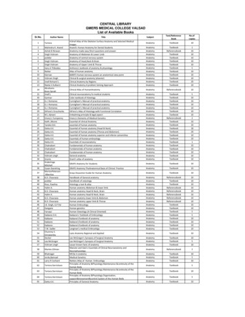
39 poddar Handbook of osteology Anatomy Textbook 10 40 Ross ,Pawlina Histology a text & atlas Anatomy Textbook 10 41 Halim A. Human anatomy Abdomen & lower limb Anatomy Referencebook 10 42 B.D. Chaurasia Human anatomy Head & Neck, Brain Anatomy Referencebook 10 43 Halim A. Human anatomy Head & Neck, Brain Anatomy Referencebook 10
Clinical Anatomy RK Zargar, Sushil Kumar 8. Human Embryology Daksha Dixit 9. Manipal Manual of Anatomy Sampath Madhyastha 10. Exam-Oriented Anatomy Shoukat N Kazi 11. Anatomy and Physiology of Eye AK Khurana, Indu Khurana 12. Surface and Radiological Anatomy A. Halim 13. MCQ in Human Anatomy DK Chopade 14. Exam-Oriented Anatomy for Dental .
HUMAN ANATOMY AND PHYSIOLOGY Anatomy: Anatomy is a branch of science in which deals with the internal organ structure is called Anatomy. The word “Anatomy” comes from the Greek word “ana” meaning “up” and “tome” meaning “a cutting”. Father of Anatomy is referred as “Andreas Vesalius”. Ph
Descriptive anatomy, anatomy limited to the verbal description of the parts of an organism, usually applied only to human anatomy. Gross anatomy/Macroscopic anatomy, anatomy dealing with the study of structures so far as it can be seen with the naked eye. Microscopic
abdomen and pelvis volume 5 8 cbs anatomy 1 25 chaurasia, b.d. bd chaurasia's human anatomy: lower limb abdomen and pelvis volume 6 8 cbs anatomy 1 26 chaurasia, b.d. bd chaurasia's human anatomy: lower limb abdomen and pelvis volume 7 8 cbs anatomy 1 27 chaurasia, b.d. bd chaurasia's human anatomy: lower limb abdomen and pelvis volume 8 8 cbs .
128 B.D.Chaurasia Human Anatomy(Lower Limb Abdomen and Pelvis)vol.II 129 B.D.Chaurasia Human Anatomy(Lower Limb Abdomen and Pelvis)vol.II 130 B.D.Chaurasia Human Anatomy(Lower Limb Abdomen and Pelvis)vol.II 131 B.D.Chaurasia Human Anatomy(Lower Limb Abdomen and Pelvis)vol.II 132 B.D.Chaurasia Human Anatomy (Head and Neck,Brain)vol.III 133 B.D .
marieb human & anatomy & physiology lab manual w/access 9780134776798 ph 13 2002 147.25 biol-213l-21683 human anatomy & physio ii lab cont mann, kj marieb human anatomy & phys lab man main vers (w/out access card) 9780134806358 pearson 12 2019 196.00 147.25 biol-213l-21684 human anatomy & physio ii lab cont roberson, s marieb human anatomy .
Pearson Benjamin Cummings Anatomy and Physiology Integrated Anatomy – Gross anatomy, or macroscopic anatomy, examines large, visible structures Surface anatomy: exterior features Regional anatomy: body areas Systemic anatomy: groups of organs working


